Local cortical pulling-force repression switches centrosomal centration and posterior displacement in C. elegans
- PMID: 18158330
- PMCID: PMC2373484
- DOI: 10.1083/jcb.200706005
Local cortical pulling-force repression switches centrosomal centration and posterior displacement in C. elegans
Abstract
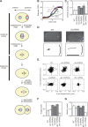
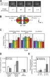
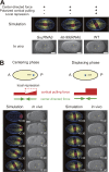
Centrosome positioning is actively regulated by forces acting on microtubules radiating from the centrosomes. Two mechanisms, center-directed and polarized cortical pulling, are major contributors to the successive centering and posteriorly displacing migrations of the centrosomes in single-cell-stage Caenorhabditis elegans. In this study, we analyze the spatial distribution of the forces acting on the centrosomes to examine the mechanism that switches centrosomal migration from centering to displacing. We clarify the spatial distribution of the forces using image processing to measure the micrometer-scale movements of the centrosomes. The changes in distribution show that polarized cortical pulling functions during centering migration. The polarized cortical pulling force directed posteriorly is repressed predominantly in the lateral regions during centering migration and is derepressed during posteriorly displacing migration. Computer simulations show that this local repression of cortical pulling force is sufficient for switching between centering and displacing migration. Local regulation of cortical pulling might be a mechanism conserved for the precise temporal regulation of centrosomal dynamic positioning.
Figures


Similar articles
-
Modeling microtubule-mediated forces and centrosome positioning in Caenorhabditis elegans embryos.Methods Cell Biol. 2010;97:437-53. doi: 10.1016/S0091-679X(10)97023-4. Methods Cell Biol. 2010. PMID: 20719284
-
Computer simulations and image processing reveal length-dependent pulling force as the primary mechanism for C. elegans male pronuclear migration.Dev Cell. 2005 May;8(5):765-75. doi: 10.1016/j.devcel.2005.03.007. Dev Cell. 2005. PMID: 15866166
-
Cell contacts orient some cell division axes in the Caenorhabditis elegans embryo.J Cell Biol. 1995 May;129(4):1071-80. doi: 10.1083/jcb.129.4.1071. J Cell Biol. 1995. PMID: 7744956 Free PMC article.
-
Mechanisms of spindle positioning.J Cell Biol. 2013 Jan 21;200(2):131-40. doi: 10.1083/jcb.201210007. J Cell Biol. 2013. PMID: 23337115 Free PMC article. Review.
-
Physical Limits on the Precision of Mitotic Spindle Positioning by Microtubule Pushing forces: Mechanics of mitotic spindle positioning.Bioessays. 2017 Nov;39(11):10.1002/bies.201700122. doi: 10.1002/bies.201700122. Epub 2017 Sep 28. Bioessays. 2017. PMID: 28960439 Free PMC article. Review.
Cited by
-
Centrosome centering and decentering by microtubule network rearrangement.Mol Biol Cell. 2016 Sep 15;27(18):2833-43. doi: 10.1091/mbc.E16-06-0395. Epub 2016 Jul 20. Mol Biol Cell. 2016. PMID: 27440925 Free PMC article.
-
End-on microtubule-dynein interactions and pulling-based positioning of microtubule organizing centers.Cell Cycle. 2012 Oct 15;11(20):3750-7. doi: 10.4161/cc.21753. Epub 2012 Aug 16. Cell Cycle. 2012. PMID: 22895049 Free PMC article. Review.
-
Choice between 1- and 2-furrow cytokinesis in Caenorhabditis elegans embryos with tripolar spindles.Mol Biol Cell. 2019 Jul 22;30(16):2065-2075. doi: 10.1091/mbc.E19-01-0075. Epub 2019 Feb 20. Mol Biol Cell. 2019. PMID: 30785847 Free PMC article.
-
Dynactin binding to tyrosinated microtubules promotes centrosome centration in C. elegans by enhancing dynein-mediated organelle transport.PLoS Genet. 2017 Jul 31;13(7):e1006941. doi: 10.1371/journal.pgen.1006941. eCollection 2017 Jul. PLoS Genet. 2017. PMID: 28759579 Free PMC article.
-
The extra-embryonic space and the local contour are crucial geometric constraints regulating cell arrangement.Development. 2022 May 1;149(9):dev200401. doi: 10.1242/dev.200401. Epub 2022 May 12. Development. 2022. PMID: 35552395 Free PMC article.
References
-
- Afshar, K., F.S. Willard, K. Colombo, C.A. Johnston, C.R. McCudden, D.P. Siderovski, and P. Gönczy. 2004. RIC-8 is required for GPR-1/2-dependent Galpha function during asymmetric division of C. elegans embryos. Cell. 119:219–230. - PubMed
-
- Albertson, D.G. 1984. Formation of the first cleavage spindle in nematode embryos. Dev. Biol. 101:61–72. - PubMed
-
- Barnard, C., F. Gilbert, and P. McGregor. 2001. Asking Questions in Biology: Key Skills for Practical Assessments and Project Work. Pearson Education, New York. 190 pp.
-
- Bringmann, H., C.R. Cowan, J. Kong, and A.A. Hyman. 2007. LET-99, GOA-1/GPA-16, and GPR-1/2 are required for aster-positioned cytokinesis. Curr. Biol. 17:185–191. - PubMed
Publication types
MeSH terms
LinkOut - more resources
Full Text Sources

