

Therapeutic B cell depletion impairs adaptive and autoreactive CD4+ T cell activation in mice
- PMID: 18093919
- PMCID: PMC2409235
- DOI: 10.1073/pnas.0709205105
Therapeutic B cell depletion impairs adaptive and autoreactive CD4+ T cell activation in mice
Abstract
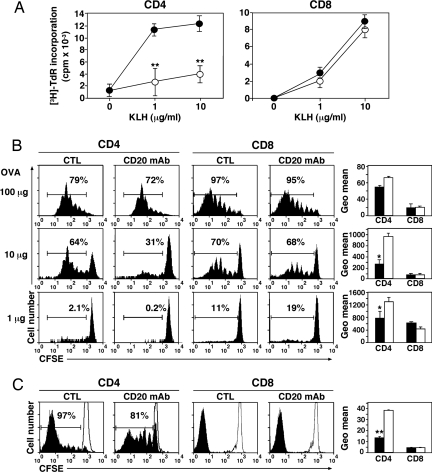
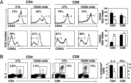
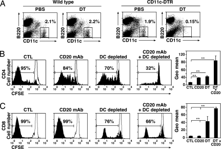
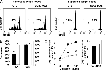
CD20 antibody depletion of B lymphocytes effectively ameliorates multiple T cell-mediated autoimmune diseases through mechanisms that remain unclear. To address this, a mouse CD20 antibody that depletes >95% of mature B cells in mice with otherwise intact immune systems was used to assess the role of B cells in CD4(+) and CD8(+) T cell activation and expansion in vivo. B cell depletion had no direct effect on T cell subsets or the activation status of CD4(+) and CD8(+) T cells in naive mice. However, B cell depletion impaired CD4(+) T cell activation and clonal expansion in response to protein antigens and pathogen challenge, whereas CD8(+) T cell activation was not affected. In vivo dendritic cell ablation, along with CD20 immunotherapy, revealed that optimal antigen-specific CD4(+) T cell priming required both B cells and dendritic cells. Most importantly, B cell depletion inhibited antigen-specific CD4(+) T cell expansion in both collagen-induced arthritis and autoimmune diabetes mouse models. These results provide direct evidence that B cells contribute to T cell activation and expansion in vivo and offer insights into the mechanism of action for B cell depletion therapy in the treatment of autoimmunity.
Conflict of interest statement
Conflict of interest statement: T.F.T. is a paid consultant for MedImmune, Inc. and a consultant and shareholder for Angelica Therapeutics, Inc. J.C.P is a paid consultant for Angelica Therapeutics, Inc.
Figures
Similar articles
-
CD8+ T-cell-dependent immunity following xenogeneic DNA immunization against CD20 in a tumor challenge model of B-cell lymphoma.Clin Cancer Res. 2005 Jan 1;11(1):370-9. Clin Cancer Res. 2005. PMID: 15671568
-
CD4+ T cell-derived IL-2 signals during early priming advances primary CD8+ T cell responses.PLoS One. 2009 Nov 10;4(11):e7766. doi: 10.1371/journal.pone.0007766. PLoS One. 2009. PMID: 19901991 Free PMC article.
-
[B cells as key contributors in determining the level of immune responses -B-cell-targeted therapy in patients with autoimmune diseases].Fukuoka Igaku Zasshi. 2005 Apr;96(4):86-92. Fukuoka Igaku Zasshi. 2005. PMID: 15991605 Review. Japanese.
-
Differential kinetics of antigen dependency of CD4+ and CD8+ T cells.J Immunol. 2014 Apr 15;192(8):3507-17. doi: 10.4049/jimmunol.1302725. Epub 2014 Mar 17. J Immunol. 2014. PMID: 24639353
-
IL-1β strikingly enhances antigen-driven CD4 and CD8 T-cell responses.Cold Spring Harb Symp Quant Biol. 2013;78:117-24. doi: 10.1101/sqb.2013.78.021246. Epub 2013 Oct 3. Cold Spring Harb Symp Quant Biol. 2013. PMID: 24092469 Review.
Cited by
-
Regulatory B cell (B10 Cell) expansion during Listeria infection governs innate and cellular immune responses in mice.J Immunol. 2013 Feb 1;190(3):1158-68. doi: 10.4049/jimmunol.1201427. Epub 2012 Dec 28. J Immunol. 2013. PMID: 23275601 Free PMC article.
-
Regulatory B cells (B10 cells) and regulatory T cells have independent roles in controlling experimental autoimmune encephalomyelitis initiation and late-phase immunopathogenesis.J Immunol. 2010 Aug 15;185(4):2240-52. doi: 10.4049/jimmunol.1001307. Epub 2010 Jul 12. J Immunol. 2010. PMID: 20624940 Free PMC article.
-
B-cell therapy in lupus nephritis: an overview.Nephrol Dial Transplant. 2019 Jan 1;34(1):22-29. doi: 10.1093/ndt/gfy267. Nephrol Dial Transplant. 2019. PMID: 30165690 Free PMC article. Review.
-
Humoral immunity in hepatitis B virus infection: Rehabilitating the B in HBV.JHEP Rep. 2021 Nov 19;4(2):100398. doi: 10.1016/j.jhepr.2021.100398. eCollection 2022 Feb. JHEP Rep. 2021. PMID: 35059620 Free PMC article. Review.
-
Insights into the heterogeneity of human B cells: diverse functions, roles in autoimmunity, and use as therapeutic targets.Immunol Res. 2009 Dec;45(2-3):144-58. doi: 10.1007/s12026-009-8096-7. Epub 2009 Apr 7. Immunol Res. 2009. PMID: 19350211 Free PMC article. Review.
References
-
- Ermann J, Fathman CG. Nat Immunol. 2001;2:759–761. - PubMed
-
- Edwards JCW, Cambridge G. Rheumatology. 2001;40:205–211. - PubMed
-
- Stasi R, Del Poeta G, Stipa E, Evangelista ML, Trawinska MM, Cooper N, Amadori S. Blood. 2007;110:2924–2930. - PubMed
-
- Edwards JCW, Cambridge G. Rheumatology. 2005;44:151–156. - PubMed
-
- Lipsky PE. Nat Immunol. 2001;2:764–766. - PubMed
Publication types
MeSH terms
Substances
Grants and funding
LinkOut - more resources
Full Text Sources
Other Literature Sources
Research Materials
