. 2007 Dec 17;179(6):1123-31.
doi: 10.1083/jcb.200703166.

Dimeric PKD regulates membrane fission to form transport carriers at the TGN

Affiliations

Dimeric PKD regulates membrane fission to form transport carriers at the TGN

Carine Bossard et al. J Cell Biol. .

Abstract

Protein kinase D (PKD) is recruited to the trans-Golgi network (TGN) through interaction with diacylglycerol (DAG) and is required for the biogenesis of TGN to cell surface transport carriers. We now provide definitive evidence that PKD has a function in membrane fission. PKD depletion by siRNA inhibits trafficking from the TGN, whereas expression of a constitutively active PKD converts TGN into small vesicles. These findings demonstrate that PKD regulates membrane fission and this activity is used to control the size of transport carriers, and to prevent uncontrolled vesiculation of TGN during protein transport.


Figures

Figure 1.
Figure 1.
Relative expression of PKD isoforms in HeLa cells and their depletion by siRNA. (A) Analysis of mRNA expression by RT-PCR shows that PKD2 and PKD3 are the only PKD isoforms expressed in HeLa cells. RT-PCR reaction without the reverse transcriptase (RT−) was used as a negative control and PCR with the corresponding PKD cDNA (C) as a positive control. (B) Quantitative real-time RT-PCR analysis was performed on RNA extracted from HeLa cells. Bars represent the mean (±SD) of the relative mRNA expression of each PKD isoform compared with the average expression of β-actin. *, P < 0.01 compared with PKD1. (C) PKD2 and PKD3 protein levels in HeLa cells transfected with the indicated siRNA were detected by immunoblot analysis after immunoprecipitation from 100 μg of cell lysate using anti-PKD2 and anti-PKD3 antibodies, respectively. (D) β-Actin expression in the lysates used for immunoprecipitation was monitored as a loading control. (E) The effect of PKD2 and PKD3 siRNA was quantified by densitometry and normalized to the expression of PKD2 and PKD3, respectively, in cells transfected with control siRNA.
Figure 2.
Figure 2.
Depletion of PKD2 or PKD3 inhibits secretion of ss-HRP. (A and B) ss-HRP and PLAP cDNAs were cotransfected in HeLa cells depleted or not of PKD2 or PKD3. 20 h after transfection, HRP activity secreted in the medium was measured by chemiluminescence. PLAP activity in cell lysates and secreted into the medium was measured as described in Materials and methods. Bars represent the mean ± SD of HRP (A) or PLAP (B) activity in the medium normalized by PLAP activity in cell lysates/total protein concentration. (C) Depletion of PKD2 or PKD3 inhibits VSV-G transport. tso45VSV-G-GFP plasmid was transfected in HeLa cells depleted or not of PKD2 or PKD3. The levels of VSV-G at the cells surface and inside the cells were determined by FACS analysis. The bars represent the relative ratio of VSV-G at the surface to the total expressed in depleted cells compared with siRNA control transfected cells ± SEM. *, P < 0.01 compared with the control. (D–F) PKD2- and PKD3-depleted HeLa cells and control cells were transfected with ss-HRP and the organization of the Golgi membranes and the localization of HRP monitored by electron microscopy. In control cells (D), the arrow indicates a single tubular profile and arrowheads indicate round profiles (presumably vesicles) in the trans-Golgi region. In contrast, in PKD2- and PKD3-depleted cells (E and F), HRP accumulates in tubular membranes. The arrows indicate multiple tubular profiles at the TGN. The open arrowheads show pearling tubules. The arrowhead in F is a clathrin-coated vesicle revealing the trans side of the Golgi stack.
Figure 3.
Figure 3.
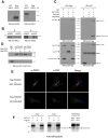
PKD2 and PKD3 dimerize and transphosphorylate. (A) Specificity of PKD2 and PKD3 antibodies. Pure recombinant Flag-PKD2 and GST-PKD3 were Western blotted with anti-PKD2 and -PKD3 antibodies. Anti-PKD2 antibody recognizes PKD2 (lane 2) and anti-PKD3 antibody recognize PKD3 (lane 3). (B) PKD2 and PKD3 interact. PKD2 or PKD3 was immunoprecipitated from HeLa cell lysates with specific antibodies and the precipitates blotted with anti-PKD2 antibody (lanes 1 and 2) or PKD3 antibody (lanes 3 and 4). (C) Exogenously expressed PKD2 and PKD3 interact. GST (lane 1) or GST-PKD2 (lanes 2 and 3) was coexpressed with Flag-HRP (lane 2) or FLAG-PKD3 (lanes 1 and 3) in HeLa cells. The cells were immunoprecipitated with anti-Flag antibody (top left) or anti-GST antibody (top right) and Western blotted with anti-GST or anti-Flag antibody, respectively. To verify that each tagged protein was expressed and immunoprecipitated, the Flag and the GST precipitates were respectively blotted with anti-Flag (bottom left) and anti-GST (bottom right). (D) PKD2 and PKD3 interact directly. Pure recombinant GST-tagged proteins were incubated in vitro with pure recombinant Flag-tagged proteins. After GST pull-down, the precipitates were Western blotted with anti-Flag antibody, followed by anti-GST antibody. (E) PKD2 and PKD3 colocalize on PKD-KD tubes. HeLa cells were cotransfected with Flag-PKD2-KD and GST-PKD3-WT (top) or with Flag-PKD2-WT and GST-PKD3-KD (bottom). The cells were visualized by fluorescence microscopy with anti-PKD2 and anti-GST antibody. (F) PKD2 and PKD3 transphosphorylate. Pure recombinant GST-PKD2-WT was incubated alone (lane 1) or mixed with either Flag-PKD2-KD (lane 3) or Flag-PKD3-KD (lane 7) for in vitro kinase assays. Similarly, GST-PKD3-WT was incubated alone (lane 4) or mixed with Flag-PKD3-KD (lane 6) or Flag-PKD2-KD (lane 8). The lack of kinase activity of Flag-PKD2-KD and Flag-PKD3-KD is shown in lanes 2 and 5, respectively.
Figure 4.
Figure 4.
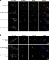
Expression of an overactivated form of PKD fragments the Golgi apparatus. (A) HeLa cells stably expressing MannII-GFP were transfected with GST-tagged PKD WT, PKD-CAAX-WT, PKD-CAAX-CA, or PKD-CAAX-KD. The localization of PKD and the organization of Golgi membranes were monitored by fluorescence microscopy with anti-GST and TGN46 antibody, respectively. (B) PKD-CAAX-CA–dependent Golgi fragmentation requires DAG. HeLa MannII-GFP cells were pretreated with fumonisin1 (FB1) 24 h before transfection with PKD constructs. 24 h after transfection, cells were fixed and stained with anti-GST and anti-TGN46 antibodies.
Figure 5.
Figure 5.
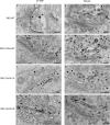
Analysis of HeLa cells transfected with PKD CAAX isoforms by electron microscopy. HeLa cells transfected with PKD-WT, PKD-CAAX-WT, PKD-CAAX-CA, or PKD-CAAX-KD alone (right) or with ST-HRP (left) were processed for electron microscopy. (A) Gold particles indicating the presence of PKD-WT reveal its presence at the TGN (where ST-HRP is also visible). Few vesicles lacking ST-HRP were also visible in sections (arrow). (B) Expression of PKD-CAAX-WT increased the number of ST-HRP–positive vesicles (arrows). (C) PKD-CAAX-CA induced an increase in the number of vesicles with ST-HRP (arrowheads); however, number of unlabeled vesicles also increases (arrows). Row of vesicles (empty arrows) between ST-positive cisterna and the rest of the stack may represent cisterna consumed by vesicles, and this also might explain why ST-positive cisterna appears to be peeling off. (D) Cells expressing PKD-CAAX-KD exhibit regular trans-Golgi cisterna (arrow) labeled with ST-HRP. Some tubular structures (presumably TGN) are also visible at the trans-face of the Golgi (arrowhead). (E) Cells expressing PKD-WT exhibit regular TGN with both tubular (arrow) and vesicles (arrowhead). PKD is detected at the TGN46-positive membranes. (F) PKD-CAAX-WT induced an increase in the number of TGN46-positive vesicles (arrows). (G) Expression of PKD-CAAX-CA induced extensive vesiculation of the Golgi apparatus. Both TGN46-positive (arrows) and TGN46-negative (arrowheads) vesicles increased in numbers under these conditions. PKD is detected at the TGN46-positive membranes. (H) Long TGN46-positive (arrows) tubular structures were detected in PKD-CAAX-KD–expressing cells without any obvious increase in number of vesicles.

Similar articles

Cited by

References

    1. Audhya, A., M. Foti, and S.D. Emr. 2000. Distinct roles for the yeast phosphatidylinositol 4-kinases, Stt4p and Pik1p, in secretion, cell growth, and organelle membrane dynamics. Mol. Biol. Cell. 11:2673–2689. - PMC - PubMed
    1. Bard, F., and V. Malhotra. 2006. The formation of TGN-to-plasma-membrane transport carriers. Annu. Rev. Cell Dev. Biol. 22:439–455. - PubMed
    1. Bard, F., L. Casano, A. Mallabiabarrena, E. Wallace, K. Saito, H. Kitayama, G. Guizzunti, Y. Hu, F. Wendler, R. Dasgupta, et al. 2006. Functional genomics reveals genes involved in protein secretion and Golgi organization. Nature. 439:604–607. - PubMed
    1. Baron, C.L., and V. Malhotra. 2002. Role of diacylglycerol in PKD recruitment to the TGN and protein transport to the plasma membrane. Science. 295:325–328. - PubMed
    1. Choy, E., V.K. Chiu, J. Silletti, M. Feoktistov, T. Morimoto, D. Michaelson, I.E. Ivanov, and M.R. Philips. 1999. Endomembrane trafficking of ras: the CAAX motif targets proteins to the ER and Golgi. Cell. 98:69–80. - PubMed

Publication types

MeSH terms

LinkOut - more resources