Assembly, organization, and function of the COPII coat
- PMID: 18060556
- PMCID: PMC2228377
- DOI: 10.1007/s00418-007-0363-x
Assembly, organization, and function of the COPII coat
Abstract
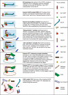
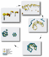
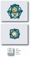
A full mechanistic understanding of how secretory cargo proteins are exported from the endoplasmic reticulum for passage through the early secretory pathway is essential for us to comprehend how cells are organized, maintain compartment identity, as well as how they selectively secrete proteins and other macromolecules to the extracellular space. This process depends on the function of a multi-subunit complex, the COPII coat. Here we describe progress towards a full mechanistic understanding of COPII coat function, including the latest findings in this area. Much of our understanding of how COPII functions and is regulated comes from studies of yeast genetics, biochemical reconstitution and single cell microscopy. New developments arising from clinical cases and model organism biology and genetics enable us to gain far greater insight in to the role of membrane traffic in the context of a whole organism as well as during embryogenesis and development. A significant outcome of such a full understanding is to reveal how the machinery and processes of membrane trafficking through the early secretory pathway fail in disease states.
Figures


Similar articles
-
ER-to-Golgi transport: COP I and COP II function (Review).Mol Membr Biol. 2003 Jul-Sep;20(3):197-207. doi: 10.1080/0968768031000122548. Mol Membr Biol. 2003. PMID: 12893528 Review.
-
Traffic COPs of the early secretory pathway.Traffic. 2000 May;1(5):371-7. doi: 10.1034/j.1600-0854.2000.010501.x. Traffic. 2000. PMID: 11208122 Review.
-
Vesicle-mediated export from the ER: COPII coat function and regulation.Biochim Biophys Acta. 2013 Nov;1833(11):2464-72. doi: 10.1016/j.bbamcr.2013.02.003. Epub 2013 Feb 15. Biochim Biophys Acta. 2013. PMID: 23419775 Free PMC article. Review.
-
Auxilin facilitates membrane traffic in the early secretory pathway.Mol Biol Cell. 2016 Jan 1;27(1):127-36. doi: 10.1091/mbc.E15-09-0631. Epub 2015 Nov 4. Mol Biol Cell. 2016. PMID: 26538028 Free PMC article.
-
The highly conserved COPII coat complex sorts cargo from the endoplasmic reticulum and targets it to the golgi.Cold Spring Harb Perspect Biol. 2013 Feb 1;5(2):a013367. doi: 10.1101/cshperspect.a013367. Cold Spring Harb Perspect Biol. 2013. PMID: 23378591 Free PMC article. Review.
Cited by
-
A cataract-causing connexin 50 mutant is mislocalized to the ER due to loss of the fourth transmembrane domain and cytoplasmic domain.FEBS Open Bio. 2012 Nov 27;3:22-9. doi: 10.1016/j.fob.2012.11.005. Print 2013. FEBS Open Bio. 2012. PMID: 23772370 Free PMC article.
-
Penta-EF-Hand Protein Peflin Is a Negative Regulator of ER-To-Golgi Transport.PLoS One. 2016 Jun 8;11(6):e0157227. doi: 10.1371/journal.pone.0157227. eCollection 2016. PLoS One. 2016. PMID: 27276012 Free PMC article.
-
The essential neutral sphingomyelinase is involved in the trafficking of the variant surface glycoprotein in the bloodstream form of Trypanosoma brucei.Mol Microbiol. 2010 Jun;76(6):1461-82. doi: 10.1111/j.1365-2958.2010.07151.x. Epub 2010 Apr 1. Mol Microbiol. 2010. PMID: 20398210 Free PMC article.
-
The Golgi Apparatus and its Next-Door Neighbors.Front Cell Dev Biol. 2022 Apr 28;10:884360. doi: 10.3389/fcell.2022.884360. eCollection 2022. Front Cell Dev Biol. 2022. PMID: 35573670 Free PMC article. Review.
-
Organisation of human ER-exit sites: requirements for the localisation of Sec16 to transitional ER.J Cell Sci. 2009 Aug 15;122(Pt 16):2924-34. doi: 10.1242/jcs.044032. Epub 2009 Jul 28. J Cell Sci. 2009. PMID: 19638414 Free PMC article.
References
-
- {'text': '', 'ref_index': 1, 'ids': [{'type': 'PubMed', 'value': '10903204', 'is_inner': True, 'url': 'https://pubmed.ncbi.nlm.nih.gov/10903204/'}]}
- Allan BB, Moyer BD, Balch WE (2000) Rab1 recruitment of p115 into a cis-SNARE complex: programming budding COPII vesicles for fusion. Science 289:444–448 - PubMed
-
- {'text': '', 'ref_index': 1, 'ids': [{'type': 'PMC', 'value': 'PMC2168100', 'is_inner': False, 'url': 'https://pmc.ncbi.nlm.nih.gov/articles/PMC2168100/'}, {'type': 'PubMed', 'value': '10601335', 'is_inner': True, 'url': 'https://pubmed.ncbi.nlm.nih.gov/10601335/'}]}
- Alvarez C, Fujita H, Hubbard A, Sztul E (1999) ER to Golgi transport: requirement for p115 at a pre-Golgi VTC stage. J Cell Biol 147:1205–1222 - PMC - PubMed
-
- {'text': '', 'ref_index': 1, 'ids': [{'type': 'PMC', 'value': 'PMC165101', 'is_inner': False, 'url': 'https://pmc.ncbi.nlm.nih.gov/articles/PMC165101/'}, {'type': 'PubMed', 'value': '12802079', 'is_inner': True, 'url': 'https://pubmed.ncbi.nlm.nih.gov/12802079/'}]}
- Alvarez C, Garcia-Mata R, Brandon E, Sztul E (2003) COPI Recruitment is modulated by a Rab1b-dependent mechanism. Mol Biol Cell 14:2116–2127 - PMC - PubMed
-
- {'text': '', 'ref_index': 1, 'ids': [{'type': 'PubMed', 'value': '11389436', 'is_inner': True, 'url': 'https://pubmed.ncbi.nlm.nih.gov/11389436/'}]}
- Antonny B, Madden D, Hamamoto S, Orci L, Schekman R (2001) Dynamics of the COPII coat with GTP and stable analogues. Nat Cell Biol 3:531–537 - PubMed
-
- {'text': '', 'ref_index': 1, 'ids': [{'type': 'PMC', 'value': 'PMC1319167', 'is_inner': False, 'url': 'https://pmc.ncbi.nlm.nih.gov/articles/PMC1319167/'}, {'type': 'PubMed', 'value': '12671686', 'is_inner': True, 'url': 'https://pubmed.ncbi.nlm.nih.gov/12671686/'}]}
- Antonny B, Gounon P, Schekman R, Orci L (2003) Self-assembly of minimal COPII cages. EMBO Rep 4:419–424 - PMC - PubMed
Publication types
MeSH terms
Substances
Grants and funding
LinkOut - more resources
Full Text Sources
Other Literature Sources
Medical
Molecular Biology Databases

