Review
. 2008 Apr;29(2):155-92.
doi: 10.1210/er.2007-0014. Epub 2007 Dec 5.

Receptor activator of nuclear factor kappaB ligand and osteoprotegerin regulation of bone remodeling in health and disease

Affiliations
Review

Receptor activator of nuclear factor kappaB ligand and osteoprotegerin regulation of bone remodeling in health and disease

Ann E Kearns et al. Endocr Rev. 2008 Apr.

Abstract

Osteoclasts and osteoblasts dictate skeletal mass, structure, and strength via their respective roles in resorbing and forming bone. Bone remodeling is a spatially coordinated lifelong process whereby old bone is removed by osteoclasts and replaced by bone-forming osteoblasts. The refilling of resorption cavities is incomplete in many pathological states, which leads to a net loss of bone mass with each remodeling cycle. Postmenopausal osteoporosis and other conditions are associated with an increased rate of bone remodeling, which leads to accelerated bone loss and increased risk of fracture. Bone resorption is dependent on a cytokine known as RANKL (receptor activator of nuclear factor kappaB ligand), a TNF family member that is essential for osteoclast formation, activity, and survival in normal and pathological states of bone remodeling. The catabolic effects of RANKL are prevented by osteoprotegerin (OPG), a TNF receptor family member that binds RANKL and thereby prevents activation of its single cognate receptor called RANK. Osteoclast activity is likely to depend, at least in part, on the relative balance of RANKL and OPG. Studies in numerous animal models of bone disease show that RANKL inhibition leads to marked suppression of bone resorption and increases in cortical and cancellous bone volume, density, and strength. RANKL inhibitors also prevent focal bone loss that occurs in animal models of rheumatoid arthritis and bone metastasis. Clinical trials are exploring the effects of denosumab, a fully human anti-RANKL antibody, on bone loss in patients with osteoporosis, bone metastasis, myeloma, and rheumatoid arthritis.


Figures

Figure 1
Figure 1
Mechanisms of action for OPG, RANKL, and RANK. RANKL is produced by osteoblasts, bone marrow stromal cells, and other cells under the control of various proresorptive growth factors, hormones, and cytokines. Osteoblasts and stromal cells produce OPG, which binds to and thereby inactivates RANKL. The major binding complex is likely to be a single OPG homodimer interacting with high affinity with a single RANKL homotrimer (17). In the absence of OPG, RANKL activates its receptor RANK, found on osteoclasts and preosteoclast precursors. RANK-RANKL interactions lead to preosteoclast recruitment, fusion into multinucleated osteoclasts, osteoclast activation, and osteoclast survival. Each of these RANK-mediated responses can be fully inhibited by OPG. [Adapted from Women’s Health 2:517–525, 2006 (266), with permission of Future Medicine Ltd.]
Figure 2
Figure 2
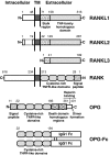
Protein structures of RANKL, RANK, OPG, and OPG-Fc. Three isoforms of RANKL are described (44), each of which possesses similar C-terminal TNF-homology domains that are required for RANK activation (39). RANKL1 and RANKL2 isoforms possess a transmembrane (TM) domain and a stalk region that contains a proteolytic site that allows for cleavage of RANKL from cell surfaces (443). RANKL3 contains a truncated stalk region that possesses a proteolytic cleavage site, the function of which is unclear because this isoform lacks a transmembrane domain and is therefore secreted in the absence of proteolysis. RANK is a transmembrane protein with a large C-terminal cyotoplasmic domain and an amino-terminal extracellular domain. Both RANK and OPG contain amino-terminal signal peptides as well as cysteine-rich TNF receptor-like domains that bind to RANKL. OPG lacks a transmembrane domain, consistent with its secretion as a soluble protein. OPG also includes two DDH regions, the roles of which are unknown. A heparin binding domain at the carboxy-terminus of OPG limits the half-life and distribution of the molecule, and this domain is also involved in dimer formation. Human OPG-Fc is a recombinant fusion protein that has been used in human clinical trials and in the majority of preclinical animal studies. This construct includes amino acids 22–194 of native OPG, comprising the minimal TNF receptor-like domains that mediate RANKL inhibition. This fragment lacks the signal peptide, DDH regions, and heparin binding domain of native OPG. The Fc fragment of human IgG1 was fused to the carboxy-terminus of this 22–194 fragment to maintain a dimeric molecule with a sustained circulating half-life. Numbers in figure represent amino acids. [Reproduced from S. Theoleyre et al.: Cytokine Growth Factor Rev 15:457–475, 2004 (15) with permission from Elsevier.]
Figure 3
Figure 3
Essential signaling pathways activated by RANKL interactions with RANK. RANKL, which is produced by osteoblasts, stromal cells, T cells, and other sources, activates RANK on the surface of osteoclasts and osteoclast precursors. RANK activation leads to the recruitment of the adaptor protein TRAF 6, leading to NF-κB activation and translocation of NF-κB to the nucleus. NF-κB increases c-Fos expression and c-Fos interacts with NFATc1 to trigger the transcription of osteoclastogenic genes. Activation of these pathways is prevented naturally by OPG, which prevents RANKL from activating RANK in the extracellular environment. RANKL-RANK interactions are also prevented by RANK-Fc (a truncated form of RANK that acts as a nonsignaling decoy receptor) and by denosumab, a fully human monoclonal antibody that binds and inhibits RANKL. [Adapted with permission from B. F. Boyce and L. Xing: Arthritis Res Ther 9:S1–S7, 2007 (66).]
Figure 4
Figure 4
PTH regulation of OPG and RANKL mRNA in parathyroidectomized (Px) rats. RNA was isolated from the distal femur of Px rats immediated after a 6-h infusion with vehicle or with different doses of human PTH 1-38. Bone mRNA was assessed by Northern blot analysis for expression of OPG and RANKL, the levels of which were standardized to the housekeeping gene GAPDH. This graphic presentation of Northern blot results depicts the relative values of OPG and RANKL mRNA at different dose levels of PTH. The highest levels of OPG mRNA were seen at the lowest levels of PTH infusion and decreased with increasing levels of PTH. In contrast, RANKL mRNA was lowest in the absence of PTH infusion and increased in a dose-dependent manner with increasing levels of PTH. These results illustrate the reciprocal regulation of OPG and RANKL in a manner that would amplify the proresorptive stimulus of PTH infusion. [Reproduced from Y. L. Ma et al.: Endocrinology 142:4047–4054, 2001 (88) with permission from The Endocrine Society.]
Figure 5
Figure 5
Regions of analysis (left panel) and representative microCT images (middle and right panels) of trabecular regions from the lumbar vertebra (bottom) and distal femur (top) of mice treated for 3 wk with either human OPG-Fc (5 mg/kg, 2/wk) or vehicle. These samples represented the median value for trabecular bone volume for the lumbar vertebra (top panels) and the distal femur (bottom panels) from each group (n = 8 per group).
Figure 6
Figure 6
Effects of RANKL inhibition via OPG on prednisolone-induced changes in bone. Slow-release prednisolone (Pred, 1 or 2 mg/kg·d) or placebo pellets (Plac) were implanted sc in normal young male rats for 45 d, during which time animals were treated with either vehicle (saline) or OPG-Fc (1 mg/kg, 3 times/wk by sc injection). A, BMD was measured by DXA on d 45 at the distal femur and proximal tibia. In vehicle-treated rats, both doses of prednisolone led to significant decreases in BMD compared with vehicle-treated rats with placebo pellets. OPG significantly increased BMD in animals treated with either dose of predisolone. B, Osteoclast surface was measured in histological sections of the proximal tibia and expressed as percentage of cancellous bone surface. High-dose prednisolone (2 mg/kg) led to a significant increase in osteoclast surface compared with Plac + vehicle (Veh) controls, and OPG treatment significantly reduced osteoclast surface in animals treated with either dose of prednisolone. Data represent means ± sd, n = 6–7 per group. *, Significantly different from Plac + Veh; ∧, significantly different from respective Pred + Veh control; P < 0.05 by ANOVA.
Figure 7
Figure 7
Theoretical mechanisms by which cancer cells might promote bone resorption by regulation of OPG and/or RANKL. An annotated description is provided in Section III.G. Tumor-mediated increases in RANKL or decreases in OPG would tend to favor bone resorption.
Figure 8
Figure 8
Radiographic and histological assessment of bone metastases in mice treated with OPG. Human MDA-231 breast cancer cells were injected into the left ventricle of female Balb/c nu/nu mice. After 3 wk, mice were x-rayed to assess radiographic osteolysis and allocated to three groups with similar levels of bone destruction. One group was killed at wk 3 as a baseline control (Wk 3 Veh). Other groups were treated with either vehicle (Veh, saline) or with OPG-Fc (3 mg/kg) every second day for 7 d, then x-rayed and killed (Wk 4). Tibiae and femurs were analyzed by histomorphometry for skeletal tumor burden and tumor-associated osteoclasts per tumor area. A, Significant radiographic damage was apparent at wk 3 in vehicle controls, and by wk 4 the total radiographic lesions area increased by 3-fold in vehicle-treated mice. No radiographic progression was observed in mice treated during wk 4 with OPG. B, Histomorphometric assessment at wk 3 indicated the presence of metastases in femurs and tibiae, and by wk 4 there was a 3-fold increase in skeletal tumor burden (% tumor area) in vehicle-treated mice. OPG treatment during wk 4 was associated with no significant change in tumor burden relative to wk 3 controls. C, OPG treatment during wk 4 was associated with a 90% decrease in osteoclast numbers per square millimeter of tumor compared with either vehicle control group. Data represent means ± sd, n = 13–15 per group. *, Significantly different from Wk 3 Veh group; ∧, significantly different from Wk 4 Veh group; P < 0.05 by ANOVA.

Similar articles

Cited by

References

    1. Compston JE, Vedi S, Kaptoge S, Seeman E 2007 Bone remodeling rate and remodeling balance are not co-regulated in adulthood: implications for the use of activation frequency as an index of remodeling rate. J Bone Miner Res 22:1031–1036 - PubMed
    1. Rodan GA, Martin TJ 2000 Therapeutic approaches to bone disease. Science 289:1508–1514 - PubMed
    1. Eriksen EF 1986 Normal and pathological remodeling of human trabecular bone: three dimensional reconstruction of the remodeling sequence in normals and in metabolic bone disease. Endocr Rev 7:379–408 - PubMed
    1. Simonet WS, Lacey DL, Dunstan C, Kelly M, Chang MS, Luthy R, Nhuyen HQ, Wooden S, Bennett L, Boone T, Shimamoto G, DeRose M, Elliot R, Colombero A, Tan HL, Trail G, Sullivan J, Davy E, Bucay N, Renshaw-Gegg L, Hughes TM, Hill D, Pattison W, Campbell P, Sander S, Van G, Tarpley J, Derby P, Lee R, Boyle WJ 1997 Osteoprotegerin: a novel secreted protein involved in the regulation of bone density. Cell 89:309–319 - PubMed
    1. Tsuda E, Goto M, Mochizuki SI, Yano K, Kobayashi F, Morinaga T, Higashio K 1997 Isolation of a novel cytokine from human fibroblasts that specifically inhibits osteoclastogenesis. Biochem Biophys Res Comm 234:137–142 - PubMed

Publication types