. 2008 Feb;19(2):633-45.
doi: 10.1091/mbc.e06-01-0088. Epub 2007 Nov 28.

Paxillin phosphorylation controls invadopodia/podosomes spatiotemporal organization

Affiliations

Paxillin phosphorylation controls invadopodia/podosomes spatiotemporal organization

Cédric Badowski et al. Mol Biol Cell. 2008 Feb.

Abstract

In Rous sarcoma virus (RSV)-transformed baby hamster kidney (BHK) cells, invadopodia can self-organize into rings and belts, similarly to podosome distribution during osteoclast differentiation. The composition of individual invadopodia is spatiotemporally regulated and depends on invadopodia localization along the ring section: the actin core assembly precedes the recruitment of surrounding integrins and integrin-linked proteins, whereas the loss of the actin core was a prerequisite to invadopodia disassembly. We have shown that invadopodia ring expansion is controlled by paxillin phosphorylations on tyrosine 31 and 118, which allows invadopodia disassembly. In BHK-RSV cells, ectopic expression of the paxillin mutant Y31F-Y118F induces a delay in invadopodia disassembly and impairs their self-organization. A similar mechanism is unraveled in osteoclasts by using paxillin knockdown. Lack of paxillin phosphorylation, calpain or extracellular signal-regulated kinase inhibition, resulted in similar phenotype, suggesting that these proteins belong to the same regulatory pathways. Indeed, we have shown that paxillin phosphorylation promotes Erk activation that in turn activates calpain. Finally, we observed that invadopodia/podosomes ring expansion is required for efficient extracellular matrix degradation both in BHK-RSV cells and primary osteoclasts, and for transmigration through a cell monolayer.


Figures
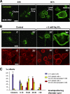
Figure 1.
Figure 1.
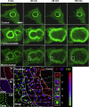
Dynamics of invadopodia in src-transformed BHK cells. (A) BHK-RSV cells were plated on glass coverslips for 24 or 48 h at 37°C, and then they were immunostained with an anti-cortactin mAb. Bar, 20 μm. (B) BHK-RSV cells plated for 24 h (control; 0 min) were treated either with the orthovanadate vehicle (H2O) (control; 30 min) or with 5 mM orthovanadate for 30 min (+Na3VO4; 30 min) or 90 min (+Na3VO4; 90 min) before immunostaining for cortactin or actin. Bar, 20 μm. (C) Size distribution of invadopodia rings in BHK-RSV cells plated for 24 h without orthovanadate (blue line) and 24-h–plated cells and treated with 5 mM orthovanadate during 30 min (yellow line) or 90 min (red line), respectively (n = 60 cells for each condition).
Figure 2.
Figure 2.
Orthovanadate-induced invadopodia ring expansion results from the coordinated processes of invadopodia assembly at the periphery and disassembly at the ring center. BHK-RSV cells plated for 24 h were transiently transfected with cDNA encoding cortactin-GFP. The expressed fusion protein was followed by time-lapse videomicroscopy for 130 min at room temperature, in the absence (control) or in the presence of 5 mM orthovanadate (+Na3VO4) by using a confocal microscope. Bar, 10 μm. Image deconvolution and zoom, using MetaMorph software revealed the individual invadopodia within the invadopodia rings. Merge of the images in false colors from 0 min (red), 80 min (blue), and 130 min (green) allowed following each invadopodia along the time course of the experiment. According to the color pattern, five regions could be defined from the center to the periphery of the ring that reflected both the spatiotemporal organization of invadopodia within the ring and the relative immobility of the individual invadopodia.
Figure 3.
Figure 3.
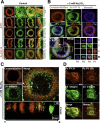
Endogenous paxillin is phosphorylated on tyrosine 31 and 118 and accumulates at the inner rim of the BHK-RSV invadopodia ring. (A) BHK-RSV cells were plated for 24 h on glass coverslips, and then they were stained for actin with phalloidin-TRITC (a) and the general phospho-tyrosine antibody 4G10 (b). Accumulation of tyrosine-phosphorylated proteins was observed at the ring center (merged images, c; zoom, d). Staining with a paxillin mAb revealed that endogenous paxillin (f) also accumulated at the center of the actin ring (e) (merge images g; zoom, h) together with phosphorylated paxillin on tyrosine 118 (PY118; j) and on tyrosine 31 (PY31; n) (i and m are actin staining). Changing the secondary antibody labeling did not modify the respective localization of paxillin (q) at the center of the ring and cortactin (r) at the periphery (merge images, s; zoom, t). Bar, 6 μm. (B) Cortactin-DsRed–transfected BHK-RSV cells were plated for 24 h on glass coverslips, and then they were treated with 5 mM Na3VO4 for 30 min. Invadopodia cores were specifically visualized by cortactin-DsRed (red). Endogenous phospho-paxillin and vinculin localizations were detected by specific monoclonal antibodies. Bar, 10 μm. Bottom, triple merged images. Zoom on four invadopodia (P1–P4) localized from the periphery (P1) to the ring center (P4) is presented. V, P, C, and M are vinculin, PY118 paxillin, cortactin-DsRed, and merge staining, respectively. Each invadopodia showed a specific composition. (C) BHK-RSV cells were transiently transfected with cortactin-DsRed, plated for 24 h, and treated subsequently with 5 mM Na3VO4 for 30 min before staining with a monoclonal anti-β3 integrin antibody. Z scanning along the white arrow (A→B) reveals an increase in the length of invadopodia core along a cross section of the ring and β3 integrins recruitment in invadopodia located at the inner rim of the ring. Bar, 10 μm. (D) BHK-RSV cells plated for 24 h were stained with anti-β3 integrin antibody and phospho-specific polyclonal PY31 or PY118 paxillin antibodies. Bar, 10 μm.
Figure 4.
Figure 4.
The inner rim of the invadopodia ring is a site of close contact with the extracellular matrix. BHK-RSV cells plated for 24 h were treated with 5 mM orthovanadate for 30 min and stained for GFP-paxillin and cortactin-DsRed. Fluorescence was visualized either by TIRF or by classical epi-illumination. For easier comparisons, the whole image was reconstituted with false colors. The ring displayed three regions from the periphery to the center: cortactin not in close contact with the matrix (gray), cortactin in contact with the matrix (red), and paxillin (blue) always in contact with the matrix.
Figure 5.
Figure 5.
Mutant YF GFP-paxillin modifies invadopodia organization. (A) Decrease in the number of invadopodia rings of YF GFP-paxillin cells compared with WT GFP-paxillin cells under control conditions (n = 101 cells; p = 0.021). (B) BHK-RSV cells were double transfected with cortactin-DsRed and WT GFP-paxillin or YF GFP-paxillin, and treated or not with 5 mM orthovanadate as described previously. Evolution of invadopodia rings was followed by time-lapse videomicroscopy (only the red channel is shown). (C) Quantification of the relative thickness of the rings (internal diameter divided by external diameter) in the presence of orthovanadate, indicating a significant modification in invadopodia rings width (data from three independent experiments; n = 54; p value between control and WT = 0.5; p value between WT and YF, <0.001).
Figure 6.
Figure 6.
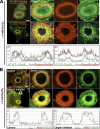
YF GFP-paxillin overexpression, or calpain inhibitor treatment, impairs invadopodia core disassembly. (A) BHK-RSV cells were double transfected with cortactin-DsRed and WT GFP-paxillin or YF GFP-paxillin and treated with 5 mM orthovanadate for 30 min. Then, cells were fixed with 3% paraformaldehyde. With WT GFP-paxillin cells, WT GFP-paxillin accumulated at the center of the rosette at sites where cortactin disassembled as shown on fluorescence intensity scans along the ring axis (bottom left). In YF GFP-paxillin cells, YF GFP-paxillin still accumulated at the center of the rosette, but cortactin did not dissociate at the rosette center, resulting in a much thicker ring and colocalization with YF GFP-paxillin (shown by fluorescence intensity scans along the ring axis; bottom right). (B) BHK-RSV cells treated with 50 μM ALLM calpain inhibitor and 5 mM orthovanadate for 30 min had thicker rings similar in structure and composition to those observed in YF GFP paxillin cells. Bar, 5 μm.
Figure 7.
Figure 7.
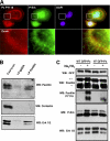
Orthovanadate-dependent Erk activation is controlled by paxillin phosphorylation on tyrosine 31 and 118. (A) In BHK-RSV cells, endogenous paxillin coimmunoprecipitates with Erk but not with cortactin. (B) In BHK-RSV cells, P-Erk is enriched invadopodia rings together with PY118 paxillin. (C) Western blotting carried out with lysates from polyclonal population of cell lines expressing WT GFP-paxillin (WT GFP-Px) or YF GFP-paxillin (YF GFP-Px), respectively, and treated or not with 5 mM orthovanadate for 30 min. *, endogenous paxillin; **, GFP paxillin. Equal protein loadings were assessed by proteins staining with Red Ponceau S (data not shown).
Figure 8.
Figure 8.
Inhibition of Erk phosphorylation by U0126 prevents invadopodia cores disassembly. (A) BHK-RSV cells were treated with 100 μM Erk inhibitor U0126. They formed thick invadopodia rings under orthovanadate treatment (5 mM; 30 min). Bar, 2 μm. (B) Statistical measures of invadopodia rings thickness in cells treated or not with U0126 after orthovanadate treatment. (C) BHK-RSV cells were treated with either DMSO or calpain inhibitor ALLM at 50 μM (1 h), orthovanadate at 5 mM (30 min), UO126 at 100 μM (1 h), UO126 at 100 μM (1 h), and then orthovanadate at 5 mM (30 min). After each treatment, cells were supplemented with CMAC t-Boc-Leu-Met (Invitrogen) at 50 μM (10 min) and fixed with 3% PBS-PFA. Cells were mounted and analyzed by fluorescence microscopy. Bar, 10 μm. (D) Western blotting showing that Erk inhibitor U0126 increased cortactin level either with or without orthovanadate treatment. Equal protein loading was checked by proteins staining on the nitrocellulose membrane with Red Ponceau S (data not shown).
Figure 9.
Figure 9.
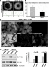
Paxillin knockdown impairs podosomes organization in primary mature osteoclasts. (A) Differential localization of actin and paxillin in the podosome belt of primary differentiated mouse osteoclasts on coverslips. The cells were fixed with 3% paraformaldehyde and stained for actin with phalloidin-TRITC and anti-paxillin antibodies. (B) siRNA paxillin performed in differentiated primary mouse osteoclasts. Western blot analysis showing the silencing of the protein paxillin (siRNA paxillin), compared with control cell treated with Oligofectamine alone (mock). (C) Differential organization of podosomes upon paxillin knockdown.
Figure 10.
Figure 10.
Extracellular matrix degradation takes place at the invadopodia/podosomes ring and is reduced by YF GFP paxillin overexpression. (A) Transfected and control BHK-RSV cells were plated 24 h on an extracellular matrix constituted by a layer of gelatin-TRITC fixed with 3% paraformaldehyde and covered by a layer of vitronectin. Left and middle, efficient matrix degradation by the control and WT GFP paxillin cells, respectively, at the level of the invadopodia rings. Right, BHK-RSV cells transfected with YF GFP-paxillin plated 24 h on the gelatin TRITC/vitronectin matrix. The ECM exhibited small and sparse patches of proteolysis corresponding to the individual disorganized invadopodia. Bar, 10 μm. (B) Differentiated primary mouse osteoclasts were plated on the gelatin-TRITC/vitronectin matrix for 24 h, fixed with 3% paraformaldehyde, and stained for actin with phalloidin-FITC. Degradation of this organic matrix is observed within the area defined by the podosome belt. (C) Quantification of the degradation ability of the extracellular matrix was established by calculating the relative degradation index DR. (data from 3 independent experiments; n = 48 cells; p < 0.001).
Figure 11.
Figure 11.
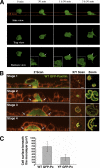
Invadopodia ring expansion promotes BHK-RSV cells transmigration through a cell monolayer. (A) 4D time-lapse videomicroscopy showing a single WT GFP-paxillin–transfected BHK-RSV cell crossing a HeLa cell monolayer (outlined by the red lines) and observed from side, top, and bottom views. The side view shows the sliding of the cell trough the cell layer, whereas the bottom view shows that in the same time at the ventral membrane, the cell formed an invadopodia ring that expanded toward the periphery of the area in contact with the matrix. (B) BHK-RSV cells transfected with WT GFP-paxillin were seeded on top of a HeLa cell monolayer in a Lab-Tek chamber. After 24 h, the cells were fixed in 3% paraformaldehyde, permeabilized with 0.1% Triton X-100 in PBS, stained for actin with TRITC-phalloidin, and observed by confocal microscopy (Z scan and X/Y scan). Stage 1, view of a BHK-RSV cell sending a membrane protrusion through the cell layer, ended by an invadopodia ring (yellow staining and arrows). Stage 2, a BHK-RSV cell crosses the cell layer. Z scan revealed an invadopodia ring (yellow staining and blue arrows) that slides under the neighboring cells. Stage 3, enlargement of the rosette pull down BHK-RSV cell under the HeLa monolayer. Stage 4, complete transmigration of a BHK-RSV cell through the cell monolayer. Bar, 10 μm. (C) Ovrexpression of YF GFP-paxillin in BHK-RSV cells diminished the average surface in contact with the matrix beneath the HeLa monolayer (n = 58; p <0.001).

Similar articles

Cited by

References

    1. Abram C. L., Seals D. F., Pass I., Salinsky D., Maurer L., Roth T. M., Courtneidge S. A. The adaptor protein fish associates with members of the ADAMs family and localizes to podosomes of Src-transformed cells. J. Biol. Chem. 2003;278:16844–16851. - PubMed
    1. Artym V. V., Zhang Y., Seillier-Moiseiwitsch F., Yamada K. M., Mueller S. C. Dynamic interactions of cortactin and membrane type 1 matrix metalloproteinase at invadopodia: defining the stages of invadopodia formation and function. Cancer Res. 2006;66:3034–3043. - PubMed
    1. Ayala I., Baldassarre M., Caldieri G., Buccione R. Invadopodia: a guided tour. Eur. J. Cell Biol. 2006;85:159–164. - PubMed
    1. Baldassarre M., Ayala I., Beznoussenko G., Giacchetti G., Machesky L. M., Luini A., Buccione R. Actin dynamics at sites of extracellular matrix degradation. Eur. J. Cell Biol. 2006;85:1217–1231. - PubMed
    1. Baldassarre M., Pompeo A., Beznoussenko G., Castaldi C., Cortellino S., McNiven M. A., Luini A., Buccione R. Dynamin participates in focal extracellular matrix degradation by invasive cells. Mol. Biol. Cell. 2003;14:1074–1084. - PMC - PubMed

Publication types

MeSH terms

LinkOut - more resources