. 2007 Dec 3;179(5):965-80.
doi: 10.1083/jcb.200702187. Epub 2007 Nov 26.

Adenovirus RIDalpha regulates endosome maturation by mimicking GTP-Rab7

Affiliations

Adenovirus RIDalpha regulates endosome maturation by mimicking GTP-Rab7

Ankur H Shah et al. J Cell Biol. .

Abstract

The small guanosine triphosphatase Rab7 regulates late endocytic trafficking. Rab7-interacting lysosomal protein (RILP) and oxysterol-binding protein-related protein 1L (ORP1L) are guanosine triphosphate (GTP)-Rab7 effectors that instigate minus end-directed microtubule transport. We demonstrate that RILP and ORP1L both interact with the group C adenovirus protein known as receptor internalization and degradation alpha (RIDalpha), which was previously shown to clear the cell surface of several membrane proteins, including the epidermal growth factor receptor and Fas (Carlin, C.R., A.E. Tollefson, H.A. Brady, B.L. Hoffman, and W.S. Wold. 1989. Cell. 57:135-144; Shisler, J., C. Yang, B. Walter, C.F. Ware, and L.R. Gooding. 1997. J. Virol. 71:8299-8306). RIDalpha localizes to endocytic vesicles but is not homologous to Rab7 and is not catalytically active. We show that RIDalpha compensates for reduced Rab7 or dominant-negative (DN) Rab7(T22N) expression. In vitro, Cu(2+) binding to RIDalpha residues His75 and His76 facilitates the RILP interaction. Site-directed mutagenesis of these His residues results in the loss of RIDalpha-RILP interaction and RIDalpha activity in cells. Additionally, expression of the RILP DN C-terminal region hinders RIDalpha activity during an acute adenovirus infection. We conclude that RIDalpha coordinates recruitment of these GTP-Rab7 effectors to compartments that would ordinarily be perceived as early endosomes, thereby promoting the degradation of selected cargo.


Figures

Figure 1.
Figure 1.
RIDα localization and interaction with RILP. (a) Model showing known RIDα membrane topology and C-terminal tail amino acid sequence used as bait in a yeast two-hybrid screen. The loop domain in the lumen has a site for signal peptidase cleavage and disulfide bond formation (Hoffman et al., 1992b). (b) Stably expressed FLAG-RIDα in CHO cells was stained for the FLAG epitope (red) and with DAPI (blue) after treatment with vehicle or cytoskeleton-disrupting drugs. Cell peripheries (green) were traced from a phase image using MetaMorph software. (c) Schematic representation of RILP with known protein-binding domains. The RIDα-binding domain partially overlaps residues in the second α-helical regions (a1 and a2) previously identified as the GTP-Rab7–binding site (Wu et al., 2005). (d) GST fusion proteins with the progressive stop codons introduced at residues highlighted in black in panel a were purified and separated by SDS-PAGE. (e) GST fusion proteins were incubated with lysates from cells overexpressing HA-RILP. Bound proteins were detected by a Western blot for the HA epitope tag. (f) GST fusion proteins were incubated with an irrelevant HA-tagged protein and analyzed as in panel e. (g) HA-RILP immobilized on an anti-HA affinity column was incubated with purified GST or full-length GST-RIDα cytoplasmic tail. Bound proteins were detected by a Western blot for GST. Bars, 10 μm.
Figure 2.
Figure 2.
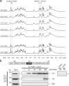
Cu2+ interacts with RIDα and enhances RIDα–RILP interaction. (a) 1D 1H NMR spectra of the amide/aromatic region of 23 C-terminal residues of RIDα illustrating the effects of metal ion (Mg2+, Zn2+, and Cu2+) binding. Aromatic side chain Hδ2 and Hɛ1 resonances of His75 and His76 (dashed lines) were previously determined (Vinogradova et al., 1998). Selective line broadening was observed for histidine side chains upon the addition of Cu2+ but not Zn+2 or Mg+2. (b) GST pull-down assays were performed as shown in Fig. 1 e with fusion proteins encoding the full-length C-terminal tail in the presence of indicated cation concentrations. Mutations of Cu2+-binding residues 75HH to 75AA or 75SS blocked in vitro interactions with RILP.
Figure 3.
Figure 3.
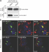
Colocalization and coimmunoprecipitation of RIDα and RILP. (a) Cell lysates from GP2-293 cells stably expressing FLAG-RIDα or FLAG-RIDα(75AA) and transfected with a plasmid encoding HA-RILP were immunoprecipitated with an anti-FLAG antibody and followed by a Western blot for FLAG to detect RIDα or HA to detect coimmunoprecipitated HA-RILP. (b) GP2-293 cells transfected with a plasmid encoding GFP-RILP were fixed and stained for the FLAG epitope tag (red) and with DAPI (blue) 12 h after transfection. Confocal microscopy demonstrates partial merging of GFP-RILP signals with FLAG-RIDα but not FLAG-RIDα(75AA). Bars, 10 μm.
Figure 4.
Figure 4.
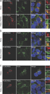
Localization of RIDα with compartment-specific markers. Stable GP2-293 cell lines were stained for the FLAG epitope (red), with DAPI (blue), and for transferrin receptor (a), RhoB (b), and furin (c; green channels). Boxed areas are enlarged on the right. Bars, 10 μm.
Figure 5.
Figure 5.
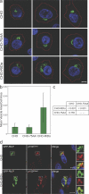
Enlargement of GFP-RILP–labeled vesicles by RIDα. (a) CHO, CHO-RIDα, or CHO-75AA cell populations transfected with a plasmid encoding GFP-RILP were fixed and stained with DAPI (blue). Images were generated by merging of the largest traced cell periphery from a phase image plane (red) with a confocal plane representing the largest area for an individual GFP-RILP–labeled vesicle (green). Three representative images of each cell type are shown. Bar, 10 μm. (b) Ratios of vesicle to cell area for every identifiable vesicle (n > 70 for each cell type) were obtained with MetaMorph software. Data are presented as mean ratios ± SDs (error bars). (c) A mixed effects ANOVA statistical model treatment of data found the difference in vesicle size between RIDα versus RIDα(75AA) or parental CHO cells to be significant (P < 0.001). (d) GFP-RILP–transfected CHO cells were stained for p150Glued (red) and with DAPI (blue). Boxed areas are enlarged on the right. Bars, 15 μM.
Figure 6.
Figure 6.
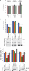
RIDα compensates for Rab7. (a) Cells incubated with 125I-LDL were assayed for surface-bound (0 h), internalized (0.5 h), and degraded LDL in the medium (6 h). Results are presented as percentages relative to parental A549 cells. (b, top) 125I-LDL degradation assay was performed in cells transfected with Rab7-specific siRNA and incubated for 24 h. Experiments were performed in triplicate on two separate occasions. Data (mean ratios ± SDs [error bars]) are plotted as percentages of mock-transfected cells and analyzed by t test. (bottom) Equal aliquots of total cell protein were Western blotted with antibodies against Rab7, actin, and RIDα. (c, top) 125I-LDL degradation assay was performed in cells transfected with GFP or GFP-tagged Rab7 constructs. (bottom) Transient protein expression was determined by Western blotting for GFP.
Figure 7.
Figure 7.
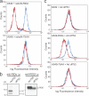
Down-regulation of cell surface EGFR and Fas. (a) A549 cells infected with retrovirus expressing RIDα or RIDα(75AA) were analyzed for EGFR surface expression by flow cytometry 24 h after infection. Data are plotted for uninfected (blue) and infected (red) cells. (b) A549 cells were infected with wild-type adenovirus (rec700) or an RIDα deletion mutant (dl753) and were metabolically labeled with [35S]Cys/Met. Cell lysates were immunoprecipitated with RIDα or RIDβ antibodies under nondenaturing conditions and were subjected to SDS-PAGE and autoradiography. (c) A549, A549-RIDα, or A549-RIDα(75AA) cells were infected with dl753 for 18 h, labeled with a FITC-conjugated antibody recognizing the extracellular domain of Fas, and subjected to FACS analysis. Data are plotted for uninfected (blue) and infected (red) cells. All experiments were performed on three separate occasions, and a representative experiment is presented.
Figure 8.
Figure 8.
ΔN-RILP impairs RIDα-mediated EGFR down-regulation. (a) GST fusion proteins were incubated with lysates from cells overexpressing GFP-RILP or GFP–ΔN-RILP. Bound proteins were detected by a Western blot for GFP. (b) A549 cells were transiently transfected with a plasmid encoding GFP or GFP–ΔN-RILP and infected with rec700. To ensure that RILP proteins do not interfere with infection, total cell lysates were analyzed for E1A expression by Western blotting. (c) A FACS profile is presented of the EGFR surface expression of ungated control cells (blue), GFP-transfected, rec700-infected, and GFP-gated cells (red), and GFP–ΔN-RILP–transfected, rec700-infected, and GFP-gated cells (green).
Figure 9.
Figure 9.
RIDα additionally interacts with ORP1L. (a) Schematic representation of ORP1L. The shortest cDNA clone identified in our screen corresponds to amino acids 755–923 in the sterol-binding domain (SD). ORP1L also has a pleckstrin homology domain (PH) and ankyrin repeats (Lehto et al., 2001). GTP-Rab7 binding has been mapped to the N terminus (Johansson et al., 2005). (b) ORP1L and Hsp60-binding domains were mapped using the GST fusion proteins shown in Fig. 1 d. Total cell lysates were immunodepleted for Hsp60 before addition to GST fusion proteins for ORP1L mapping. (c) ORP1L binding to the full-length GST fusion protein was determined in the presence of indicated cation concentrations.
Figure 10.
Figure 10.
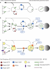
Role of RIDα in receptor down-regulation. Model depicting EGFR endocytic trafficking under resting conditions, after ligand stimulation, or during RIDα expression. Major trafficking routes are represented by thicker black arrows, and minor trafficking routes are indicated by thinner gray arrows. (a) During resting conditions, a majority of EGFRs internalized at a low basal rate recycle back to the cell surface. (b) During ligand stimulation, EGFR internalization rates and GTP-Rab7 recruitment to EGFR-containing vesicles are enhanced. GTP-Rab7 binds RILP and ORP1L (dashed arrows), which subsequently recruit and activate motor proteins that induce microtubule-dependent displacement toward the MTOC. (c) RIDα associates with EGFR-containing vesicles in the early endosomal network under resting conditions and recruits GTP-Rab7 effectors RILP and ORP1L, leading to microtubule-dependent displacement toward the MTOC. RIDα also enhances the degradation of LDL after its release from internalized LDL receptors and acts cooperatively with RIDβ to down-regulate Fas. Although RIDα and RIDβ are enriched in endosomes and plasma membrane, respectively, they can also form physical complexes that are important for some aspects of E3 function.

Similar articles

Cited by

References

    1. Agarraberes, F.A., and J.F. Dice. 2001. A molecular chaperone complex at the lysosomal membrane is required for protein translocation. J. Cell Sci. 114:2491–2499. - PubMed
    1. Austin, C.D., A.M. De Maziere, P.I. Pisacane, S.M. van Dijk, C. Eigenbrot, M.X. Sliwkowski, J. Klumperman, and R.H. Scheller. 2004. Endocytosis and sorting of ErbB2 and the site of action of cancer therapeutics trastuzumab and geldanamycin. Mol. Biol. Cell. 15:5268–5282. - PMC - PubMed
    1. Brown, M.S., and J.L. Goldstein. 1975. Regulation of the activity of the low density lipoprotein receptor in human fibroblasts. Cell. 6:307–316. - PubMed
    1. Brown, V., R.A. Brown, A. Ozinsky, J.R. Hesselberth, and S. Fields. 2006. Binding specificity of Toll-like receptor cytoplasmic domains. Eur. J. Immunol. 36:742–753. - PMC - PubMed
    1. Bucci, C., R.G. Parton, I.H. Mather, H. Stunnenberg, K. Simons, B. Hoflack, and M. Zerial. 1992. The small GTPase rab5 functions as a regulatory factor in the early endocytic pathway. Cell. 70:715–728. - PubMed

Publication types

MeSH terms