. 2008 Jan;19(1):368-77.
doi: 10.1091/mbc.e07-08-0801. Epub 2007 Nov 14.

Distinct Dgrip84 isoforms correlate with distinct gamma-tubulins in Drosophila

Affiliations

Distinct Dgrip84 isoforms correlate with distinct gamma-tubulins in Drosophila

Christiane Wiese. Mol Biol Cell. 2008 Jan.

Abstract

Gamma-tubulin is an indispensable component of the animal centrosome and is required for proper microtubule organization. Within the cell, gamma-tubulin exists in a multiprotein complex containing between two (some yeasts) and six or more (metazoa) additional highly conserved proteins named gamma ring proteins (Grips) or gamma complex proteins (GCPs). gamma-Tubulin containing complexes isolated from Xenopus eggs or Drosophila embryos appear ring-shaped and have therefore been named the gamma-tubulin ring complex (gammaTuRC). Curiously, many organisms (including humans) have two distinct gamma-tubulin genes. In Drosophila, where the two gamma-tubulin isotypes have been studied most extensively, the gamma-tubulin genes are developmentally regulated: the "maternal" gamma-tubulin isotype (named gammaTub37CD according to its location on the genetic map) is expressed in the ovary and is deposited in the egg, where it is thought to orchestrate the meiotic and early embryonic cleavages. The second gamma-tubulin isotype (gammaTub23C) is ubiquitously expressed and persists in most of the cells of the adult fly. In those rare cases where both gamma-tubulins coexist in the same cell, they show distinct subcellular distributions and cell-cycle-dependent changes: gammaTub37CD mainly localizes to the centrosome, where its levels vary only slightly with the cell cycle. In contrast, the level of gammaTub23C at the centrosome increases at the beginning of mitosis, and gammaTub23C also associates with spindle pole microtubules. Here, we show that gammaTub23C forms discrete complexes that closely resemble the complexes formed by gammaTub37CD. Surprisingly, however, gammaTub23C associates with a distinct, longer splice variant of Dgrip84. This may reflect a role for Dgrip84 in regulating the activity and/or the location of the gamma-tubulin complexes formed with gammaTub37CD and gammaTub23C.


Figures
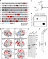
Figure 1.
Figure 1.
Analysis of Drosophila γTub23C and γTub37CD and isotype-specific antibodies. (A) Sequence alignment between γTub37CD and γTub23C reveals that nonconserved differences between the two isotypes are scattered throughout the primary sequence. Light gray, identities; medium gray, conserved changes; red, differences that change the character of the amino acid; blue, amino acid insertions in γTub23C relative to γTub37CD; the transparent amino acids near the C-termini of the molecules are not represented in the structural model. (B) A hypothetical structural surface model of γTub23C (generated by the structure prediction program SwissModel; Peitsch, 1995; Guex and Peitsch, 1997; Schwede et al., 2003) is based on the crystal structure for human γ-tubulin (Aldaz et al., 2005). To highlight the residues not conserved between the isotypes, the backbone of the residues whose character is significantly different between γTub23C and γTub37CD are colored in red and the M-loop insertion in γTub23C that is not present in γTub37CD is colored in blue, as in A. Snapshot images of different faces of the molecule are shown in each panel, as indicated in the upper left-hand corner. The orientation of the snapshots are based on the observation that γ-tubulin can roughly be modeled as a cube, and on the orientation of α and β tubulin within the microtubule lattice, as illustrated in (C). The snapshots illustrate two important points: first, the differences between the two γ-tubulins are not randomly distributed throughout the molecule but map mainly to the surfaces, presumably leaving the core of the structure largely unaffected (see also Supplementary Figure S1). Second, the differences map to all six faces of the cube. Corresponding ribbon models can be found in the online supplementary material, Figure S1. (D) Dot-blot analysis of affinity-purified antibodies. Aqueous solutions of each peptide were spotted onto a piece of nitrocellulose membrane, and the membrane was probed with each antibody, as indicated. The antibodies do not cross-react with each other. (E) Western blot of total extracts from embryos (emb, lanes 1 and 3) or S2 cells (S2 cells, lanes 2 and 4) probed with antibodies to γTub37CD (lanes 1 and 2) or γTub23C (lanes 3 and 4). Positions of molecular weight markers are indicated on the right. (F) Western blot of purified γ-tubulin complexes. Lane 1, γTub23C complexes probed with γTub37CD antibodies; lane 2, γTub37CD complexes probed with γTub23C antibodies. The ∼40-kDa band recognized by the γTub37C antibodies in embryo extracts probably shares an epitope with γTub37CD, as this band cross-reacts with antisera produced by several different rabbits (our unpublished observation); however, this protein apparently does not coimmunoprecipitate with the γTuRC and thus did not interfere with the analysis of the purified complex.
Figure 2.
Figure 2.
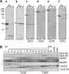
Drosophila tissue culture cells express a slower migrating version of Dgrip84. (A) Western blot analysis of γTuRC components in embryos, S2 cells, and Kc cells, as indicated above the lanes. Probes are indicated below the blots and are as follows: (a) γTub37CD, (b) γTub23C, (c) Dgrip98/GCP3, (d) Dgrip128/GCP5, (e) Dgrip163/GCP6, and (f) Dgrip84/GCP2. The embryonic “cognate” proteins are indicated by arrowheads on the left of each panel. Asterisks in f denote two bands (∼100 and ∼75 kDa) in tissue culture cells that are recognized by the Dgrip84 antibody. Note the absence in the tissue culture cells of a band corresponding to γTub37CD in panel a, and the absence of a band corresponding to embryonic Dgrip84 in panel f. (B) Sucrose gradient analysis of S2 cell extracts reveals that the slower migrating protein recognized by the Dgrip84 antibodies migrates similarly to γTub23C. Sucrose gradient fractions were probed for γTuRC components as indicated on the right. The positions of γTuSC and γTuRC are indicated below the blot. Asterisks on the left denote the two proteins in tissue culture cell extracts that are recognized by the Dgrip84 antibody. Only the ∼100-kDa protein migrates like the embryonic Dgrip84 (compare with Figure 3).
Figure 3.
Figure 3.
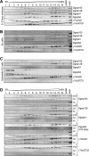
Sucrose gradient analysis of γ-tubulins and Dgrips in embryos and tissue culture cells. (A) Embryo extracts, 0–8 h, were analyzed on 5–40% sucrose gradients. Gradient fractions were probed for Dgrips and γ-tubulins as indicated on the right. (B and C) Extracts made from Kc cells (B) or S2 cells (C) were analyzed on sucrose gradients as described for A. D. Individual panels from A to C were regrouped to allow a more direct comparison for each γTuRC component between the migration patterns in embryos and tissue culture cells. γTub23C migrates differently in embryos, but in S2 and Kc cells resembles the behavior of γTub37CD. For Dgrip84, only the ∼100-kDa bands are shown in S2 and Kc cells.
Figure 4.
Figure 4.
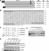
Dgrip84 isoforms in Drosophila. (A) Diagram of the relationships between different Dgrip84 isoforms. The position of the 33-amino acid C-terminal insertion and the 74-amino acid N-terminal extension are indicated. Theoretical molecular weights, number of amino acids, and pI are indicated on the right for each isoform (A–D). Isoforms A–C are currently present in the database (Accession no. NP_728264, NP_728265, and NP_523409, respectively). The isoform found in S2 cells (see below) has the extension but not the insertion. We named this the “D” isoform. (B) Amino acid sequence of Dgrip84 isoform A. The N-terminal extension and C-terminal insertion are indicated by gray shading and a box, respectively. (C) Gene structure of the Dgrip84 isoforms. Numbers below the diagram indicate the number of nucleotides in each exon (top row) or intron (bottom row). (D) Western blot of staged embryos. Drosophila embryos were collected for 4 h and were then aged for the times indicated above the blots. Maximum age was 24 h. Embryos were then homogenized, loaded onto SDS-PAGE gels, and Western blotted. The blots were probed with antibodies to Dgrip163, γTub37CD, γTub23C, or Dgrip84, as indicated on the right. The positions of the slower migrating (“C”) and faster-migrating (“D”) Dgrip84 bands are indicated on the left. (E) Northern blot of staged embryos probed with a Dgrip84 probe common to all isoforms. Embryos were collected and aged as in D. Early embryos (0–4 h) express a shorter mRNA than later embryos or S2 cells. To determine the identity of the isoforms, we performed RT-PCR and sequenced the relevant regions of cDNAs recovered form early embryos or S2 cells (see Supplementary Figure S3). This analysis shows that early embryos express the “C” isoform, whereas S2 cells express the “D” isoform. The reduction in signal for the older embryos (starting with 12–16-h embryos) may be due to sample degradation, although our loading control (rp49; DeZazzo et al., 2000) remained unchanged.
Figure 5.
Figure 5.
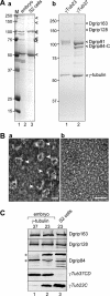
Dgrip84-D is a component of the γTuRC. (A) Comparison between protein complexes immunoprecipitated with various antibodies. Panel a, Coomassie-stained gel of proteins that coimmunoprecipitate with Dgrip84 antibodies from embryo (lane 2) or S2 cell (lane 3) extracts. γTuRC proteins are highlighted by arrowheads on the right, positions of molecular-weight markers are indicated on the left. Panel b, γ-tubulin complexes isolated from S2 cells using antibodies to γTub23C (lane 1), or from 0-to 8-h embryos using antibodies to γTub37CD (lane 2). Complexes isolated from S2 cells with antibodies to Dgrip84 or γTub23C resemble each other, as do complexes isolated from embryos by antibodies to Dgrip84 or γTub37CD. Complexes isolated from S2 cells lack a band corresponding to the embryonic Dgrip84; however, a new band appears above Dgrip91. (B) Negative-staining electron microscopy of γTub23C complexes isolated from S2 cells. Arrowheads in the left panel point to individual ring complexes. Scale bars, 25 nm in the left panel, 100 nm in the right panel. (C) Western blot of proteins that copurify with γTub37CD from early (0–8 h) embryos (lane 1), with γTub23C from late (8–16 h) embryos (lane 2), or with γTub23C from S2 cells (lane 3). Antibodies to Dgrips used to probe the Western blot are indicated on the right. The asterisks mark the two Dgrip84 isoforms that copurify with γTub37CD or γTub23C. The multiple bands in lane 3 might represent additional Dgrip84 isoforms or degradation products.
Figure 6.
Figure 6.
γTub23C migrates as a large complex in early embryos that is distinct from the embryonic γTuRC. (A) Coomassie gel of proteins that precipitate from early embryos with antibodies to γTub23C (lane 2) or γTub37CD (lane 3). Positions of γTuRC components are indicated on the right. Lane 1, molecular-weight markers (sizes of markers are indicated on the left). (B) Sucrose gradient analysis of extracts made from 0- to 8-h Drosophila embryos. Western blot of 0–8 h embryo extracts fractionated on a sucrose gradient probed with antibodies specific for each γ-tubulin isotype or Dgrips 163, 128, 98, and 84, as indicated on the right. The positions of the γTuRC (fractions 13–15), the γTuSC (fractions 6–8), and the peak fractions for γTub23C (fractions 11 and 12) are indicated below the blot. The gel is loaded such that the lighter sucrose fractions (starting at 5%) are on the left, and the heavier fractions (up to 40%) are on the right. Fraction numbers are indicated above the blots. Load, 1 μl of starting material was loaded as a control. (C) γTub23C and γTub37CD can be distinguished by their sensitivity to polyethyleneglycol (PEG). PEG (2.5%) was added to extracts, and precipitated proteins were separated from soluble proteins by centrifugation. Supernatant (S) and resuspended pellet (P) fractions were then fractionated on sucrose gradients, run on a SDS-PAGE gel, and blotted for γTub23C or γTub37CD as indicated on the right.

Similar articles

Cited by

References

    1. Anders A., Lourenco P. C., Sawin K. E. Noncore components of the fission yeast γ-tubulin complex. Mol. Biol. Cell. 2006;17:5075–5093. - PMC - PubMed
    1. Andersen J. S., Wilkinson C. J., Mayor T., Mortensen P., Nigg E. A., Mann M. Proteomic characterization of the human centrosome by protein correlation profiling. Nature. 2003;426:570–574. - PubMed
    1. Aldaz H., Rice L. M., Stearns T., Agard D. A. Insights into microtubule nucleation from the crystal structure of human γ-tubulin. Nature. 2005;435:523–527. - PubMed
    1. Barbosa V., Yamamoto R. R., Henderson D. S., Glover D. Mutation of a Drosophila γ-tubulin ring complex subunit encoded by discs degenerate-4 differentially disrupts centrosomal protein localization. Genes Dev. 2000;14:3126–3139. - PMC - PubMed
    1. Barbosa V., Gatt M., Rebollo E., Gonzalez C., Glover D. M. Drosophila dd4 mutants reveal that γTuRC is required to maintain juxtaposed half spindles in spermatocytes. J. Cell Sci. 2003;116:929–941. - PubMed

Publication types

MeSH terms

LinkOut - more resources