. 2007 Nov 12:8:48.
doi: 10.1186/1471-2121-8-48.

Filamin repeat segments required for photosensory signalling in Dictyostelium discoideum

Affiliations

Filamin repeat segments required for photosensory signalling in Dictyostelium discoideum

Sarah J Annesley et al. BMC Cell Biol. .

Abstract

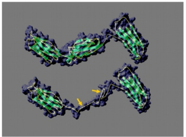
Background: Filamin is an actin binding protein which is ubiquitous in eukaryotes and its basic structure is well conserved - an N-terminal actin binding domain followed by a series of repeated segments which vary in number in different organisms. D. discoideum is a well established model organism for the study of signalling pathways and the actin cytoskeleton and as such makes an excellent organism in which to study filamin. Ddfilamin plays a putative role as a scaffolding protein in a photosensory signalling pathway and this role is thought to be mediated by the unusual repeat segments in the rod domain.

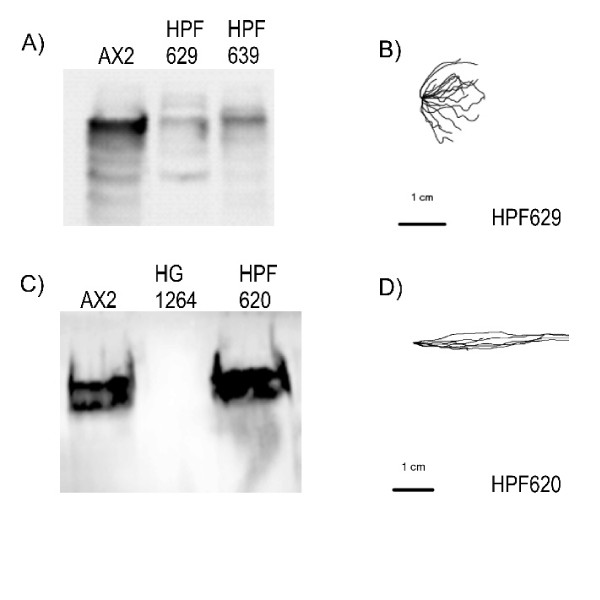
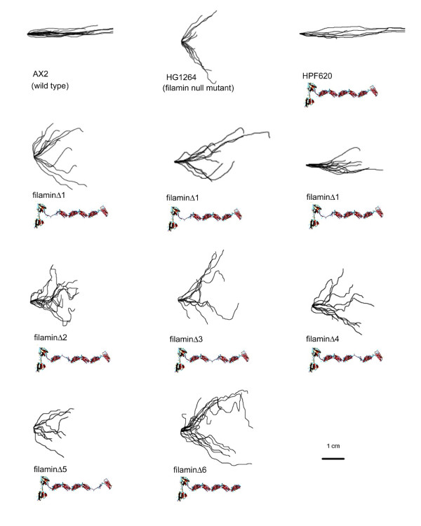
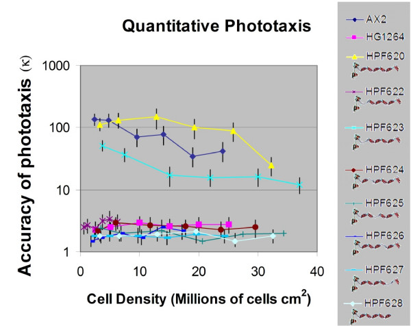
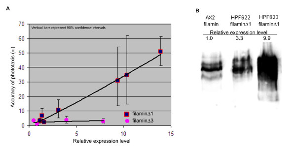
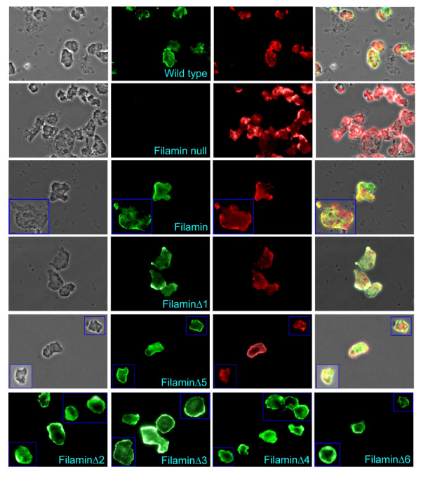
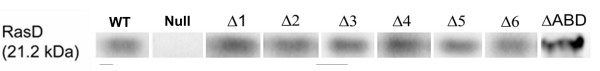
Results: To study the role of filamin in phototaxis, a filamin null mutant, HG1264, was transformed with constructs each of which expressed wild type filamin or a mutant filamin with a deletion of one of the repeat segments. Transformants expressing the full length filamin to wild type levels completely rescued the phototaxis defect in HG1264, however if filamin was expressed at lower than wild type levels the phototaxis defect was not restored. The transformants lacking any one of the repeat segments 2-6 retained defective phototaxis and thermotaxis phenotypes, whereas transformants expressing filaminDelta1 exhibited a range of partial complementation of the phototaxis phenotype which was related to expression levels. Immunofluorescence microscopy showed that filamin lacking any of the repeat segments still localised to the same actin rich areas as wild type filamin. Ddfilamin interacts with RasD and IP experiments demonstrated that this interaction did not rely upon any single repeat segment or the actin binding domain.

Conclusion: This paper demonstrates that wild type levels of filamin expression are essential for the formation of functional photosensory signalling complexes and that each of the repeat segments 2-6 are essential for filamins role in phototaxis. By contrast, repeat segment 1 is not essential provided the mutated filamin lacking repeat segment 1 is expressed at a high enough level. The defects in photo/thermosensory signal transduction caused by the absence of the repeats are due neither to mislocalisation of filamin nor to the loss of RasD recruitment to the previously described photosensory signalling complex.


Figures

Figure 1
Figure 1
Complementation of filamin null mutant HG1264 depends upon the level of filamin expression. (A) Western blot of proteins from AX2 and two filamin mutant transformants, HPF629 and HPF639 ectopically expressing filamin. The two transformants expressed filamin at a lower level than that observed in the wild type. (B) The slug trails of HPF629 were digitised and this transformant displayed a defective phototaxis phenotype as observed in the filamin mutant from which this strain was derived, HG1264. The light source is to the right of the figure. (C) Western blot using the antifilamin antibody against total cellular protein from AX2 (lane 1, wild type), HG1264 (lane 2, filamin null mutant) and HPF620 (lane 3, filamin mutant ectopically expressing filamin). No filamin can be detected in the filamin mutant whereas filamin is expressed in HPF620 to a level comparable to that of the wild type. (D) The slug trails of HPF620 were digitised and this transformant displayed a wild type phototaxis phenotype. The light source is to the right of the figure.
Figure 2
Figure 2
Prediction of the structure of filaminΔ5. Structural prediction of a region of filaminΔ5 corresponding to segments 4 through 6 of the full length filamin (amino acids 547 to 838, including the first two β strands of segment 5) but with residues 672 through 743 (most of segment 5) deleted (DDB0201554). The prediction was made using the SwissModel first approach method accessed through the ExPASy Proteomics Server [20-25]. The resulting prediction was based on known D. discoideum and human filamin structures. The structures for repeat segments 4–6 have been determined by Popowitcz et al. [19] and as the repeats are very similar it is believed that repeat segments 1–3 would contain a similar structure to repeats 4 and 5. Therefore this figure uses the deletion of repeat segment 5 as an example which could be extrapolated for all the other repeat segments. The top panel is the known structure [19] of repeat segments 4 through to 6, the bottom panel shows the predicted structure for the corresponding region of filaminΔ5. This figure illustrates that by deleting all but the first two β strands of a repeat segment, it is possible to correctly fold and maintain the correct distance between the repeat segments that flank the deleted region. The gold arrows indicate the position of the sequences corresponding to the first two β strands of segment 5 in the wild type filamin structure.
Figure 3
Figure 3
Qualitative phototaxis by strains lacking repeat segments. Digitised slug trails of wild type AX2, the filamin null mutant HG1264, the rescued strain HPF620 and the strains lacking one of the repeat segments. Slug trails were plotted from a common origin so that the source of light is to the right of the figures. Wild type slugs migrate directly towards the light source whereas filamin mutant slugs show disoriented, bimodal phototaxis with two preferred directions either side of the light source. The rescued strain HPF620 displays wild type phototaxis whereas the strains lacking repeat segments 2–6 display mutant phototaxis and transformants lacking the first repeat show phototaxis phenotypes ranging from wild type to mutant.
Figure 4
Figure 4
Quantitative phototaxis of strains lacking repeat segments. Phototaxis at defined cell densities was performed on wild type AX2, the filamin null mutant HG1264, the rescued strain HPF620 and the strains expressing filamin lacking one of the repeat segments as indicated by the cartoon under the strain name. The trails were digitised and analysed using directional statistics to determine their accuracies of phototaxis (κ) at the defined cell densities. Vertical bars represent 90% phototaxis than the wild type AX2, the original mutant (HG1264) displayed poor accuracies, as did all of the strains lacking repeat segments 2–6. Two transformants expressing filamin with a deletion of repeat one showed accuracies of phototaxis of approximately 1% (HPF622) or 50% (HPF623) of the wild type values.
Figure 5
Figure 5
Quantitative thermotaxis of strains lacking repeat segments. Quantitative measurements of thermotaxis by slugs of wild type AX2, filamin null mutant HG1264, the rescued strain HPF620, two transformants expressing filaminΔ1 HPF622 and HPF623 and HPF627 a transformant expressing filaminΔ5. All transformants expressing filmainΔ2–6 showed similar thermotaxis phenotypes to HPF627 which was included as an example. Temperatures follow the previous convention of being designated 1 through 8 in arbitrary units. Separate calibration experiments have shown that these correspond to temperatures at the surface of the centre of each plate ranging from 14°C in 2°C steps to 28°C, with a temperature gradient at the agar surface of 0.2°C/cm. The slug trails were digitised and analysed using directional statistics [3]. Positive values indicate the accuracy of positive thermotaxis (κ) towards the warmth, negative values indicate the accuracy of negative thermotaxis (κ) towards the cold and a value of zero indicates there was no preferred direction. Vertical bars are 90% confidence limits. AX2, HPF620 and HPF623 all display high accuracies of positive thermotaxis at T4 and T5 and low positive accuracies of thermotaxis at the cooler and hotter temperatures with AX2 displaying negative accuracies of thermotaxis at T1. HG1264, HPF622 and HPF627 displayed small accuracies of thermotaxis at all temperature points. It is normal for the accuracy of thermotaxis by wild type AX2 slugs to decline above and below the growth temperature and either approach zero or even switch to negative thermotaxis at T1 [16, 39]. At some of the higher temperatures slugs of several of the strains did not migrate so thermotaxis could not be measured.
Figure 6
Figure 6
Transformants expressing filaminΔ1 show increased accuracy of phototaxis in response to increased expression levels. (A) The expression level of mutant filamin protein in several transformants was quantitated and normalised against wild type levels. As the level of filaminΔ1 expression increased, so too did the accuracy of phototaxis. Transformants expressing any of the other mutant filamin protein (filaminΔ3 used as an example in this figure) did not show this correlation increased expression had no effect on the accuracy of phototaxis. Vertical bars represent 90% confidence limits. (B) Western blot detecting filamin in wild type (AX2), and filaminΔ1 in two transformants HPF622 and HPF623. The amount of filamin protein detected was normalised against AX2 expression so that the level of filamin expressed in AX2 was 1. The level of filaminΔ1 expressed in HPF622 was 3.3 and these cells showed a relatively low accuracy of phototaxis whereas HPF623 cells expressed filaminΔ1 at 9.9 and showed a higher degree of accuracy of phototaxis.
Figure 7
Figure 7
Filamin and mutated filamin localise to the same actin rich areas. Vegetative amoebae were viewed under a fluorescence microscope. For all but the last row, the first column contains phase contrast images, the second column contains the corresponding fields detecting green immunofluorescence using Alexa Fluor 488-conjugated anti-rabbit IgG and rabbit polyclonal antifilamin antibodies while the third column contains the corresponding field detecting actin stained with Texas Red-X phalloidin. The fourth column is an overlay of all three images and the yellow colour depicts regions where filamin and actin colocalise. Fluorescence images were obtained using an Olympus BX61TRF microscope under epifluorescence using blue light for excitation and either green or red emission filters. Images were obtained in grey scale and the colour balance adjusted post hoc to leave only the red or green channel as appropriate. As seen in column 2 filamin is found throughout the cells but is concentrated at the cell cortex. No filamin is detected in the filamin mutant HG1264. Strains expressing filaminΔ1 (which showed partial complementation of the phototaxis phenotype) and filaminΔ5 (which did not) were used as examples, as all strains expressing mutant filamins behaved in the same fashion. Column 3 illustrates that actin was found throughout the cell and was concentrated at the cell cortex. Comparison of columns 2 and 3 and examination of column 4 shows that in all strains tested filamin colocalises predominantly with actin at the cell cortex. The last row contains images of cells from the other strains tested expressing the various mutant filamins and depicts the localisation of filamin by immunofluorescence. As in all the other strains mutant filamin is concentrated at the cell cortex.
Figure 8
Figure 8
RasD binding does not depend on any single repeat in the rod domain nor the actin binding domain. Western blot using the antiRasD antibody to detect RasD in immunoprecipitations using protein extract from HPF616 (lane 1, WT) (AX2 ectopically expressing RasD), HPF631 (lane 2, Null) (HG1264 ectopically expressing RasD) and strains HPF632-638 (lanes 3–10) (HG1264 ectopically expressing RasD and one of filaminΔ1–6 or filaminΔABD as indicated). RasD is detected in all IP samples with the exception of the null mutant HPF631, indicating that filamin is required to pull down RasD and RasD interacts with filamin regardless of which portion of the scaffolding protein is absent.

Similar articles

Cited by

References

    1. Van Troys M, Vandekeerckhove J, Ampe C. Structural modules in actin-binding proteins: towards a new classification. Biochimica et Biophysica Acta. 1999;1448:323–348. - PubMed
    1. Noegel AA, Rapp S, Lottspeich F, Schleicher M, Stewart M. The Dictyostelium gelation factor shares a putative actin binding site with alpha-actinins and dystrophin and also has a rod domain containing six 100-residue motifs that appear to have a cross-beta conformation. J Cell Biol. 1989;109:607–618. doi: 10.1083/jcb.109.2.607. - DOI - PMC - PubMed
    1. Fisher PR, Smith E, Williams KL. An extracellular chemical signal controlling phototactic behaviour by D. discoideum slugs. Cell. 1981;23:799–807. doi: 10.1016/0092-8674(81)90444-X. - DOI - PubMed
    1. Feng Y, Walsh CA. A versatile molecular scaffold for cell motility and signalling. Nature Cell Biol. 2004;6:1034–1038. doi: 10.1038/ncb1104-1034. - DOI - PubMed
    1. Zigmond SH, Joyce M, Borleis J, Bokoch GM, Devreotes PN. Regulation of actin polymerisation in cell free systems by GTPγS and Cdc42. J Cell Biol. 1997;138:363–374. doi: 10.1083/jcb.138.2.363. - DOI - PMC - PubMed

Publication types

MeSH terms

LinkOut - more resources