. 2007 Nov 13;104(46):18235-40.
doi: 10.1073/pnas.0705856104. Epub 2007 Nov 8.

An influenza virus replicon system in yeast identified Tat-SF1 as a stimulatory host factor for viral RNA synthesis

Affiliations

An influenza virus replicon system in yeast identified Tat-SF1 as a stimulatory host factor for viral RNA synthesis

Tadasuke Naito et al. Proc Natl Acad Sci U S A. .

Abstract

Influenza viruses infect vertebrates, including mammals and birds. Influenza virus reverse-genetics systems facilitate the study of the structure and function of viral factors. In contrast, less is known about host factors involved in the replication process. Here, we developed a replication and transcription system of the negative-strand RNA genome of the influenza virus in Saccharomyces cerevisiae, which depends on viral RNAs, viral RNA polymerases, and nucleoprotein (NP). Disruption of SUB2 encoding an orthologue of human RAF-2p48/UAP56, a previously identified viral RNA synthesis stimulatory host factor, resulted in reduction of the viral RNA synthesis rate. Using a genome-wide set of yeast single-gene deletion strains, we found several host factor candidates affecting viral RNA synthesis. We found that among them, Tat-SF1, a mammalian homologue of yeast CUS2, was a stimulatory host factor in influenza virus RNA synthesis. Tat-SF1 interacted with free NP, but not with NP associated with RNA, and facilitated formation of RNA-NP complexes. These results suggest that Tat-SF1 may function as a molecular chaperone for NP, as does RAF-2p48/UAP56. This system has proven useful for further studies on the mechanism of influenza virus genome replication and transcription.


Conflict of interest statement

The authors declare no conflict of interest.

Figures

Fig. 1.
Fig. 1.
Replication and transcription of the influenza virus genome in yeast cells. (A) Yeast spheroplasts were mock-transfected (lane 1) or transfected with vRNP (0.1, 0.3, 1, and 2 μg of NP equivalents for lanes 2, 3, 4, and 5, respectively) purified from virions. At 48 h posttransfection (hpt), total yeast RNA was extracted and subjected to reverse transcription. PCR was then performed with primer sets specific for negative- (vRNA) and positive-sense RNA (mRNA or cRNA) of segment 5 and ACT1 mRNA. Amplified double-stranded DNAs were subjected to 7% PAGE and visualized by ethidium bromide. (B) Yeast cells transfected with vRNP were incubated in the absence (lanes 1 and 2) or presence of 3 mg/ml (lane 3) and 10 mg/ml (lane 4) cycloheximide (CHX). RT-PCR was performed with primers specific for segment 3 RNA and ACT1 mRNA. (C) Viral protein synthesis in vRNP-transfected yeast and mammalian cells. HeLa cells or yeast cells were transfected with 3 μl of RNP (450 ng of NP equivalents). After transfection, cells were incubated in the absence (lanes 1–3 and 5–7) or presence of 100 μg/ml (lane 4) and 3 mg/ml (lane 8) CHX. We used the previously described method (40) for the preparation of total protein from yeast cells. HeLa cell (20 μg) or yeast cell lysates (22.5 μg) were loaded for each lane. Western blot analyses were carried with anti-NP or -β-actin antibodies as a control. Coomassie brilliant blue (CBB) staining is also shown as a control for the preparation of yeast lysates because of the lack of an appropriate antibody. The molecular mass marker is shown in lane 9. (D–L) vRNP-transfected yeast cells were immunostained at 24 hpt. NP and DNA were stained with anti-NP antibody (D, G, and J) and DAPI (E, H, and K), respectively. Overlay of NP and DAPI staining panels (F, I, and L). Yeast cells were incubated in the absence (D–F and J–L) or presence (G–I) of 5 mg/ml CHX. Before transfection, vRNPs were treated with RNase A for 10 min at 37°C (J–L). (M–O) The complementation experiment of segment 5-depleted vRNP. (M) Digestion of segment 5 vRNA. The vRNP (3 μg of NP equivalents) was mixed with 300 ng of an oligonucleotide corresponding to part of segment 5 vRNA (segment 5 digestion; see SI Text) in the presence of 0.4 M NaCl for 5 min at 37°C and then treated (lane 1) or mock-treated (lane 2) with 30 units of RNase H for 5 min at 37°C. Purified RNA was loaded onto a 3.2% polyacrylamide gel containing 7.7 M urea and visualized by silver staining. Asterisks indicate bands possibly corresponding to digested fragments. (N) Western blot analysis of induced viral proteins. Control (lanes 1 and 2), NP (lanes 3 and 4), and PB2 (lanes 5 and 6). (O) RT-PCR analysis of viral RNAs. vRNP and vRNP devoid of segment 5 vRNA (RNase H digestion) were transfected into yeast cells transformed with pYES2 (lanes 1–3), pYES2-NP (lanes 4–6), and pYES2-PB2 (lanes 7–9). Yeast cells were incubated for 24 h in medium containing galactose. RT-PCR analysis was performed with primer sets specific for segment 3 cRNA, mRNA, and ADH1 mRNA.
Fig. 2.
Fig. 2.
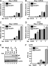
Effect of Tat-SF1 knockdown on virus infection. (A) Total RNA was prepared from HeLa cells transfected with control siRNA or siTat-SF1 and superinfected with influenza virus. Real-time RT-PCR was carried out with primer sets specific for M1 mRNA, M2 mRNA, and β-actin mRNA. (B) Representation of M1 and M2 mRNA generated from segment 7. (C) The ratio of the amount of M2 mRNA to that of M1 mRNA in HeLa cells transfected with control siRNA or siTat-SF1 and those infected with influenza virus. (D–G) HeLa cells were transfected with control siRNA or siTat-SF1. At 72 hpt, cells were transfected with pCAGGS-FLAG-rTat-SF1 and pCAGGS-empty plasmids. After 24-h incubation, cells were superinfected with influenza virus. Total RNA was prepared from cells at 3 h postinfection (hpi). Real-time RT-PCR was carried out with primer sets specific for endogenous Tat-SF1 (D Left), exogenous FLAG-rTat-SF1 (D Right), segment 7 RNAs (E, cRNA; F, mRNA; G, vRNA), and β-actin mRNA. The results are normalized as the ratio to the level of β-actin mRNA. Error bars show standard deviation. (H) Western blot analyses of viral proteins. Mock- (lanes 1, 3, 5, and 7) or siTat-SF1 siRNA-transfected (lanes 2, 4, 6, and 8) HeLa cells were infected with influenza virus at an moi of 5. Western blot analyses were carried with anti-NP, -PB1, or -β-actin antibodies. (I) Single-step virus growth. Mock- or siTat-SF1 siRNA-transfected HeLa cells were infected with influenza virus at an moi of 0.1. Virus titer was examined by plaque assay at the indicated times after infection.
Fig. 3.
Fig. 3.
Tat-SF1 as a stimulatory host factor involved in virus RNA synthesis. (A–D) Viral RNA synthesis in the presence of a protein synthesis inhibitor. HeLa cells expressing FLAG-Tat-SF1 (Tat-SF1) or control (Neo) HeLa cells were infected with influenza virus in the absence (A–C) or presence (D) of 100 μg/ml CHX. Real-time RT-PCR was carried out with primer sets for segment 3 cRNA (A), segment 3 mRNA (B and D), segment 3 vRNA (C), and β-actin mRNA. Error bars show standard deviation. (E) Western blot analyses of viral proteins. HeLa cells expressing FLAG-Tat-SF1 (lanes 2, 4, and 6) or control HeLa cells (lanes 1, 3, and 5) were infected with influenza virus at an moi of 1. Western blot analyses were carried out with anti-NP, -PB1, or -β-actin antibodies. (F) Single-step virus growth. HeLa cells expressing FLAG-Tat-SF1 or control HeLa cells were infected with influenza virus at an moi of 0.1. Virus titer was examined as described.
Fig. 4.
Fig. 4.
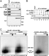
Stimulatory activity of Tat-SF1 in vitro. (A) Interaction between Tat-SF1 and viral proteins. Immunoprecipitation assays were carried out by anti-myc antibody-conjugated agarose beads using purified His-myc-Tat-SF1 (lanes 6–8) and either vRNP (lanes 4 and 7) or mnRNP (lanes 5 and 8). Affinity beads were washed with immunoprecipitation buffer containing 300 mM KCl. Western blot analyses were carried out with anti-NP, -PB1, -PB2, -PA, or -myc antibodies. Input (20%) is shown in lanes 1 and 2. (B) Dissociation of NP-Tat-SF1 complexes by the addition of RNA. An NP-Tat-SF1 complex was reconstituted by mixing purified His-myc-Tat-SF1 (lanes 3 and 5–7) and mnRNP (lanes 4–7). The mixtures were further incubated in the presence of v53-mer RNA [20 ng (lane 6) and 200 ng (lane 7)]. After immunoprecipitation assays using anti-myc antibody-conjugated agarose, Western blot analyses were carried with anti-NP or -myc antibodies. Input (20%) is shown in lane 1. (C) Tat-SF1-mediated NP-RNA complex formation. 32P-labeled RNA probe (v53 mer RNA) mixed with recombinant NP was incubated in the presence of BSA (lanes 1–9) or recombinant Tat-SF1 (lanes 10–18), and v53 mer RNA probe mixed with recombinant Tat-SF1 was incubated in the absence of recombinant NP (lanes 19–27). After sedimentation through a 15–35% glycerol density gradient, fractions were collected from the top of the tube. An aliquot of each fraction was analyzed by PAGE on a 6% polyacrylamide gel, and the RNA was visualized by autoradiography. The unbound RNA probe and NP-RNA complexes are indicated by arrowheads.

Similar articles

Cited by

References

    1. Bullock PA. Crit Rev Biochem Mol Biol. 1997;32:503–568. - PubMed
    1. Lindenbach BD, Evans MJ, Syder AJ, Wolk B, Tellinghuisen TL, Liu CC, Maruyama T, Hynes RO, Burton DR, McKeating JA, Rice CM. Science. 2005;309:623–626. - PubMed
    1. Prentice E, McAuliffe J, Lu X, Subbarao K, Denison MR. J Virol. 2004;78:9977–9986. - PMC - PubMed
    1. Scholle F, Girard YA, Zhao Q, Higgs S, Mason PW. J Virol. 2004;78:11605–11614. - PMC - PubMed
    1. Ahlquist P, Noueiry AO, Lee WM, Kushner DB, Dye BT. J Virol. 2003;77:8181–8186. - PMC - PubMed

LinkOut - more resources