Structural studies of neuropilin/antibody complexes provide insights into semaphorin and VEGF binding
- PMID: 17989695
- PMCID: PMC2099469
- DOI: 10.1038/sj.emboj.7601906
Structural studies of neuropilin/antibody complexes provide insights into semaphorin and VEGF binding
Abstract
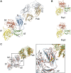
Neuropilins (Nrps) are co-receptors for class 3 semaphorins and vascular endothelial growth factors and important for the development of the nervous system and the vasculature. The extracellular portion of Nrp is composed of two domains that are essential for semaphorin binding (a1a2), two domains necessary for VEGF binding (b1b2), and one domain critical for receptor dimerization (c). We report several crystal structures of Nrp1 and Nrp2 fragments alone and in complex with antibodies that selectively block either semaphorin or vascular endothelial growth factor (VEGF) binding. In these structures, Nrps adopt an unexpected domain arrangement in which the a2, b1, and b2 domains form a tightly packed core that is only loosely connected to the a1 domain. The locations of the antibody epitopes together with in vitro experiments indicate that VEGF and semaphorin do not directly compete for Nrp binding. Based upon our structural and functional data, we propose possible models for ligand binding to neuropilins.
Figures


Similar articles
-
Neuropilin structure governs VEGF and semaphorin binding and regulates angiogenesis.Angiogenesis. 2008;11(1):31-9. doi: 10.1007/s10456-008-9097-1. Epub 2008 Feb 19. Angiogenesis. 2008. PMID: 18283547 Review.
-
Structural and functional relation of neuropilins.Adv Exp Med Biol. 2002;515:55-69. doi: 10.1007/978-1-4615-0119-0_5. Adv Exp Med Biol. 2002. PMID: 12613543 Review.
-
VEGF-A121a binding to Neuropilins - A concept revisited.Cell Adh Migr. 2018 May 4;12(3):204-214. doi: 10.1080/19336918.2017.1372878. Epub 2017 Nov 2. Cell Adh Migr. 2018. PMID: 29095088 Free PMC article. Review.
-
Neuropilins lock secreted semaphorins onto plexins in a ternary signaling complex.Nat Struct Mol Biol. 2012 Dec;19(12):1293-9. doi: 10.1038/nsmb.2416. Epub 2012 Oct 28. Nat Struct Mol Biol. 2012. PMID: 23104057 Free PMC article.
-
Structure of the semaphorin-3A receptor binding module.Neuron. 2003 Aug 14;39(4):589-98. doi: 10.1016/s0896-6273(03)00502-6. Neuron. 2003. PMID: 12925274
Cited by
-
Receptor complexes for each of the Class 3 Semaphorins.Front Cell Neurosci. 2012 Jul 5;6:28. doi: 10.3389/fncel.2012.00028. eCollection 2012. Front Cell Neurosci. 2012. PMID: 22783168 Free PMC article.
-
Architecture of the Sema3A/PlexinA4/Neuropilin tripartite complex.Nat Commun. 2021 May 26;12(1):3172. doi: 10.1038/s41467-021-23541-x. Nat Commun. 2021. PMID: 34039996 Free PMC article.
-
CMT2N-causing aminoacylation domain mutants enable Nrp1 interaction with AlaRS.Proc Natl Acad Sci U S A. 2021 Mar 30;118(13):e2012898118. doi: 10.1073/pnas.2012898118. Proc Natl Acad Sci U S A. 2021. PMID: 33753480 Free PMC article.
-
Semaphorins in angiogenesis and tumor progression.Cold Spring Harb Perspect Med. 2012 Jan;2(1):a006718. doi: 10.1101/cshperspect.a006718. Cold Spring Harb Perspect Med. 2012. PMID: 22315716 Free PMC article. Review.
-
Cryo-electron Microscopic Analysis of Single-Pass Transmembrane Receptors.Chem Rev. 2022 Sep 14;122(17):13952-13988. doi: 10.1021/acs.chemrev.1c01035. Epub 2022 Jun 17. Chem Rev. 2022. PMID: 35715229 Free PMC article. Review.
References
-
- Antipenko A, Himanen JP, van Leyen K, Nardi-Dei V, Lesniak J, Barton WA, Rajashankar KR, Lu M, Hoemme C, Puschel AW, Nikolov DB (2003) Structure of the semaphorin-3A receptor binding module. Neuron 39: 589–598 - PubMed
-
- Beckmann G, Bork P (1993) An adhesive domain detected in functionally diverse receptors. Trends Biochem Sci 18: 40–41 - PubMed
-
- Chen H, Bagri A, Zupicich JA, Zou Y, Stoeckli E, Pleasure SJ, Lowenstein DH, Skarnes WC, Chedotal A, Tessier-Lavigne M (2000) Neuropilin-2 regulates the development of selective cranial and sensory nerves and hippocampal mossy fiber projections. Neuron 25: 43–56 - PubMed
-
- Chen H, Chedotal A, He Z, Goodman CS, Tessier-Lavigne M (1997) Neuropilin-2, a novel member of the neuropilin family, is a high affinity receptor for the semaphorins Sema E and Sema IV but not Sema III. Neuron 19: 547–559 - PubMed
Publication types
MeSH terms
Substances
Associated data
- Actions
- Actions
- Actions
- Actions
- Actions
- Actions
- Actions
LinkOut - more resources
Full Text Sources
Other Literature Sources
Molecular Biology Databases
Miscellaneous

