. 2008 Jan;28(1):108-21.
doi: 10.1128/MCB.01402-07. Epub 2007 Oct 29.

STAGA recruits Mediator to the MYC oncoprotein to stimulate transcription and cell proliferation

Affiliations

STAGA recruits Mediator to the MYC oncoprotein to stimulate transcription and cell proliferation

Xiaohui Liu et al. Mol Cell Biol. 2008 Jan.

Abstract

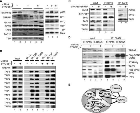
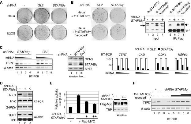
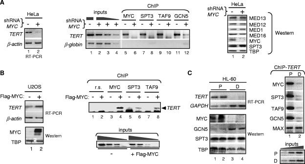
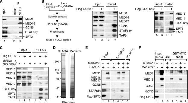
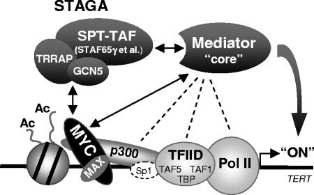
Activation of eukaryotic gene transcription involves the recruitment by DNA-binding activators of multiprotein histone acetyltransferase (HAT) and Mediator complexes. How these coactivator complexes functionally cooperate and the roles of the different subunits/modules remain unclear. Here we report physical interactions between the human HAT complex STAGA (SPT3-TAF9-GCN5-acetylase) and a "core" form of the Mediator complex during transcription activation by the MYC oncoprotein. Knockdown of the STAF65gamma component of STAGA in human cells prevents the stable association of TRRAP and GCN5 with the SPT3 and TAF9 subunits; impairs transcription of MYC-dependent genes, including MYC transactivation of the telomerase reverse transcriptase (TERT) promoter; and inhibits proliferation of MYC-dependent cells. STAF65gamma is required for SPT3/STAGA interaction with core Mediator and for MYC recruitment of SPT3, TAF9, and core Mediator components to the TERT promoter but is dispensable for MYC recruitment of TRRAP, GCN5, and p300 and for acetylation of nucleosomes and loading of TFIID and RNA polymerase II on the promoter. These results suggest a novel STAF65gamma-dependent function of STAGA-type complexes in cell proliferation and transcription activation by MYC postloading of TFIID and RNA polymerase II that involves direct recruitment of core Mediator.


Figures

FIG. 1.
FIG. 1.
Knockdown of STAF65γ affects the integrity of STAGA but not TFIID complexes in human cells. (A) STAF65γ-knockdown HeLa S3 cell lines. Western blot analyses were performed with whole-cell extracts from parental HeLa S3 cells (−) (lanes 1 and 2); two STAF65γ-knockdown HeLa S3 cell lines stably transfected with the STAF65γ shRNA vector pSUPER-STAF65γ828 (+) (clone 1 in lanes 3 and clone 4 in lane 4); and two control HeLa S3 cell lines (lanes 5 and 6) stably transfected with a different shRNA vector, pSUPER-STAF65γ218 (c), which retained normal STAF65γ protein levels. The indicated antibodies were used to probe different stripes of the blots (panels) shown. (B and C) Whole-cell extracts (input) of STAF65γ-knockdown cells (+) (clone 1) and control HeLa S3 cells stably transfected with the empty pSUPER vector (−) were immunoprecipitated with either preimmune rabbit serum (IP: r.s.) or TBP (IP: TBP), TAF5 (IP: TAF5), SPT3 (IP: SPT3), or TAF9 (IP: TAF9) antisera, and complexes were analyzed by SDS-polyacrylamide gel electrophoresis and Western blotting with specific antibodies to the indicated proteins. (D) Whole-cell extracts (input) of parental (−) and STAF65γ-knockdown (+) fh:SPT3 and fh:GCN5 cell lines were immunoprecipitated with the Flag antibody (IP: FLAG) and analyzed by Western blotting with antibodies to the indicated proteins. (E) Diagram summarizing the interaction results of panels C and D. Binary associations are shown as straight lines between two subunits (except for the direct interaction of STAF65γ with TAF10). Solid lines interrupted by STAF65γ indicate a strong dependency on STAF65γ for the corresponding binary association. Dashed lines indicate a partial dependency on STAF65γ. The large circle highlights SPT-TAF subunits.
FIG. 2.
FIG. 2.
STAF65γ is required for proliferation of HeLa cells and expression of MYC target genes and mediates MYC activation of TERT transcription. (A) Colony formation assays with HeLa and U2OS cells transfected with the empty pSUPER vector (−) or the shRNA-expressing vectors pSUPER-GL2 (GL2) and pSUPER-STAF65γ828 (STAF65γ). A representative result of three experiments done in duplicate is shown. (B) Colony formation assay as in panel A but with two HeLa cell lines stably expressing ectopic Flag-tagged STAF65γ (top) or STAF65γ “recoded” (bottom), as indicated. Right panels are Western blots probed with the Flag antibody of total extracts (input, lanes 1 to 4) and anti-Flag immunoprecipitated proteins (IP: Flag, lanes 5 to 8) from these two cell lines after transient transfection with pSUPER (−) or pSUPER-STAF65γ828 (+). Arrows point to the Flag-STAF65γ proteins. IgH, immunoglobulin heavy chain. (C) Knockdown of STAF65γ in HeLa cells specifically reduces TERT, CAD CDK4, and HSP60 mRNA levels. HeLa cells were transiently transfected with pSUPER-STAF65γ828 (STAF65γ) or pSUPER-GL2 (GL2). TERT (two alternative splicing variants) and β-actin transcripts were analyzed by RT-PCR of serial dilutions of total cDNA (mRNA); a negative image of an ethidium bromide-stained agarose gel is shown (left panel; RT-PCR). Whole-cell extracts from the above transfected cells (shRNA: STAF65γ and GL2) and from untransfected cells (−) were also analyzed by Western blotting with the indicated antibodies (middle panel, Western blot, lanes 1 to 3). The bar graph shows the quantitation of TERT, CAD, CDK4, and HSP60 transcripts in transfected cells by RT-PCR and serial dilution of total cDNA (mRNA). RT-PCR signals in STAF65γ shRNA-expressing cells (black bars) and in GL2 control-transfected cells (white bars) were normalized to β-actin signals at each dilution, and the normalized values for GL2 cells were arbitrarily set to 100% (white bars). (D) RT-PCR and Western blot analyses of whole-cell extracts from parental HeLa S3 (−), STAF65γ-knockdown HeLa S3 (+; clone 1), and stably transfected control HeLa S3 (c) cell lines shown in Fig. 1A. (E) Role of endogenous STAF65γ in TERT promoter activation by MYC. HeLa cells were transiently transfected with a TERT(−211/+41)-luciferase reporter gene construct and the pCMV-β-galactosidase reference gene and with expression vectors for Flag-MYC and the STAF65γ shRNA (+ and ++) or with corresponding empty vectors (−), as indicated. Relative luciferase activities (normalized to β-galactosidase) are the means ± standard deviation from three independent experiments performed in duplicate. Corresponding whole-cell extracts of transfected cells (duplicates) were analyzed by Western blotting (right panel; Western) with anti-Flag (Flag-MYC) and anti-TBP antibodies. (F) A “recoded” Flag-STAF65γ restores endogenous TERT transcription in STAF65γ-knockdown cells. Adherent HeLa cell lines stably transfected with the empty pSUPER vector (lanes 1 and 2) or with the pSUPER-STAF65γ828 (shRNA STAF65γ) vector (clone 7; lanes 3 to 8) were transiently transfected with the empty pIRESneo vector (lanes 1 to 4) or increasing amounts of pIRESneo-fh:STAF65γ “recoded”: 2 μg (lanes 5 and 6) and 4 μg (lanes 7 and 8). TERT and β-actin transcripts were analyzed by RT-PCR.
FIG. 3.
FIG. 3.
MYC recruits STAGA to the endogenous TERT promoter in human cells. (A) MYC knockdown decreases TERT transcription and recruitment of STAGA to the TERT promoter. MYC-knockdown (+ shRNA MYC) and control (−) HeLa cells were analyzed by RT-PCR (left panel) and by ChIP with MYC, SPT3, TAF9, and GCN5 antibodies, as indicated (middle panel). Lanes 1 to 4 show PCRs with serial dilutions of input chromatin (inputs). The TERT promoter region between −226 and +120 (containing the two E-boxes at −187 and +28) was amplified with specific primers (22). A region of the β-globin gene third intron was amplified as a nonspecific/background reference. Negative images of ethidium bromide-stained agarose gels are shown. A Western blot of whole-cell extracts from parental (−) and MYC-knockdown (+) HeLa S3 cells was probed with specific antibodies to the indicated proteins (right panel; Western). (B) Overexpression of Flag-MYC induces TERT transcription and STAGA binding to the endogenousTERT promoter in U2OS cells. Cells were transiently transfected with empty (−) or Flag-MYC (+) expression vectors and analyzed by RT-PCR and Western blotting (with MYC and TBP antibodies), and by ChIP, as described above. Preimmune rabbit serum (r.s.) was used in the control ChIP assays (lanes 1 and 2). (C) Proliferating (P) HL-60 cells were induced to differentiate (D), transcripts for TERT and GAPDH were analyzed by RT-PCR (top panel), and whole-cell extracts were analyzed by Western blotting with the indicated antibodies (bottom panel). Right panels show ChIP analyses of the TERT promoter with chromatin from P and D cells using antibodies to MYC, SPT3, TAF9, GCN5, and MAX.
FIG. 4.
FIG. 4.
STAF65γ is required for MYC-dependent recruitment of selected STAGA and core Mediator components to the endogenous TERT promoter in human cells. (A to D) Equivalent amounts of chromatin from control pSUPER-transfected (−), STAF65γ-knockdown (shRNA STAF65γ + in panels A to C) or MYC-knockdown (shRNA MYC + in panel D) HeLa S3 cell lines were used in ChIP experiments with specific antibodies or preimmune rabbit serum (r.s.), as indicated. AcH3 and AcH4 are anti-acetylated histone H3 and H4 antibodies. PCR analyses were performed with primers specific for the TERT and GAPDH promoters and for a region in the third intron of the β-globin gene, as in Fig. 3.
FIG. 5.
FIG. 5.
Efficient interaction of MYC with core Mediator requires STAF65γ and an intact TAD. (A) STAF65γ-dependent association of STAGA and core Mediator subunits with endogenous MYC/MAX in HeLa cells. MAX was immunoprecipitated with a specific antibody (IP: MAX) from whole-cell extracts (input) of control pSUPER-transfected (−) and STAF65γ-knockdown (+) HeLa S3 cell lines, and associated proteins were analyzed by Western blotting with specific antibodies to the indicated proteins. (B) HEK293 cells were transfected with an empty pCbS vector (−) or pCbS-Flag-MYC (+), and whole-cell extracts (input) were immunoprecipitated with the Flag antibody (IP: FLAG) and analyzed by Western blotting. (C) Western blot as in panel B, but with HEK293 cells transfected with pCbS-Flag-MYC (WT) or Δ1-110 (Δ110) together with pCbS-HA-MAX. The top stripe (MED12 + 13) was probed with both MED12 and MED13 antibodies. The lack of MAX signal in “input” (lanes 1 to 3) is due to the short exposure used to prevent saturation of the signals in IP: FLAG lanes 5 and 6. (D and E) GST-pulldown experiments were performed with equivalent amounts of GST, GST-VP16, GST-MYC, or GST-MYC(1-263) resins (as indicated) and with nuclear extracts (NE) from parental HeLa S3 cells (D) and pSUPER-transfected (−) or STAF65γ-knockdown (+) HeLa S3 cell lines (E). Input nuclear extracts (input) and proteins bound to GST, GST-VP16, and GST-MYC were analyzed by Western blotting.
FIG. 6.
FIG. 6.
Physical interaction of STAGA and Mediator in human cells and in vitro. (A) HeLa S3 nuclear proteins were immunoprecipitated (IP) with a MED1-specific antibody (lane 3) or protein G-agarose (mock, lane 2) and analyzed by Western blotting with specific antibodies to the indicated proteins. (B) Nuclear extracts (input) of HeLa S3 (−) and derivative cell lines expressing (+) Flag-GCN5 or Flag-STAF65γ were immunoprecipitated with the FLAG antibody M2 resin, and complexes were eluted with excess Flag peptide (Eluted), as indicated in the scheme to the left. Proteins were analyzed by Western blotting. (C) Nuclear extracts of HeLa S3 cells (lanes 1 and 4), fh:SPT3 cells (lanes 2 and 5), and fh:SPT3 cells expressing the STAF65γ shRNA (lanes 3 and 6) were immunoprecipitated with the Flag antibody resin (IP:FLAG; lanes 4 to 6) and analyzed by Western blotting. The weak (or undetectable) signals for STAF65γ in input lanes (B and C) are due to the use of a short autoradiographic exposure (same gel/exposure for input and IP lanes) to prevent saturation of signals in the IP lanes. STAF65γ levels in fh:SPT3 cells ± shRNA vector are shown in Fig. 1D (lanes 1 and 2). (D) Immunoaffinity-purified STAGA (Flag-SPT3; lane 1) and Mediator (Flag-Nut2; lane 2) complexes resolved by SDS-polyacrylamide gel electrophoresis and silver stained. Positions of size markers in kDa are shown. (E) Direct interaction of purified human STAGA and Mediator complexes in vitro. STAGA and Mediator complexes (shown in panel D) either alone or combined (as indicated by + and − signs) were immunoprecipitated with a MED1-specific antibody (IP: MED1) or with protein G agarose (IP: mock) and analyzed by Western blotting with the indicated antibodies. Elution buffer containing the Flag peptide was used instead of the corresponding complex (−) in lanes 3 and 4. (F) In vitro pull-down assays were performed with immobilized GST and GST-MYC(1-263) and affinity-purified Flag-SPT3 (STAGA) and/or Flag-Nut2 (Mediator) complexes, as indicated (− and + signs). Forty percent of each input complex (lanes 1 and 2) and GST/GST-MYC-bound proteins (lanes 3 to 7) was analyzed by Western blotting with antibodies to the indicated subunits.
FIG. 7.
FIG. 7.
Model for STAGA-dependent transcription activation of TERT by MYC involving recruitment and/or stabilization of a core Mediator complex at the promoter. Double arrows indicate direct interactions between MYC, STAGA, and Mediator described here and previously (44). Dashed lines indicate previously reported interactions of Mediator with p300, TFIID, and Pol II (see references and and references therein), which could also take place on the TERT promoter but are not sufficient for the stable recruitment of core Mediator in the absence of either MYC or the STAF65γ-dependent SPT-TAF subunits of STAGA. The binding of Sp1/3 to TERT GC-boxes (dashed oval Sp1) is expected but was not tested here. “(Ac)” indicates acetylation of nucleosomal histones H3 and H4, which is independent of the STAF65γ/SPT-TAF components of STAGA (see also Discussion).

Similar articles

Cited by

References

    1. Andrau, J. C., L. van de Pasch, P. Lijnzaad, T. Bijma, M. G. Koerkamp, J. van de Peppel, M. Werner, and F. C. Holstege. 2006. Genome-wide location of the coactivator mediator: binding without activation and transient Cdk8 interaction on DNA. Mol. Cell 22179-192. - PubMed
    1. Baek, H. J., Y. K. Kang, and R. G. Roeder. 2006. Human Mediator enhances basal transcription by facilitating recruitment of TFIIB during preinitiation complex assembly. J. Biol. Chem. 28115172-15181. - PubMed
    1. Barlev, N. A., A. V. Emelyanov, P. Castagnino, P. Zegerman, A. J. Bannister, M. A. Sepulveda, F. Robert, L. Tora, T. Kouzarides, B. K. Birshtein, and S. L. Berger. 2003. A novel human Ada2 homologue functions with Gcn5 or Brg1 to coactivate transcription. Mol. Cell. Biol. 236944-6957. - PMC - PubMed
    1. Belotserkovskaya, R., D. E. Sterner, M. Deng, M. H. Sayre, P. M. Lieberman, and S. L. Berger. 2000. Inhibition of TATA-binding protein function by SAGA subunits Spt3 and Spt8 at Gcn4-activated promoters. Mol. Cell. Biol. 20634-647. - PMC - PubMed
    1. Bhaumik, S. R., and M. R. Green. 2001. SAGA is an essential in vivo target of the yeast acidic activator Gal4p. Genes Dev. 151935-1945. - PMC - PubMed

Publication types

MeSH terms

LinkOut - more resources