Review
. 2007 Nov;7(11):834-46.
doi: 10.1038/nrc2256.

Models, mechanisms and clinical evidence for cancer dormancy

Affiliations
Review

Models, mechanisms and clinical evidence for cancer dormancy

Julio A Aguirre-Ghiso. Nat Rev Cancer. 2007 Nov.

Abstract

Patients with cancer can develop recurrent metastatic disease with latency periods that range from years even to decades. This pause can be explained by cancer dormancy, a stage in cancer progression in which residual disease is present but remains asymptomatic. Cancer dormancy is poorly understood, resulting in major shortcomings in our understanding of the full complexity of the disease. Here, I review experimental and clinical evidence that supports the existence of various mechanisms of cancer dormancy including angiogenic dormancy, cellular dormancy (G0-G1 arrest) and immunosurveillance. The advances in this field provide an emerging picture of how cancer dormancy can ensue and how it could be therapeutically targeted.


Figures
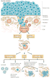
Figure 1
Figure 1. Tumour dormancy as a component of cancer progression
Tumour cells carrying genetic or epigenetic changes enabling motile and invasive properties can degrade the basement membrane and invade the underlying stroma. Invading tumour cells interact with fibroblasts or immune cells and the stromal matrix. Tumour cells (in cooperation with stromal cells) can degrade the extracellular matrix (ECM) and the vascular walls and intravasate (through either arterial or lymphatic routes). Tumour cells that arrest in the vasculature of the bone marrow can proliferate or remain dormant. Although the bone can be a target organ, it might also serve as a transit site from which cells can again disseminate, through as yet unknown mechanisms, to their final destination (that is, lungs, liver and so on, where they form metastases). Tumour cells in the bone marrow are not yet a metastasis but have the potential to become secondary lesions and they can also carry the information about the future progression of the disease,. Tumour cells can arrest in lymph nodes or in the target organ vasculature, where they can extravasate into the organ parenchyma. At this stage (this can also happen in the bone marrow or lymph nodes) intra- or extra-vascularly lodged tumour cells have four possible fates: they die (the vast majority of cells undergo apoptosis), they can enter a state of quiescence or dormancy, either as a single solitary cell or as a micrometastatic lesion that underwent a proliferative expansion and cannot recruit a vascular bed, or they can resume proliferation (growing micrometastases).
Figure 2
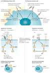
Figure 2. Manifestation of cancer dormancy
a Tumour cells that have accumulated genetic and epigenetic changes that provide a growth advantage (solid blue line) form a primary tumour. After a treatment that results in tumour regression, residual disease can be detectable for long periods thereafter (dashed blue line). After this time, the tumour mass can increase again, but now in distant secondary organs (dashed red lines). b During the dormancy stage, sub-clinical disease might be due to dormant cells that have entered a G0–G1 arrest (cellular dormancy) and these cells might develop mechanisms to evade immune system recognition and eradication. c Angiogenic dormancy results from the balance between pro- and anti-angiogenic factors (such as vascular endothelial growth factor (VEGF) and thrombospondin (TSP), respectively). Genetic alterations in the pathways that maintain angiogenic dormancy or an exogenous angiogenic ‘spike’ might restore tumour growth. Oncogenic Ras can induce VEGF and repress TSP. By contrast, the stress-activated kinase p38 and the tumour suppressor p53 can induce TSP or repress VEGF. Loss of function of p53 and/or p38 might tip the balance towards enhanced angiogenesis. d Immunosurveillance. Proliferating tumour cells are kept at low numbers (sub-clinical) by an active immune system. This can be due to cytotoxic CD8+ T lymphocytes or anti-idiotypic antibodies against the B-cell receptor that arrest the tumour cells. An interruption of this state of dormancy might be due to tumour cell escape from immune system control by downregulation of specific tumour-associated antigens or by expression of co-stimulatory molecules that induce apoptosis of cytotoxic CD8+ T lymphocytes. It is unclear whether these forms of dormancy are mutually exclusive, although they are probably not, how long they last or whether they occur at different times. It is possible that cellular dormancy most frequently precedes the immunosurveillance or the angiogenic dormancy phase.
Figure 3
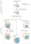
Figure 3. Signals that regulate cellular tumour dormancy
a A matching microenvironment and set of receptors allows metastatic cells to adapt and remodel their microenvironment to integrate growth-promoting signals. As an example (middle panel), signals derived from fibronectin (FN) and transduced by the uPAR (metastasis-associated urokinase receptor)–α5β1-integrin complex, focal adhesion kinase (FAK) and epidermal growth factor receptor (EGFR) can result in extracellular signal-regulated kinase (ERK) activation and p38 inactivation in an expanding tumour. In this scenario metastases might arise from disseminated tumour cells that acquire additional genetic abnormalities, re-activation of uPAR and mitogenic signalling (ERBB2 or EGFR) or from the expansion of tumour stem cells (not depicted). b Loss of a surface receptor (for example, uPAR, α5β1 integrin or EGFR) that transduces growth signals from the microenvironment (for example, fibronectin) results in stress signalling (low FAK–Ras–ERK, and high CDC42 (cell division cycle 42)–p38 activity; middle panel), which in turn might lead to dormancy. This is one example to illustrate the theme of crosstalk between the microenvironment and/or receptor signalling in cellular dormancy (for others see REFS 20,21,29,56,84,124,125). It is likely that other unidentified pathways are also involved. The bottom panels illustrate the possibility that some of the molecules that are found in experimental models (such as that shown in the middle panels) may also regulate the fate of disseminated tumour cells. Those disseminated tumour cells that are cytokeratin (CK) positive, have low uPAR expression and reduced ERBB2 signalling, might be dormant and negative for proliferation markers. Re-expression of uPAR and/or ERBB2 could allow these cells to escape dormancy.
Figure 4
Figure 4. An integrated view of cancer metastasis dormancy
Tumour cells that survive dissemination lodge in the target organ parenchyma. This new microenvironment most probably determines the fate of the disseminated tumour cells and could account for most of the dormancy time (Time #1). If the cells are not genetically progressed it is possible that they are unable to grow autonomously or transduce growth signals from the microenvironment, instead entering a quiescence-like phenotype. Stress from dissemination might contribute to activating growth arrest programmes. Even with genetic alterations, stress and/or microenvironment signals might impose a growth-suppressive programme. For tumour stem cells, a quiescent state might be a natural response to a microenvironment that lacks recruitment signals. Normal differentiated cells can remain growth arrested for years and solitary cells are found years after surgery, suggesting that a prolonged tumour cell arrest might be plausible. Upon exit from quiescence, tumour cells can fully progress into overt lesions. It is possible that before becoming overt lesions, dormancy might continue (Time #2) owing to the immune system preventing tumour expansion. The immune system can control pathogens during a lifetime. Therefore, it might prevent tumour mass expansion for long periods. After exit from quiescence or evasion of the immune system a tumour cell mass can enter angiogenic dormancy. Differentiated tissues such as the retinal pigment epithelium, which produces angiogenesis inhibitors (pigment epithelium-derived factor 126), can maintain the vasculature from expanding for long periods and therefore prevent diseases such as macular degeneration. However, it is still unclear how long (Time #3) this mechanism can be maintained in a genetically unstable proliferative tumour cell population, which probabilistically should be prone to accumulating new genetic alterations that activate the angiogenic switch.

Similar articles

Cited by

References

    1. Pantel K, Brakenhoff RH. Dissecting the metastatic cascade. Nature Rev Cancer. 2004;4:448–456. - PubMed
    1. Schmidt-Kittler O, et al. From latent disseminated cells to overt metastasis: genetic analysis of systemic breast cancer progression. Proc Natl Acad Sci U S A. 2003;100:7737–7742.First evidence that breast cancer cells can disseminate in a far less progressed genomic state than previously thought. Genomic aberrations that make them metastatic are acquired after this step.

    1. Karrison TG, Ferguson DJ, Meier P. Dormancy of mammary carcinoma after mastectomy. J Natl Cancer Inst. 1999;91:80–85. - PubMed
    1. Pfitzenmaier J, et al. Telomerase activity in disseminated prostate cancer cells. BJU Int. 2006;97:1309–1313. - PubMed
    1. Weckermann D, et al. Disseminated cytokeratin positive tumor cells in the bone marrow of patients with prostate cancer: detection and prognostic value. J Urol. 2001;166:699–703. - PubMed

Publication types

MeSH terms

Substances