. 2008 Jan;28(1):358-75.
doi: 10.1128/MCB.01214-07. Epub 2007 Oct 22.

Dominant TEL1-hy mutations compensate for Mec1 lack of functions in the DNA damage response

Affiliations

Dominant TEL1-hy mutations compensate for Mec1 lack of functions in the DNA damage response

Veronica Baldo et al. Mol Cell Biol. 2008 Jan.

Abstract

Eukaryotic genome integrity is safeguarded by two highly conserved protein kinases that are called ATR and ATM for humans and Mec1 and Tel1 for Saccharomyces cerevisiae. Although they share sequence similarities and substrates, these protein kinases perform different specialized functions. In particular, Mec1 plays a key role in the DNA damage checkpoint response, whereas Tel1 primarily is involved in telomere homeostasis, and its checkpoint function is masked by the prevailing activity of Mec1. In order to understand how this specificity is achieved, we searched for TEL1 mutations able to compensate for the lack of Mec1 functions. Here, we describe seven independent dominant TEL1-hy alleles that are able to suppress, to different extents, both the hypersensitivity to genotoxic agents and the checkpoint defects of Mec1-deficient cells. Most of these alleles also cause telomere overelongation. In vitro kinase activity was increased compared to that of wild-type Tel1 in the Tel1-hy385, Tel1-hy394, Tel1-hy680, and Tel1-hy909 variants, but its activity was not affected by the TEL1-hy184 and TEL1-hy628 mutations and was slightly reduced by the TEL1-hy544 mutation. Thus, the phenotypes caused by at least some Tel1-hy variants are not simply the consequence of improved catalytic activity. Further characterization shows that Tel1-hy909 not only can sense and signal a single double-stranded DNA break, unlike wild-type Tel1, but also contributes more efficiently than Tel1 to single-stranded DNA accumulation at double-strand ends, thus enhancing Mec1 signaling activity. Moreover, it causes unscheduled checkpoint activation in unperturbed conditions and upregulates the checkpoint response to small amounts of DNA lesions. Finally, Tel1-hy544 can activate the checkpoint more efficiently than wild-type Tel1, while it causes telomere shortening, indicating that the checkpoint and telomeric functions of Tel1 can be separable.


Figures

FIG. 1.
FIG. 1.
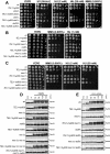
Hypersensitivity to genotoxic agents and cell viability of mec1Δ cells carrying the TEL1-hy alleles. (A) Serial dilutions of wild-type (W303; TEL1 MEC1 SML1), TEL1 mec1Δ sml1Δ (YLL490), TEL1-hy184 mec1Δ sml1Δ, TEL1-hy385 mec1Δ sml1Δ, TEL1-hy394 mec1Δ sml1Δ, TEL1-hy544 mec1Δ sml1Δ, TEL1-hy628 mec1Δ sml1Δ, TEL1-hy680 mec1Δ sml1Δ, and TEL1-hy909 mec1Δ sml1Δ strains, exponentially growing in YEPD, were spotted on plates with or without MMS or HU at the indicated concentrations. (B) Serial dilutions (1:10) of wild-type (W303; TEL1/TEL1 MEC1/MEC1 SML1/SML1), TEL1/TEL1 mec1Δ/mec1Δ sml1Δ/sml1Δ, and TEL1-hy/TEL1 mec1Δ/mec1Δ sml1Δ/sml1Δ diploid strains, exponentially growing in YEPD, were spotted on plates with or without MMS or HU at the indicated concentrations. (C) Serial dilutions of wild-type (W303; TEL1 MEC1 SML1), TEL1 mec1Δ, TEL1-hy184 mec1Δ, TEL1-hy385 mec1Δ, TEL1-hy394 mec1Δ, TEL1-hy544 mec1Δ, TEL1-hy628 mec1Δ, TEL1-hy680 mec1Δ, and TEL1-hy909 mec1Δ strains, all carrying a wild-type copy of the SML1 gene and the MEC1 gene on a centromeric URA3 plasmid, were spotted on SC-Ura (SC without uracil) plates and on SC plates with 5-FOA.
FIG. 2.
FIG. 2.
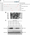
Tel1-hy amino acid changes and in vitro kinase activity. (A) The C-terminal part of wild-type Tel1, spanning amino acid residues 1700 to 2787, is schematically depicted in the top part, with a gray box highlighting the C-terminal domain (between amino acids 2460 and 2755) that is shared by a number of PI3-like kinases, including mammalian ATM, ATR, DNA-PK, and MTOR; S. pombe Tel1 and Rad3; and S. cerevisiae Tel1, Mec1, and Tor1. Black lines inside the gray block indicate the G2611, D2612, N2616, and D2631 amino acid residues that have been shown to be essential for Tel1 kinase activity (25). Black horizontal lines represent the same C-terminal region in the Tel1-hy variants indicated on the left, with the corresponding amino acid changes listed on the right. Vertical lines indicate the position of the amino acid changes, with red lines highlighting changes affecting conserved amino acid residues based on a ClustalW alignment of the whole Tel1 and human ATM amino acid sequences. (B) ClustalW alignment of Tel1 amino acid sequence between residues 2679 and 2708, spanning the conserved PI3 kinase domain, with the corresponding sequences of PI3-like kinases indicated on the left. Identical amino acids are indicated by black boxes, and similar ones are indicated by different shades of gray. An asterisk indicates the position corresponding to the N2692D alteration in Tel1-hy385. (C) Immunoprecipitation and in vitro kinase assays of HA-tagged Tel1 and Tel1-hy proteins. The strains used were TEL1-hy184-HA (YLL2104), TEL1-hy385-HA (YLL1974), TEL1-hy394-HA (YLL1975), TEL1-hy544-HA (YLL1976), TEL1-hy628-HA (YLL1977), TEL1-hy680-HA (YLL1978), TEL1-hy909-HA (YLL1979), TEL1-HA (DMP4690/9A), and TEL1 (YLL490), all carrying the deletions of both MEC1 and SML1. Kinase assays and Western blot analysis (see Materials and Methods) were performed on equal amounts of anti-HA immunoprecipitates of protein extracts from exponentially growing untreated cells. Products of a kinase reaction using γ-32P-labeled ATP were analyzed by SDS-polyacrylamide gel electrophoresis (kinase assay). All of the immunoprecipitates also were subjected to Western blot analysis using anti-HA antibodies (western blot).
FIG. 3.
FIG. 3.
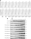
G1/S DNA damage checkpoint in TEL1-hy mec1Δ cells. Cell cultures of wild-type (W303; TEL1 MEC1), TEL1 mec1Δ sml1Δ (YLL490), TEL1-hy184 mec1Δ sml1Δ, TEL1-hy385 mec1Δ sml1Δ, TEL1-hy394 mec1Δ sml1Δ, TEL1-hy544 mec1Δ sml1Δ, TEL1-hy628 mec1Δ sml1Δ, TEL1-hy680 mec1Δ sml1Δ, and TEL1-hy909 mec1Δ sml1Δ strains, logarithmically growing in YEPD at 25°C, were synchronized in G1 with α-factor, UV irradiated (30 J/m2), and released from α-factor at time zero in YEPD at 25°C. Samples were withdrawn at the indicated times after α-factor release to analyze the DNA content by a fluorescence-activated cell sorter in unirradiated (A, top) and UV-irradiated (A, bottom) cell cultures, as well as Rad53 phosphorylation in UV-irradiated cell cultures by Western blot analysis with anti-Rad53 antibodies (B). Time zero corresponds to cell samples withdrawn immediately before UV irradiation and release from α-factor. exp, exponentially growing cells.
FIG. 4.
FIG. 4.
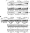
Bleomycin-induced DNA damage checkpoint in TEL1-hy mec1Δ cells. (A) Exponentially growing (exp) cell cultures of wild-type (W303; TEL1 MEC1), TEL1 mec1Δ sml1Δ (YLL490), TEL1-hy184 mec1Δ sml1Δ, TEL1-hy385 mec1Δ sml1Δ, TEL1-hy394 mec1Δ sml1Δ, TEL1-hy544 mec1Δ sml1Δ, TEL1-hy628 mec1Δ sml1Δ, TEL1-hy680 mec1Δ sml1Δ, and TEL1-hy909 mec1Δ sml1Δ strains were synchronized in G2 with nocodazole (15 μg/ml) (noc) and resuspended in YEPD containing nocodazole in the presence of 20 mU/ml bleomycin (+noc +bleo). Protein extracts prepared from cell samples collected at the indicated times after the addition of bleomycin were subjected to Western blot analysis with anti-Rad53 antibodies. (B) Exponentially growing cell cultures of wild-type (W303; TEL1 MEC1 XRS2), TEL1 xrs2-11 mec1Δ sml1Δ (DMP4973/10C), TEL1 MEC1 xrs2-11 sml1Δ (DMP4971/9D), TEL1-hy184, TEL1-hy385, TEL1-hy394, TEL1-hy544, TEL1-hy628, TEL1-hy680, and TEL1-hy909 strains, all carrying deletions of MEC1, SML1, and the xrs2-11 allele, were synchronized with nocodazole (noc) and resuspended in YEPD containing nocodazole in the presence of 20 mU/ml bleomycin (+noc +bleo). Protein extracts prepared from cell samples collected at the indicated times were subjected to Western blot analysis with anti-Rad53 antibodies.
FIG. 5.
FIG. 5.
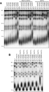
Telomere length in TEL1-hy mutants. (A) Genomic DNA was prepared from wild-type (W303; TEL1), TEL1 mec1Δ sml1Δ (YLL490), and TEL1 sml1Δ (YLL488) cell cultures and from cultures of cells carrying the TEL1-hy184, TEL1-hy385, TEL1-hy394, TEL1-hy544, TEL1-hy628, TEL1-hy680, and TEL1-hy909 alleles, either alone or together with the deletion of MEC1 and/or SML1, exponentially growing in YEPD for more than 80 generations. DNA was digested with XhoI and hybridized with a poly(GT) telomere-specific probe. (B) Genomic DNA was prepared from wild-type (W303; TEL1) and TEL1 mre11Δ (YLL936) cell cultures and from cultures of cells carrying the TEL1-hy184, TEL1-hy385, TEL1-hy394, TEL1-hy544, TEL1-hy628, TEL1-hy680, and TEL1-hy909 alleles, as well as either the mre11Δ or MRE11 allele, exponentially growing in YEPD for more than 80 generations. DNA was digested with XhoI and hybridized with a poly(GT) telomere-specific probe.
FIG. 6.
FIG. 6.
Tel1-hy909 requires Rad53 and its mediators, Rad9 and Mrc1, for DNA damage response. (A) Serial dilutions of wild-type (wt; W303), TEL1-hy909, ddc2Δ sml1Δ (DMP2995/1B), TEL1-hy909 ddc2Δ sml1Δ (DMP4967/2D), ddc1Δ (DMP2056/7B), TEL1-hy909 ddc1Δ (DMP4712/1D), rad53Δ sml1Δ (YLL509), and TEL1-hy909 rad53Δ sml1Δ (DMP4713/7B) cell cultures, exponentially growing in YEPD, were spotted on plates with or without MMS or HU at the indicated concentrations. One YEPD plate was exposed at the indicated UV dose. (B) Serial dilution of wild-type, TEL1-hy909, mec1Δ sml1Δ (YLL490), TEL1-hy909 mec1Δ sml1Δ, rad9Δ (DMP1911/1C), TEL1-hy909 rad9Δ (DMP4711/3C), rad9Δ mec1Δ sml1Δ (DMP4726/6C), and TEL1-hy909 rad9Δ mec1Δ sml1Δ (DMP4726/12A) cell cultures, exponentially growing in YEPD, were spotted on plates with or without MMS or HU at the indicated concentrations. (C) Serial dilution of wild-type, TEL1-hy909, mec1Δ sml1Δ (YLL490), TEL1-hy909 mec1Δ sml1Δ, mrc1Δ (YLL1310), TEL1-hy909 mrc1Δ (DMP4714/2D), rad9Δ mrc1Δ sml1Δ (DMP4742/4C), and TEL1-hy909 rad9Δ mrc1Δ sml1Δ (DMP4732/1A) cell cultures, exponentially growing in YEPD, were spotted on plates with or without MMS or HU at the indicated concentrations. (D) Exponentially growing (exp) cell cultures of wild-type, TEL1-hy909, mec1Δ sml1Δ (YLL490), TEL1-hy909 mec1Δ sml1Δ, rad9Δ (DMP1911/1C), TEL1-hy909 rad9Δ (DMP4711/3C), mrc1Δ (YLL1310), mec1Δ mrc1Δ sml1Δ (DMP4774/16C), TEL1-hy909 mec1Δ mrc1Δ sml1Δ (DMP4774/41D), and TEL1-hy909 mec1Δ mrc1Δ rad9Δ sml1Δ (DMP4831/34D) strains were arrested in G1 with α-factor (αf) and released into the cell cycle in YEPD containing 0.01% MMS (+MMS). Protein extracts prepared from cell samples collected at the indicated times after MMS addition were subjected to Western blot analysis with anti-Rad53 antibodies. (E) Exponentially growing cell cultures of wild-type, TEL1-hy909, mec1Δ sml1Δ (YLL490), TEL1-hy909 mec1Δ sml1Δ, rad9Δ (DMP1911/1C), TEL1-hy909 rad9Δ (DMP4711/3C), rad9Δ mec1Δ sml1Δ (DMP4726/6C), and TEL1-hy909 rad9Δ mec1Δ sml1Δ (DMP4726/12A) strains were arrested in G2 with nocodazole (noc) and resuspended in YEPD containing nocodazole in the presence of 3 μg/ml phleomycin (+phleo +noc). Protein extracts prepared from cell samples collected at the indicated times after phleomycin addition were subjected to Western blot analysis with anti-Rad53 antibodies.
FIG. 7.
FIG. 7.
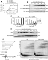
TEL1-hy909 cell response to a single irreparable DSB. (A and B) Cell cultures of wild-type (JKM139; TEL1 MEC1), TEL1 mec1Δ sml1Δ (DMP4894/10A), and TEL1-hy909 mec1Δ sml1Δ (YLL1939) strains, exponentially growing in YEP+lac (time zero), were transferred to YEP+lac+gal to induce HO expression. Samples were collected at the indicated times after galactose addition to determine the percentage of mononucleate large-budded cells (A) and for Western blot analysis of protein extracts with anti-Rad53 antibodies (B). (C) YEP+lac G1-arrested cell cultures of wild-type (JKM139; TEL1) and isogenic TEL1-hy909 (DMP4740/10B) strains, both expressing wild-type MEC1, were spotted on galactose-containing plates, which were incubated at 30°C (time zero). At the indicated time points, 200 cells for each strain were analyzed to determine the frequency of single cells and of cells forming microcolonies of two, four, or more than four cells. (D) Cell cultures of strains shown in panel C, exponentially growing in YEP+lac (time zero), were transferred to YEP+lac+gal to induce HO expression. Protein extracts from aliquots withdrawn at the indicated times after galactose addition were subjected to Western blot analysis with anti-Rad53 antibodies. (E) YEP+lac nocodazole-arrested cell cultures of wild-type JKM139 and isogenic TEL1-hy909 (DMP4740/10B) strains (time zero) were transferred to YEP+lac+gal to induce HO expression in the presence of nocodazole. (E, left) Schematic representation of the region immediately centromere distal to the MAT HO site and of the DSB and 5′- to 3′-resection products detectable with the indicated ssRNA probe after alkaline gel electrophoresis of SspI (S)-digested DNA. The probe is specific for the MAT locus and reveals a 1.1-kb fragment from the uncut MAT locus. When HO cuts the MAT locus, a smaller 0.9-kb HO-cut fragment is produced. 5′ to 3′ resection progressively eliminates SspI sites, generating larger ssDNA SspI fragments (r1 through r7) detected by the probe. (E, right) Genomic DNA prepared from samples taken at the indicated times during the experiment was digested with SspI and run on an alkaline agarose gel, followed by gel blotting and hybridization with the ssRNA probe shown on the left. wt, wild type.
FIG. 8.
FIG. 8.
Checkpoint activation in TEL1-hy909 MEC1 cells during an unperturbed cell cycle or in response to mild genotoxic treatments. (A) Growth rate of asynchronous wild-type (W303; TEL1) and TEL1-hy909 cell cultures growing exponentially at 25°C. (B and C) Exponentially growing cell cultures of wild-type (W303; TEL1), TEL1-hy909, rad9Δ (DMP1911/1C), and TEL1-hy909 rad9Δ (DMP4711/3C) strains, all expressing wild-type MEC1, were arrested in G1 with α-factor (0) and released from the pheromone block in YEPD at 21°C. When 95% of cells had budded after release, 3 μg/ml α-factor was added back to all cultures. Samples were collected at the indicated times after α-factor release to analyze the DNA content by a fluorescence-activated cell sorter (B) and to determine the kinetics of nuclear division after propidium iodide staining (C). wt, wild type. (D) Cell cultures of exponentially growing (exp) wild-type (W303; TEL1) and TEL1-hy909 strains were UV irradiated (10 or 20 J/m2) or were resuspended in YEPD containing 50 mM HU (+HU), 0.005% MMS (+MMS), or 0.5 μg/ml phleomycin (+phleo). Protein extracts prepared from cell samples collected at the indicated times after genotoxic treatment were subjected to Western blot analysis with anti-Rad53 antibodies. (E) Serial dilution of wild type (W303; TEL1) and TEL1-hy909 cell cultures, exponentially growing in YEPD, were spotted on plates with or without phleomycin, MMS, and HU at the indicated concentrations. One YEPD plate was exposed to the indicated UV dose.
FIG. 9.
FIG. 9.
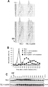
Prolonged UV-induced checkpoint activation in TEL1-hy909 cells expressing functional Mec1. Exponentially growing (exp) cell cultures of wild-type (W303; TEL1) and TEL1-hy909 strains, both expressing wild-type MEC1, were arrested in G1 with α-factor (0) and released from the pheromone block in YEPD or were UV irradiated (30 J/m2) prior to the release in YEPD. When 95% of cells had budded after release, 3 μg/ml α-factor was added back to all cultures. Samples of untreated and UV-treated cell cultures were collected at the indicated times after α-factor release to analyze the DNA content by a fluorescence-activated cell sorter (A) and to determine the kinetics of nuclear division after propidium iodide staining (B) and Rad53 phosphorylation by Western blot analysis with anti-Rad53 antibodies (C).

Similar articles

Cited by

References

    1. Adams, K. E., A. L. Medhurst, D. A. Dart, and N. D. Lakin. 2006. Recruitment of ATR to sites of ionising radiation-induced DNA damage requires ATM and components of the MRN protein complex. Oncogene 253894-3904. - PMC - PubMed
    1. Alcasabas, A. A., A. J. Osborn, J. Bachant, F. Hu, P. J. Werler, K. Bousset, K. Furuya, J. F. Diffley, A. M. Carr, and S. J. Elledge. 2001. Mrc1 transduces signals of DNA replication stress to activate Rad53. Nat. Cell Biol. 3958-965. - PubMed
    1. Bakkenist, C. J., and M. B. Kastan. 2003. DNA damage activates ATM through intermolecular autophosphorylation and dimer dissociation. Nature 421499-506. - PubMed
    1. Bi, X., S. C. Wei, and Y. S. Rong. 2004. Telomere protection without a telomerase; the role of ATM and Mre11 in Drosophila telomere maintenance. Curr. Biol. 141348-1353. - PubMed
    1. Chakhparonian, M., D. Faucher, and R. J. Wellinger. 2005. A mutation in yeast Tel1p that causes differential effects on the DNA damage checkpoint and telomere maintenance. Curr. Genet. 48310-322. - PubMed

Publication types

MeSH terms