. 2007 Dec;75(12):5586-96.
doi: 10.1128/IAI.01020-07. Epub 2007 Oct 1.

Chlamydia pneumoniae inclusion membrane protein Cpn0585 interacts with multiple Rab GTPases

Affiliations

Chlamydia pneumoniae inclusion membrane protein Cpn0585 interacts with multiple Rab GTPases

Claudio Cortes et al. Infect Immun. 2007 Dec.

Abstract

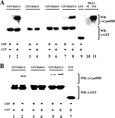
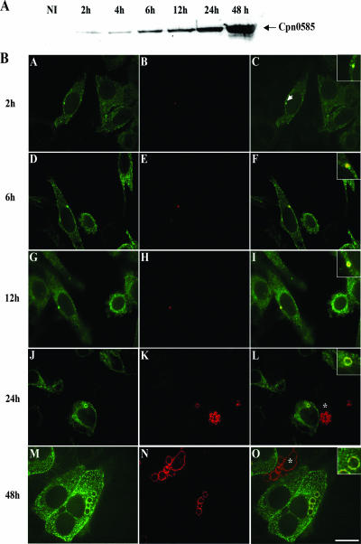
Chlamydiae are intracellular bacteria that develop within a membrane-bound vacuole called an inclusion. To ensure that the inclusion is a safe niche for chlamydial replication, chlamydiae exploit a number of host cell processes, including membrane-trafficking pathways. Recently, several Rab GTPases were found to associate with the inclusions of various chlamydial species. Here we report that Cpn0585, a Chlamydia pneumoniae inclusion membrane protein (Inc), interacts with multiple Rab GTPases. The results from yeast two-hybrid experiments revealed that an amino-terminally truncated form of Cpn0585 (Cpn0585(102-651)) interacts with Rab1, Rab10, and Rab11 but not with Rab4 or Rab6. Cpn0585-Rab GTPase interactions are direct and GTP dependent as shown in glutathione S-transferase pull-down assays using native and recombinant Cpn0585. In C. pneumoniae-infected HEp-2 cells transfected with enhanced green fluorescent protein (EGFP)-tagged Rab GTPases, the colocalization with Cpn0585 at the inclusion membrane was partial for EGFP-Rab1 and EGFP-Rab10, but extensive for wild-type EGFP-Rab11A and the constitutively active GTPase-deficient EGFP-Rab11AQ70L. Moreover, Cpn0585 colocalized with EGFP-Rab11AQ70L as early as 2 h postinfection. Upon delivery into live C. pneumoniae-infected cells, Cpn0585(628-651)-specific antibodies bound to the inclusion membrane, demonstrating that the Rab GTPase-interacting domain of Cpn0585 faces the host cell cytosol. Finally, ectopic expression of Cpn0585(102-651) partially inhibited the development of C. pneumoniae inclusions in EGFP. but not in EGFP-Rab11AQ70L-expressing HEp-2 cells. Collectively, these data suggest that Cpn0585 is involved in the recruitment of Rab GTPases to the inclusion membrane and that interfering with this function may adversely impact the fitness of the C. pneumoniae inclusion for chlamydial replication.


Figures

FIG. 1.
FIG. 1.
Schematic representation of Cpn0585: predicted secondary structures, similarity with Rab GTPase-interacting proteins, and location of CD8+ T-cell epitopes. As determined with the Kyte-Doolittle and Berger algorithms (6, 28), Cpn0585 contains a bilobed hydrophobic region located near its N terminus (black boxes) and six segments with high predicted probability (55 to 93%) to form coiled-coil structures (gray boxes; CC1 to CC6). Two CD8+ T-cell epitopes map to a region of Cpn0585 downstream the bilobed hydrophobic domain (striped boxes; residues 101 to 115 and residues 459 to 466) (; Cortes and Wizel, unpublished). Using BLAST 2 sequence alignment (55), the C-terminal region of Cpn0585 (residues 102 to 651) shares 40 to 45% sequence similarity and 20 to 25% sequence identity with the indicated Rab-binding proteins. The diagram depicts the regions of Rab-binding proteins that are similar to Cpn0585. The Rab binding domains of FIP3 (residues 695 to 756) (15), GM130 (residues 536 to 616) (60), and golgin-84 (residues 220 to 615) (14) are within the regions that bear similarity with Cpn0585 (black segments).
FIG. 2.
FIG. 2.
Screening Cpn0585-Rab GTPase interactions with the yeast two-hybrid assay. (A) S. cerevisiae strain AH109 containing the indicated pGBKT7-Rab GTPase constructs were mated with S. cerevisiae strain Y187 expressing pGAD-Cpn0585102-651. To identify possible interacting clones, diploids selected on plates lacking Leu and Trp (LW) were diluted in sterile water and transferred to plates lacking Leu, Trp, His, and Ade (LWHA). The diploid containing pGADT7-Cpn0585102-651 and pGBKT7-53 was used as a negative control. A positive interaction was detected by using the diploid containing pGADT7-T (simian virus 40 large T-antigen) and pGBKT7-53. (B) S. cerevisiae strain Y187 expressing pGADT7-Cpn0585102-651 was mated with S. cerevisiae strain AH109 expressing wild-type pGBKT7-Rab11B, GTP-constitutively active pGBKT7-Rab11BQ70L, or the GDP-dominant-negative pGBKT7-Rab11BS25N. Diploids were selected and screened as described for panel A. (C) Expression of GAL4AD-Cpn0585102-651 (arrow) was determined by immunoblotting of yeast cells transformed with pGADT7-Cpn0585102-651 (lane 1) or pGADT7 alone (lane 2) using anti-HA antibody.
FIG. 3.
FIG. 3.
Cpn0585 interacts with GST-Rab1A and GST-Rab11A in a GTP-dependent manner. GST and GST-Rab GTPase fusion proteins were expressed in E. coli and purified by affinity chromatography using glutathione-Sepharose 4B beads as described in Materials and Methods. (A) Glutathione-bound GST-Rab1A, Rab4A, Rab6A, and Rab11A were loaded with GTPγS or GDP and incubated overnight with 500 μg of precleared cell lysate protein from C. pneumoniae-infected HEp-2 cells. Beads were washed, and bound protein was eluted and separated by SDS-PAGE. Lanes loaded with 40 μg of protein extracts prepared from C. pneumoniae-infected (INF) and uninfected (NI) HEp-2 cells were used as controls. Immunoblot analysis was performed using anti-Cpn0585 antibodies and, after the membrane was stripped, with anti-GST antibodies. (B) GTPγS- or GDP-loaded GST-Rab1A, -Rab6A, and -Rab11A fusion proteins were incubated with 270 ng of rCpn0585 and processed for GST pull-down assays as described in panel A. The data shown are representative of two experiments.
FIG. 4.
FIG. 4.
Colocalization of EGFP-Rab GTPases with Cpn0585 in C. pneumoniae-infected HEp-2 cells. HEp-2 cells transiently expressing the indicated EGFP-tagged Rab GTPases (D to R) or EGFP (A to C) were infected with C. pneumoniae at an MOI of 5, and at 48 h postinfection the cells were fixed, stained with anti-Cpn0585 antibodies, and viewed under a confocal laser-scanning microscope. The EGFP (green) (A, D, G, J, M, and P) and anti-Cpn0585 stain (red) (B, E, H, K, N, and Q) signals were merged (C, F, I, L, O, and R). EGFP-Rab1, Rab10, and Rab11A colocalize with Cpn0585 at various regions along the inclusion membrane (F, O, and R). EGFP-Rab4A associates with the inclusion but does not colocalize with Cpn0585 (I). EGFP-Rab6A does not associate with the C. pneumoniae inclusion and is included as a negative control (J to L). In the panels with merged images, asterisks indicate cells that were not transfected with the EGFP constructs but that were infected with C. pneumoniae. In the insets, the arrows and arrowheads indicate areas with or without colocalization between Cpn0585 and EGFP-Rab GTPases, respectively. Bar, 10 μm. The data are representative of three experiments.
FIG. 5.
FIG. 5.
Cpn0585: kinetics of expression and colocalization with Rab11A in C. pneumoniae-infected cells. (A) Expression of Cpn0585 at various time points after C. pneumoniae infection. A total of 40 μg of protein from C. pneumoniae-infected HEp-2 cell detergent-solubilized extracts prepared at each of the indicated time points after C. pneumoniae infection was separated by SDS-PAGE and then transferred to a nitrocellulose membrane. Immunoblotting with rabbit anti-Cpn0585 antibodies detected a protein of an estimated molecular mass of 77 kDa as early as 2 h postinfection. Extracts from uninfected HEp-2 cells (NI) were used as a negative control. (B) HEp-2 cells transiently expressing EGFP-Rab11AQ70L were infected with C. pneumoniae at an MOI of 20, and at the indicated time points after infection the cells were fixed, stained with anti-Cpn0585 antibodies, and analyzed by laser scanning confocal microscopy. The signals from EGFP-Rab11AQ70L (green; A, D, G, J, and M) and anti-Cpn0585 stain (red; B, E, H, K, and N) were merged (C, F, I, L, and O). In the insets of panels with the merged images, the colocalization of Cpn0585 and EGFP-Rab11AQ70L is clearly noted as early as 2 h postinfection (arrow in panel C). Asterisks indicate C. pneumoniae-infected cells without EGFP-Rab11AQ70L expression. Bar, 10 μm. The data are representative of three experiments.
FIG. 6.
FIG. 6.
The C-terminal region of Cpn0585 containing the Rab binding domain faces the cytosol of C. pneumoniae-infected cells. Rabbit polyclonal antibodies anti-Cpn0585 (A to D) or anti-EBs (F to I) were introduced into live HEp-2 cells infected for 24 h with C. pneumoniae by using PULSin reagent under the conditions described in Materials and Methods. After 24 h, cells were fixed and stained with an Alexa Fluor 594-conjugated F(ab′)2-goat anti-rabbit IgG. Prior to mounting coverslips onto glass slides, cells were incubated for 10 min with 1 μM SYTOX Green, which stains the nucleic acid of C. pneumoniae bacteria and HEp-2 cells. The signals from SYTOX Green (green; B and G) and Alexa Fluor 594 (red; A and F) were merged (C and H). C. pneumoniae-infected cells that incorporated (arrow) or not (arrowheads) anti-Cpn0585antibodies after PULSin treatment were evident in all experiments. To show that the anti-Cpn0585 and anti-EB antibodies were immunoreactive, a conventional indirect immunofluorescence assay was carried out in parallel using HEp-2 cells that had been infected for 48 h with C. pneumoniae. After fixation and permeabilization, the cells were stained with the anti-Cpn0585 (E) or anti-EB (J) antibodies, followed by the Alexa Fluor 594 F(ab′)2-goat anti-rabbit IgG secondary antibody. Merged signals from antibody (red) and SYTOX Green (green) stainings are shown (E and J). Note the peripheral staining of the inclusion membrane with anti-Cpn0585 (red) and the merged staining of EBs with both anti-EB and SYTOX Green (yellow). The data are from a representative of three experiments.
FIG. 7.
FIG. 7.
Modulation of C. pneumoniae inclusion development by the ectopic expression of Cpn0585102-651 and the overexpression of Rab11. (A) Triplicate cultures of HEp-2 cells transiently expressing EGFP or EGFP- Cpn0585102-651 were infected with C. pneumoniae at an MOI of 1, and 48 h later the cells were fixed and stained with anti-C. pneumoniae MOMP (monoclonal antibody RR402). The numbers of normal and aberrant C. pneumoniae inclusions in 25 HPF/well were counted. (B) Representative normal (arrowheads) and aberrant (arrows) inclusions in HEp-2 cells infected for 48 h with C. pneumoniae expressing EGFP (upper panel) or EGFP-Cpn0585102-651 (lower panel). The signals from EGFP (green) and anti-MOMP (red) were merged. (C) Triplicate cultures of HEp-2 cells transiently cotransfected with the indicated pcDNA3.1- and pEGFP-based plasmids were infected with C. pneumoniae at an MOI of 1, and 48 h postinfection the cells were fixed and stained with anti-C. pneumoniae MOMP. The numbers of C. pneumoniae inclusions in 25 HPF/well were counted. Values represent the mean ± the standard deviation of three experiments. *, Differences versus numbers of C. pneumoniae inclusions counted in EGFP-expressing cells (A) or in cells cotransfected with empty pcDNA3.1 and the indicated EGFP-based construct (B) are significant (P < 0.01 [Student t test]).

Similar articles

Cited by

References

    1. Altschul, S. F., T. L. Madden, A. A. Schaffer, J. Zhang, Z. Zhang, W. Miller, and D. J. Lipman. 1997. Gapped BLAST and PSI-BLAST: a new generation of protein database search programs. Nucleic Acids Res. 25:3389-3402. - PMC - PubMed
    1. Alzhanov, D., J. Barnes, D. E. Hruby, and D. D. Rockey. 2004. Chlamydial development is blocked in host cells transfected with Chlamydophila caviae incA. BMC Microbiol. 4:24. - PMC - PubMed
    1. Bannantine, J. P., R. S. Griffiths, W. Viratyosin, W. J. Brown, and D. D. Rockey. 2000. A secondary structure motif predictive of protein localization to the chlamydial inclusion membrane. Cell Microbiol. 2:35-47. - PubMed
    1. Beatty, W. L. 2006. Trafficking from CD63-positive late endocytic multivesicular bodies is essential for intracellular development of Chlamydia trachomatis. J. Cell Sci. 119(Pt. 2):350-359. - PubMed
    1. Belland, R., D. M. Ojcius, and G. I. Byrne. 2004. Chlamydia. Nat. Rev. Microbiol. 2:530-531. - PubMed

Publication types

MeSH terms

LinkOut - more resources