SH3 binding motif 1 in influenza A virus NS1 protein is essential for PI3K/Akt signaling pathway activation
- PMID: 17881440
- PMCID: PMC2169092
- DOI: 10.1128/JVI.01427-07
SH3 binding motif 1 in influenza A virus NS1 protein is essential for PI3K/Akt signaling pathway activation
Abstract
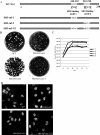
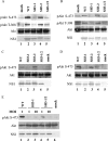
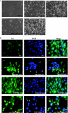
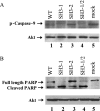
Recent studies have demonstrated that influenza A virus infection activates the phosphatidylinositol 3-kinase (PI3K)/Akt signaling pathway by binding of influenza NS1 protein to the p85 regulatory subunit of PI3K. Our previous study proposed that two polyproline motifs in NS1 (amino acids 164 to 167 [PXXP], SH3 binding motif 1, and amino acids 213 to 216 [PPXXP], SH3 binding motif 2) may mediate binding to the p85 subunit of PI3K. Here we performed individual mutational analyses on these two motifs and demonstrated that SH3 binding motif 1 contributes to the interactions of NS1 with p85beta, whereas SH3 binding motif 2 is not required for this process. Mutant viruses carrying NS1 with mutations in SH3 binding motif 1 failed to interact with p85beta and induce the subsequent activation of PI3K/Akt pathway. Mutant virus bearing mutations in SH3 binding motif 2 exhibited similar phenotype as the wild-type (WT) virus. Furthermore, viruses with mutations in SH3 binding motif 1 induced more severe apoptosis than did the WT virus. Our data suggest that SH3 binding motif 1 in NS1 protein is required for NS1-p85beta interaction and PI3K/Akt activation. Activation of PI3K/Akt pathway is beneficial for virus replication by inhibiting virus induced apoptosis through phosphorylation of caspase-9.
Figures


Similar articles
-
Influenza A virus NS1 protein activates the PI3K/Akt pathway to mediate antiapoptotic signaling responses.J Virol. 2007 Apr;81(7):3058-67. doi: 10.1128/JVI.02082-06. Epub 2007 Jan 17. J Virol. 2007. PMID: 17229704 Free PMC article.
-
Influenza A virus NS1 protein activates the phosphatidylinositol 3-kinase (PI3K)/Akt pathway by direct interaction with the p85 subunit of PI3K.J Gen Virol. 2007 Jan;88(Pt 1):13-18. doi: 10.1099/vir.0.82419-0. J Gen Virol. 2007. PMID: 17170431
-
Mechanism of influenza A virus NS1 protein interaction with the p85beta, but not the p85alpha, subunit of phosphatidylinositol 3-kinase (PI3K) and up-regulation of PI3K activity.J Biol Chem. 2008 Aug 22;283(34):23397-409. doi: 10.1074/jbc.M802737200. Epub 2008 Jun 5. J Biol Chem. 2008. PMID: 18534979
-
A new player in a deadly game: influenza viruses and the PI3K/Akt signalling pathway.Cell Microbiol. 2009 Jun;11(6):863-71. doi: 10.1111/j.1462-5822.2009.01309.x. Epub 2009 Mar 12. Cell Microbiol. 2009. PMID: 19290913 Free PMC article. Review.
-
Influenza A viruses and PI3K: are there time, place and manner restrictions?Virulence. 2012 Jul 1;3(4):411-4. doi: 10.4161/viru.20932. Epub 2012 Jun 22. Virulence. 2012. PMID: 22722241 Free PMC article. Review. No abstract available.
Cited by
-
Marek's Disease Virus Activates the PI3K/Akt Pathway Through Interaction of Its Protein Meq With the P85 Subunit of PI3K to Promote Viral Replication.Front Microbiol. 2018 Oct 23;9:2547. doi: 10.3389/fmicb.2018.02547. eCollection 2018. Front Microbiol. 2018. PMID: 30405592 Free PMC article.
-
Avian influenza virus NS1: A small protein with diverse and versatile functions.Virulence. 2013 Oct 1;4(7):583-8. doi: 10.4161/viru.26360. Epub 2013 Sep 17. Virulence. 2013. PMID: 24051601 Free PMC article. No abstract available.
-
Molecular basis of mammalian transmissibility of avian H1N1 influenza viruses and their pandemic potential.Proc Natl Acad Sci U S A. 2017 Oct 17;114(42):11217-11222. doi: 10.1073/pnas.1713974114. Epub 2017 Sep 5. Proc Natl Acad Sci U S A. 2017. PMID: 28874549 Free PMC article.
-
Influenza virus differentially activates mTORC1 and mTORC2 signaling to maximize late stage replication.PLoS Pathog. 2017 Sep 27;13(9):e1006635. doi: 10.1371/journal.ppat.1006635. eCollection 2017 Sep. PLoS Pathog. 2017. PMID: 28953980 Free PMC article.
-
Characterization of a novel mutation in NS1 protein of influenza A virus induced by a chemical substance for the attenuation of pathogenicity.PLoS One. 2015 Mar 20;10(3):e0121205. doi: 10.1371/journal.pone.0121205. eCollection 2015. PLoS One. 2015. PMID: 25793397 Free PMC article.
References
-
- Bornholdt, Z. A., and B. V. Prasad. 2006. X-ray structure of influenza virus NS1 effector domain. Nat. Struct. Mol. Biol. 13:559-560. - PubMed
-
- Cardone, M. H., N. Roy, H. R. Stennicke, G. S. Salvesen, T. F. Franke, E. Stanbridge, S. Frisch, and J. C. Reed. 1998. Regulation of cell death protease caspase-9 by phosphorylation. Science 282:1318-1321. - PubMed
-
- Carpenter, C. L., K. R. Auger, M. Chanudhuri, M. Yoakim, B. Schaffhausen, S. Shoelson, and L. C. Cantley. 1993. Phosphoinositide 3-kinase is activated by phosphopeptides that bind to the SH2 domains of the 85-kDa subunit. J. Biol. Chem. 268:9478-9483. - PubMed
-
- Datta, S. R., A. Brunet, and M. E. Greenberg. 1999. Cellular survival: a play in three Akts. Genes Dev. 13:2905-2927. - PubMed
Publication types
MeSH terms
Substances
LinkOut - more resources
Full Text Sources
Other Literature Sources
Research Materials

