. 2007 Nov;6(11):1979-91.
doi: 10.1128/EC.00249-07. Epub 2007 Sep 14.

Novel membrane-bound eIF2alpha kinase in the flagellar pocket of Trypanosoma brucei

Affiliations

Novel membrane-bound eIF2alpha kinase in the flagellar pocket of Trypanosoma brucei

Maria Carolina S Moraes et al. Eukaryot Cell. 2007 Nov.

Abstract

Translational control mediated by phosphorylation of the alpha subunit of the eukaryotic initiation factor 2 (eIF2alpha) is central to stress-induced programs of gene expression. Trypanosomatids, important human pathogens, display differentiation processes elicited by contact with the distinct physiological milieu found in their insect vectors and mammalian hosts, likely representing stress situations. Trypanosoma brucei, the agent of African trypanosomiasis, encodes three potential eIF2alpha kinases (TbeIF2K1 to -K3). We show here that TbeIF2K2 is a transmembrane glycoprotein expressed both in procyclic and in bloodstream forms. The catalytic domain of TbeIF2K2 phosphorylates yeast and mammalian eIF2alpha at Ser51. It also phosphorylates the highly unusual form of eIF2alpha found in trypanosomatids specifically at residue Thr169 that corresponds to Ser51 in other eukaryotes. T. brucei eIF2alpha, however, is not a substrate for GCN2 or PKR in vitro. The putative regulatory domain of TbeIF2K2 does not share any sequence similarity with known eIF2alpha kinases. In both procyclic and bloodstream forms TbeIF2K2 is mainly localized in the membrane of the flagellar pocket, an organelle that is the exclusive site of exo- and endocytosis in these parasites. It can also be detected in endocytic compartments but not in lysosomes, suggesting that it is recycled between endosomes and the flagellar pocket. TbeIF2K2 location suggests a relevance in sensing protein or nutrient transport in T. brucei, an organism that relies heavily on posttranscriptional regulatory mechanisms to control gene expression in different environmental conditions. This is the first membrane-associated eIF2alpha kinase described in unicellular eukaryotes.


Figures

FIG. 1.
FIG. 1.
T. brucei encodes three potential eIF2α kinases. The alignment of the catalytic domains of eIF2α kinases is shown. Identical residues are indicated in dark gray, and conserved residues are indicated in light gray. Subdomains I through XI are indicated above the sequences, as are the functional regions comprising the catalytic and activation loops. The kinase inserts between subdomains IV and V are indicated by dashes, with the number of residues in parentheses. The dots indicate conserved residues among kinases in general. Arrowheads indicate residues specific to the eIF2α kinases, with the boxed arrowhead indicating the substrate specificity determinant in PKR. The boxed arrow in subdomain VIII marks the residue in PKR that is autophosphorylated in the active protein. Sc, S. cerevisiae; Dm, D. melanogaster; Nc, N. crassa; Hs, H. sapiens; Sp, S. pombe; Rn, R. norvegicus; Tg, T. gondii; Ce, C. elegans. GeneDB accession numbers: TbK1, Tb11.02.5050; TbK2, Tb04.1H19.980; TbK3, Tb06.5F5.660. GenBank accession numbers: ScGCN2, NP_010569; DmGCN2, AAC47516; NcGCN2, CAA62973; HsGCN2, Q9P2K8; CePEK, Q19192; DmPEK, AAF61200; HsPEK, AAI26355; SpHRI, Q9UTE5; HsPKR, P19525; TgKA, AY518936.
FIG. 2.
FIG. 2.
Domain organization and expression of TbeIF2 kinases. (A) Schematics of the domain organization of the TbeIF2 kinases. Amino acid residue numbers are indicated above the bars, with the residues of the kinase domains (KD) corresponding to the sequences presented in Fig. 1. In TbeIF2K1, RWD indicates the ring finger, WD domain, and HRS indicates the sequence with similarity to histidinyl-tRNA synthetase. (B) Expression of the mRNA for each kinase. RT-PCR products obtained from procyclic (P) and bloodstream (B) forms, using the oligonucleotide pairs BC436-BC437 for TbeIF2K1, BC438-BC441 for TbeIF2K2, and BC446-BC450 for TbeIF2K3, with the expected sizes of 0.5, 1.2, and 1.4 kb, respectively; the controls represent the same DNase-treated RNA preparations used for RT-PCR, subjected to PCR with the primers for TbeIF2K1. (C) Expression of TbeIF2K2. Immunoblot of whole cells of bloodstream (B) and procyclic (P) parasites with anti-His6-TbeIF2K235-449 serum or with preimmune serum; the lane labeled “c” contains the purified His6-TbeIF2K235-449 protein that was used for immunization. The arrow indicates the TbeIF2K2 protein. Molecular weight standards (103) are indicated on the left panel.
FIG. 3.
FIG. 3.
TbeIF2K2 is a glycosylated membrane associated protein. (A) Partial fractionation. Procyclic forms were lysed by freeze-thawing and centrifuged as described in Materials and Methods. The soluble supernatant was separated (lane 1); the pellet was solubilized in the same buffer containing 1% Triton X-100, in the same volume as the supernatant. After centrifugation, the supernatant was separated (lane 2), and the pellet was solubilized in the same volume of buffer containing 8 M urea (lane 3). Identical volumes of the three samples were subjected to immunoblot analysis with anti-TbeIF2K235-449 antibodies (TbK2) and with anti-TbeIF2α serum (Tb-eIF2α). (B) Endoglycosidase H treatment. Detergent-solubilized membrane fractions of procyclic parasites were treated with endoglycosidase H (+) or incubated under the same conditions without endoglycosidase H (−) and then subjected to immunoblotting with anti-TbeIF2K235-449 antibodies (TbK2).
FIG. 4.
FIG. 4.
Localization of TbeIF2K2 in procyclic cells. (A) Affinity-purified anti-TbeIF2K235-449 antibodies. Immunoblot of membrane and soluble fractions obtained from procyclic parasites using monospecific purified antibodies used in the immunofluorescence assays. (B) Immunofluorescence of procyclic parasites. Immunofluorescence using affinity-purified anti-TbeIF2K235-449 antibodies (αTbK2), followed by anti-rabbit IgG-fluorescein isothiocyanate. Nuclei were labeled with DAPI. The specificity of the signal was ascertained by preincubation of the antibody solution with the purified His6-TbeIF2K235-449 protein, prior to addition to the cells (bottom row).
FIG. 5.
FIG. 5.
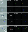
Localization of TbeIF2K2 in bloodstream cells. Bloodstream trypanosomes were incubated for 30 min with biotinyl TL at 5°C to allow flagellar pocket binding (A to D and E to H) or at 15°C to allow binding and uptake into the endosomal compartment (I to L, M to P, and Q to T) as described in Materials and Methods. Fixed and permeabilized cells were then stained with fluorescent streptavidin to detect bound or internalized TL (red), and as indicated with purified anti-TbeIF2K2 antibodies (αTbK2) and or anti-p67 (middle panels). (A to H) Cells stained with anti-TbeIF2K2 (green) and anti-p67 (red). The discrete positioning of the anterior lysosome and the posterior flagellar pocket allow simultaneous imaging of these organelles in the same channel. (I to P) Cells stained with anti-TbeIF2K2 (green). (Q to T) Cells stained with anti-p67 (green). Merged DAPI/differential interference contrast images are presented in the leftmost panels with kinetoplast (k) and nucleus (n) labeled, and merged three-channel fluorescent images are presented in rightmost panels. The positions of the lysosome (Lys), endosome (Endo), and flagellar pocket (FP) are indicated. The arrowhead in panel H indicates a region of discrete TbeIF2K2 signal, presumably the endosome, that does not colocalize with the nearby flagellar pocket. Bars, 5 μm.
FIG. 6.
FIG. 6.
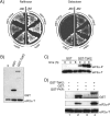
TbeIF2K2 phosphorylates specifically residue Ser51 in yeast and mammalian eIF2α. (A) GST-TbeIF2K2KD (GST-TbK2) was expressed from the inducible GAL1 promoter in yeast strain J80, which contains the wild-type copy of the gene encoding eIF2α, and in strain J82, which contains the mutant eIF2αSer51Ala. The growth of two independent transformants expressing GST-TbK2 is shown on raffinose- (left) and on galactose (right)-containing medium. As controls, the growth of the same strains expressing GST from the empty vector is shown (GST). (B) The expression of the proteins was determined by immunoblotting total cell extracts (5 μg of total protein) prepared from the same isolates as described above of strain J82 and of a strain without plasmid (⊘), grown on galactose, using antibodies to GST. As a control, the blot was probed with antibodies against total yeast eIF2α (eIF2α-T) after removal of the first antibodies (bottom panel). (C) The phosphorylation of the endogenous eIF2α was analyzed in strain J80 carrying the vector (GST) or the GST-TbeIF2K2KD expressing plasmid (GST-TbK2) after 4 or 8 h of induction with galactose, by immunoblotting with antibodies directed to the phosphorylated form of eIF2α (eIF2α-P) and normalized with antibodies to total eIF2α (eIF2α-T). (D) In vitro phosphorylation assays were performed with mammalian eIF2α purified from E. coli and with GST-TbeIF2K2KD immunoprecipitated from an extract of strain J82 grown in galactose, using as a control GST immunoprecipitated from the same strain carrying the empty vector. As a control for the efficiency of phosphorylation, we used GST-PKRKD purified from E. coli. Phosphorylation was detected by immunoblots with antibodies specific for eIF2α-P and normalized with antibodies against total mammalian eIF2α (eIF2-T). The amounts of the GST fusion proteins in the reactions were determined by immunoblots of the upper portion of the gel using anti-GST antibodies (GST).
FIG. 7.
FIG. 7.
eIF2α in trypanosomatids. (A) Alignment of the N-terminal half of T. brucei and L. major eIF2α and other characterized eIF2α proteins. A line above the sequences indicates the unstructured loop encompassing the phosphorylated residue, indicated with an arrowhead. The structural domains of eIF2α are indicated with lines below the sequences. (B) Immunoblot of total cell extract of intact bloodstream (B) and procyclic (P) form parasites using anti-TbeIF2α serum; as a control, the lane labeled rTb2α contains the His6-TbeIF2α protein purified from E. coli.
FIG. 8.
FIG. 8.
TbeIF2α substitutes for yeast eIF2α. (A) Growth of yeast cells expressing TbeIF2α. Derivatives of strain H1643 expressing the indicated eIF2α proteins from the GAL1 promoter were grown on plates containing synthetic medium supplemented with galactose in the presence or absence of 5-FOA. (B) Expression of TbeIF2α125-419 in yeast. Total cell extracts (50 μg) of two isolates carrying TbeIF2α125-419 and one isolate expressing yeast eIF2α selected from the 5-FOA plates and of control H1643 cells carrying only the vector were subjected to immunoblotting with anti-TbeIF2α serum; after stripping the first antibodies, the same membrane was incubated with antibodies against yeast eIF2α. The bottom panel shows the filter stained with Ponceau S.
FIG. 9.
FIG. 9.
In vitro phosphorylation of TbeIF2α. Purified His6-TbeIF2α was used in in vitro reactions with purified GCN2 (left panels) and purified PKR (upper right panels) and with immunoprecipitated GST-TbeIF2K2KD (bottom right panels). Controls were purified yeast GST-eIF2α and GST-eIF2αSer51Ala for the GCN2 and PKR assays and purified His6-TbeIF2αThr169Ala for the TbeIF2K2 assay. Exposure times are indicated as “short” or “long.” The total protein was visualized on the gels by Coomassie R250 stain.
FIG. 10.
FIG. 10.
Modifications of TbeIF2K2 in bloodstream parasites. (A) Phosphorylation of TbeIF2K2. Membrane-enriched (mb) and soluble (sol) fractions (5 μg of total protein) of bloodstream (B) and procyclic (P) parasites were subjected to immunoblots with anti-TbeIF2K2 antibodies and with anti-BiP serum (left panels) (BiP partitions to both soluble and membrane enriched fractions). Membrane-enriched fractions from bloodstream (B) and procyclic (P) parasites were treated with calf intestinal alkaline phosphatase (CIAP), and subjected to immunoblot with anti-TbeIF2K2 antibodies (right panel). (B) TbeIF2K2 in high-density cultures of bloodstream forms. Membrane (mb) and soluble (sol) fractions (10 μg of total protein) from bloodstream forms grown to late logarithmic phase (2.4 × 10⁶ cells/ml), and 12 h (2.0 × 10⁶ cells/ml) or 24 h (4.0 × 10⁵ cells/ml) later were subjected to immunoblotting with anti-TbeIF2K2 antibodies (TbK2) or anti-BiP (BiP) serum. The indicated number of cells corresponds to live parasites as determined by their motility at each time point.

Similar articles

Cited by

References

    1. Alexander, D. L., K. J. Schwartz, A. E. Balber, and J. D. Bangs. 2002. Developmentally regulated trafficking of the lysosomal membrane protein p67 in Trypanosoma brucei. J. Cell Sci. 115:3253-3263. - PubMed
    1. Bangs, J. D., L. Uyetake, M. J. Brickman, A. E. Balber, and J. C. Boothroyd. 1993. Molecular cloning and cellular localization of a BiP homologue in Trypanosoma brucei: divergent ER retention signals in a lower eukaryote. J. Cell Sci. 105:1101-1113. - PubMed
    1. Berriman, M., E. Ghedin, C. Hertz-Fowler, G. Blandin, H. Renauld, D. C. Bartholomeu, N. J. Lennard, E. Caler, N. E. Hamlin, B. Haas, U. Bohme, L. Hannick, M. A. Aslett, J. Shallom, L. Marcello, L. Hou, B. Wickstead, U. C. Alsmark, C. Arrowsmith, R. J. Atkin, A. J. Barron, F. Bringaud, K. Brooks, M. Carrington, I. Cherevach, T. J. Chillingworth, C. Churcher, L. N. Clark, C. H. Corton, A. Cronin, R. M. Davies, J. Doggett, A. Djikeng, T. Feldblyum, M. C. Field, A. Fraser, I. Goodhead, Z. Hance, D. Harper, B. R. Harris, H. Hauser, J. Hostetler, A. Ivens, K. Jagels, D. Johnson, J. Johnson, K. Jones, A. X. Kerhornou, H. Koo, N. Larke, S. Landfear, C. Larkin, V. Leech, A. Line, A. Lord, A. Macleod, P. J. Mooney, S. Moule, D. M. Martin, G. W. Morgan, K. Mungall, H. Norbertczak, D. Ormond, G. Pai, C. S. Peacock, J. Peterson, M. A. Quail, E. Rabbinowitsch, M. A. Rajandream, C. Reitter, S. L. Salzberg, M. Sanders, S. Schobel, S. Sharp, M. Simmonds, A. J. Simpson, L. Tallon, C. M. Turner, A. Tait, A. R. Tivey, S. Van Aken, D. Walker, D. Wanless, S. Wang, B. White, O. White, S. Whitehead, J. Woodward, J. Wortman, M. D. Adams, T. M. Embley, K. Gull, E. Ullu, J. D. Barry, A. H. Fairlamb, F. Opperdoes, B. G. Barrell, J. E. Donelson, N. Hall, C. M. Fraser, et al. 2005. The genome of the African trypanosome Trypanosoma brucei. Science 309:416-422. - PubMed
    1. Bertolotti, A., Y. Zhang, L. M. Hendershot, H. P. Harding, and D. Ron. 2000. Dynamic interaction of BiP and ER stress transducers in the unfolded-protein response. Nat. Cell Biol. 2:326-332. - PubMed
    1. Brecht, M., and M. Parsons. 1998. Changes in polysome profiles accompany trypanosome development. Mol. Biochem. Parasitol. 97:189-198. - PubMed

Publication types

MeSH terms

LinkOut - more resources