. 2007 Dec 15;110(13):4268-77.
doi: 10.1182/blood-2007-04-087775. Epub 2007 Sep 6.

HGAL, a lymphoma prognostic biomarker, interacts with the cytoskeleton and mediates the effects of IL-6 on cell migration

Affiliations

HGAL, a lymphoma prognostic biomarker, interacts with the cytoskeleton and mediates the effects of IL-6 on cell migration

Xiaoqing Lu et al. Blood. .

Abstract

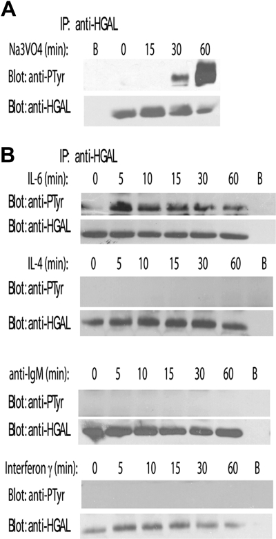
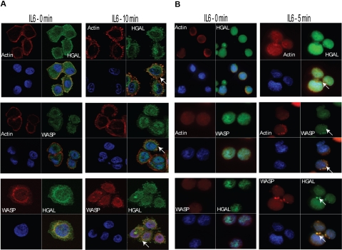
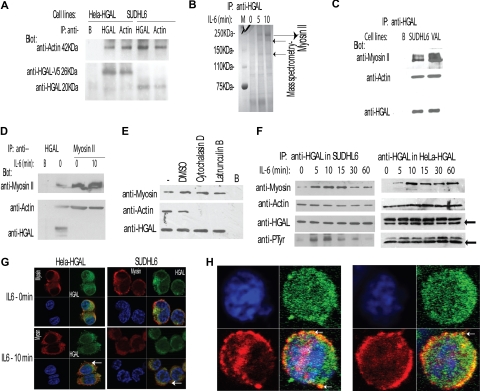
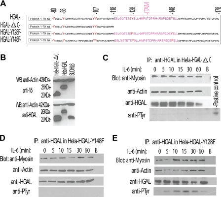
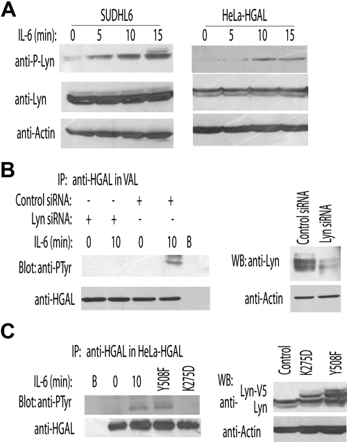
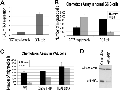
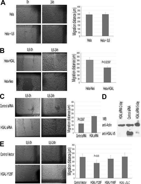
HGAL is a newly identified germinal center (GC)-specific gene whose expression by the tumor cells correlates with a favorable prognosis in patients with diffuse large B-cell and classical Hodgkin lymphomas. The function of HGAL is unknown. Previous studies demonstrated that HGAL is dispensable for GC formation, immunoglobulin gene class-switch recombination, and somatic hypermutation. Herein, we identify a role for HGAL in the regulation of cell motility. We demonstrate that IL-6 induces the phosphorylation of the C-terminal tyrosine residue of the HGAL protein via the Lyn kinase, and promotes its relocalization from the cytoplasm to podosome-like structures. Further, IL-6-induced HGAL phosphorylation increases its interaction with myosin II and is associated with inhibition of cell migration. Knockdown of endogenous HGAL ameliorates IL-6-induced inhibition of cell migration, whereas overexpression of HGAL imparts inhibitory effects of IL-6 on cell migration. Taken together, our results suggest that HGAL is involved in negative regulation of lymphocyte migration, thus constraining lymphocytes to the GC. Inhibition of lymphocyte migration might contribute to the less aggressive clinical behavior of HGAL-expressing lymphomas.


Figures

Figure 1
Figure 1
Pervanadate and IL-6 induce tyrosine phosphorylation of HGAL protein. SUDHL6 lymphoma cells were incubated for up to 60 minutes with pervanadate (1 mM) (A) or stimulated with IL-6 (10 ng/mL), IL-4 (10 ng/mL), anti-IgM (100 ng/mL), and IFNγ (1 μM) (B). Cellular lysates were extracted at the indicated time points, immunoprecipitated with anti-HGAL antibody, and blotted for phosphotyrosine (PTyr) and HGAL. Unconjugated beads served as a control (B). Representative blots of 3 independent experiments are shown.
Figure 2
Figure 2
IL-6 stimulation induces colocalization of HGAL with actin and WASP proteins in podosomes. Unstimulated or IL-6–stimulated Hela-HGAL (A) and SUDHL6 (B) cells were fixed with 4% paraformaldehyde and stained with anti-HGAL antibody (green), anti-WASP antibody (green or red, as indicated), rhodamine-labeled phalloidin (red), and DAPI (blue), and were visualized on a Carl Zeiss LSM510 microscope. Arrows indicate podosome-like structures. Representative photographs of 3 independent experiments are shown (see “Immunofluorescence microscopy” for image acquisition information).
Figure 3
Figure 3
HGAL interacts with actin and myosin II proteins, and the interaction with the myosin is increased by IL-6–induced HGAL phosphorylation. (A) Cellular lysates of unstimulated HeLa-HGAL and SUDHL6 cells were immunoprecipitated with either anti-HGAL or anti-actin antibodies and blotted with the indicated antibodies. Unconjugated beads served as a control (“B”). The size of HGAL in the HeLa cells is 26 kDa, since the cloned protein is a fusion protein with V5 tag. (B) Cellular lysates were extracted from unstimulated and IL-6–stimulated SUDHL6 cells, immunoprecipitated with anti-HGAL antibody, resolved by SDS-PAGE, and stained with Coomassie blue. Protein bands of 250 kDa, 230 kDa, and 140 kDa (→) were excised and analyzed by mass spectrometry analysis and sequencing that revealed that all the 3 proteins represent myosin II, the expected size of which is 250 kDa. (C) Cellular lysates were extracted from unstimulated SUDHL6 and VAL lymphoma cell lines, immunoprecipitated with anti-HGAL antibody, and blotted with indicated antibodies. Unconjugated beads served as a control (“B”). (D) Cellular lysates were extracted from unstimulated or IL-6–stimulated SUDHL6 cells and immunoprecipitated with either anti-HGAL or antimyosin antibodies, resolved on the SDS-PAGE, and blotted with the indicated antibodies. (E) HeLa-HGAL cells were treated with cytochalasin D (5 μM) and latrunculin B (5 μM) for 30 minutes, respectively, to inhibit actin polymerization and disrupt actin filaments. The whole-cell lysates were extracted, immunoprecipitated with anti-HGAL antibody, and blotted with the indicated antibodies. (F) SUDHL6 and HeLa-HGAL cells were stimulated with IL-6 for up to 60 minutes. At the indicated time points, cellular lysates were extracted, immunoprecipitated with anti-HGAL antibody, and blotted with the indicated antibodies. Arrows indicate immunoglobulin light chains. (G) Unstimulated or IL-6–stimulated Hela-HGAL (left) and SUDHL6 (right) cells were fixed with 4% paraformaldehyde, stained with anti-HGAL antibody (green), antimyosin antibody (red), and DAPI (blue), and visualized with the Carl Zeiss LSM510 microscope. (H) HGAL and myosin II colocalization in podosome-like structures of IL-6–stimulated SUDHL6 cells, as visualized with the confocal Carl Zeiss LSM510 microscope (see “Immunofluorescence microscopy” for detailed image acquisition information). Colors as described in panel G. In panels A,C-E, representative blots of 3 independent experiments are shown.
Figure 4
Figure 4
IL-6 induces phosphorylation of the C-terminal tyrosine residue of the HGAL protein that mediates increased interaction with myosin protein. (A) Schematic representation of HGAL protein, its deletion mutant missing the C-terminal ITAM (HGAL-ΔC), and substitution mutants in which C-terminal tyrosine residues of the ITAM were substituted with phenylalanine (HGAL-Y128F and HGAL-Y148F). (B) Mutant missing the C-terminal ITAM (HGAL-ΔC) can be expressed in the cellular context. Whole-cell lysates were extracted from SUDHL6, HeLa-HGAL, and HeLa cells transiently transfected with the HGAL-ΔC vector and blotted with the indicated antibodies. (C-E) HeLa cells, transiently transfected with HGAL-ΔC (C), HGAL-Y148F (D), or HGAL-Y128F (E) vectors, were stimulated with IL-6 (10 ng/mL) for up to 60 minutes. Cellular lysates were extracted at the specified time points, immunoprecipitated with anti-HGAL antibody, resolved on SDS-PAGE, and blotted with the indicated antibodies. In panels C-E, representative blots of 3 independent experiments are shown.
Figure 5
Figure 5
Lyn kinase mediates IL-6–induced tyrosine phosphorylation of the HGAL protein. (A) VAL and HeLa-HGAL cells were treated with IL-6 for up to 15 minutes. The whole-cell lysates were extracted, separated by SDS-PAGE, and detected with the indicated antibodies. (B) VAL cells were transfected with either siRNA for Lyn or scrambled control, respectively. At 48 hours after transfection, the cells were stimulated with IL-6 (10 ng/mL) for 10 minutes and cellular lysates were extracted, immunoprecipitated with anti-HGAL antibody, separated by SDS-PAGE, and immunoblotted with antiphosphotyrosine or anti-HGAL antibodies. (C) HeLa-HGAL cells were transiently transfected with plasmids encoding for Lyn-Y508F and Lyn-K275D. At 48 hours after ransfection, whole-cell lysates were prepared, immunoprecipitated with anti-HGAL antibody, separated by SDS-PAGE, and immunoblotted with antiphosphotyrosine or anti-HGAL antibodies. Unstimulated and IL-6–stimulated HeLa-HGAL cells served as controls. In panels A-C, representative blots of 2 independent experiments are shown.
Figure 6
Figure 6
HGAL mediates the inhibitory effects of IL-6 on cell migration. (A) CD77+ GC B cells and CD77 cells were enriched from normal tonsils as described in “RNA isolation and reverse transcription reaction.” RNA was extracted and HGAL RNA expression was measured by real-time reverse transcription–PCR in triplicates as described in “Real-time PCR measurement of HGAL mRNA expression.” (B) Enriched CD77+ GC B cells and CD77 tonsilar cells were evaluated in triplicate in IL-6 chemotaxis assay, as described in “Materials and methods.” Means and standard deviations of 2 independent experiments are demonstrated. (C) VAL lymphoma cells were transfected with control or HGAL siRNA. At 48 hours after siRNA transfection, the cells were used for IL-6 chemotaxis assay performed in triplicate, as described in “Chemotaxis and wound assays.” Means and standard deviations of 3 independent experiments are demonstrated. (D) Western blot analysis of HGAL protein in siRNA-transfected VAL lymphoma cells used for chemotaxis assays shown in panel C.
Figure 7
Figure 7
HGAL modulates IL-6 effects on cell migration: wound assay in HeLa-HGAL cells. Wound assays were performed as described in “Chemotaxis and wound assays.” (A) IL-6 effect on HeLa cell migration in wound assay. (B) IL-6 effect on migration of HeLa-HGAL and HeLA-Neo cells. HGAL expression significantly decreased migration of the cells. (C) siRNA knock-down of HGAL in the HeLa-HGAL cells markedly reversed the HGAL-mediated, IL-6–induced inhibition of cell migration. (D) Western blot analysis of HGAL protein expression in HeLa-HGAL cells following siRNA knockdown. (E) IL-6 effect on migration of HeLa-HGAL mutants. Left panel exhibits results of a representative wound assay in HeLa-HGAL Y128F and control cells. Right panel summarizes the effect of IL-6 on migration of HeLa-HGAL-Y128F, HGAL-128F, and HGAL-ΔC mutants. P value represents difference between HeLa-HGAL Y128F and control cells. Means and standard deviations of 3 independent experiments are demonstrated. See “Immunofluorescence microscopy” for image acquisition information.

Similar articles

Cited by

References

    1. Lossos IS, Alizadeh AA, Rajapaksa R, Tibshirani R, Levy R. HGAL is a novel interleukin-4-inducible gene that strongly predicts survival in diffuse large B-cell lymphoma. Blood. 2003;101:433–440. - PubMed
    1. Pan Z, Shen Y, Du C, et al. Two newly characterized germinal center B-cell-associated genes, GCET1 and GCET2, have differential expression in normal and neoplastic B cells. Am J Pathol. 2003;163:135–144. - PMC - PubMed
    1. Schenten D, Egert A, Pasparakis M, Rajewsky K. M17, a gene specific for germinal center (GC) B cells and a prognostic marker for GC B-cell lymphomas, is dispensable for the GC reaction in mice. Blood. 2006;107:4849–4856. - PMC - PubMed
    1. Natkunam Y, Hsi E, Aoun P, et al. Expression of the human germinal center–associated lymphoma (HGAL) protein identifies a subset of classical Hodgkin lymphoma of germinal center derivation and improved survival. Blood. 2007;109:298–305. - PMC - PubMed
    1. Burdin N, Galibert L, Garrone P, Durand I, Banchereau J, Rousset F. Inability to produce IL-6 is a functional feature of human germinal center B lymphocytes. J Immunol. 1996;156:4107–4113. - PubMed

Publication types