. 2007 Sep 15;179(6):3792-803.
doi: 10.4049/jimmunol.179.6.3792.

IFN-alpha is not sufficient to drive Th1 development due to lack of stable T-bet expression

Affiliations

IFN-alpha is not sufficient to drive Th1 development due to lack of stable T-bet expression

Hilario J Ramos et al. J Immunol. .

Abstract

During inflammatory immune responses, the innate cytokine IL-12 promotes CD4+ Th-1 development through the activation of the second messenger STAT4 and the subsequent expression of T-bet. In addition, type I IFN (IFN-alphabeta), secreted primarily during viral and intracellular bacterial infections, can promote STAT4 activation in human CD4+ T cells. However, the role of IFN-alphabeta in regulating Th1 development is controversial, and previous studies have suggested a species-specific pathway leading to Th1 development in human but not mouse CD4+ T cells. In this study, we found that although both IFN-alpha and IL-12 can promote STAT4 activation, IFN-alpha failed to promote Th1 commitment in human CD4+ T cells. The difference between these innate signaling pathways lies with the ability of IL-12 to promote sustained STAT4 tyrosine phosphorylation, which correlated with stable T-bet expression in committed Th1 cells. IFN-alpha did not promote Th1 development in human CD4+ T cells because of attenuated STAT4 phosphorylation, which was insufficient to induce stable expression of T-bet. Further, the defect in IFN-alpha-driven Th1 development was corrected by ectopic expression of T-bet within primary naive human CD4+ T cells. These results indicate that IL-12 remains unique in its ability to drive Th1 development in human CD4+ T cells and that IFN-alpha lacks this activity due to its inability to promote sustained T-bet expression.


Figures

Figure 1
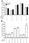
Figure 1. IL-12, but not IFN-α, promotes Th1 development in highly purified naïve human CD4+ T cells
Purified human CD4+/CD45RA+ T cells were activated with plate-bound anti-CD3 and anti-CD28 in the presence of IL-2, anti-IL-4, and the indicated cytokines and neutralizing antibodies, where “+” indicates addition of cytokine and “−” indicates addition of neutralizing anti-cytokine antibody. On day 3, cells were diluted into fresh medium containing IL-2 and rested to day 7. A, Parallel cultures were stimulated in the absence (open bars) or presence (closed bars) of neutralizing anti-hIL-4 antibody. Cells were restimulated for 4 hours in the presence of PMA + ionomycin. Intracellular cytokine staining was performed with antibodies specific for hCD4 and hIFN-γ. Data were gated on live cell populations and expressed as a percentage of CD4+/IFN-γ+ cells. B, Total RNA was isolated from cells that were resting (open bars) or restimulated (closed bars) with plate-bound anti-CD3 for 2 hours. Analysis of IFN-γ transcript levels was performed by qPCR, and transcript levels were normalized to GAPDH. Data were normalized relative to non-stimulated (Control) cells activated under neutralizing conditions. C, Cells were rested (open bars) or restimulated for 24 hours with plate-bound anti-CD3 (closed bars). Cell culture supernatants were analyzed for the presence of IFN-γ (upper panel), IL-4 (middle panel), and IL-5 (lower panel) by cytometric bead array. D, Purified CD4+/CD45RA+ T cells from 3 separate donors (D1, D2, and D3) were stimulated as in (C) under neutralizing conditions (“Control”) or with IL-12 + anti-IFNAR2 (“IL-12”) or anti-IL-12 + IFN-αA (“IFN-αA”). On day 7, cells were restimulated and stained for CD4 and intracellular IFN-γ as described above. Data were gated on live cell populations.
Figure 2
Figure 2. IL-12, but not IFN-α, promotes Th1 development in human PBMC cultures
A, Human PBMCs were stimulated with plate-bound anti-CD3 + anti-CD28 in the presence of IL-2 and the indicated cytokines and/or neutralizing antibodies, where “+” indicates addition of cytokine, and “−” indicates addition of neutralizing anti-cytokine antibody. Parallel cultures were stimulated in the absence (open bars) or presence (closed bars) of neutralizing anti-hIL-4 antibody. B, Human PBMCs were activated with plate-bound anti-CD3 and anti-CD28 in the presence of IL-2 and the indicated cytokines and neutralizing antibodies, where “+” indicates addition of cytokine, “−” indicates addition of neutralizing anti-cytokine antibody, and “o” indicates that the cytokine was not manipulated. On day 3, cells were diluted 1:8 into fresh medium containing IL-2 and rested to day 7. Cells were restimulated for 4 hours in the presence of PMA + ionomycin. Intracellular cytokine staining was performed with antibodies specific for hCD4 and hIFN-γ. Data were gated on live cell populations and expressed as a percentage of CD4+/IFN-γ+ cells.
Figure 3
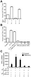
Figure 3. Type I interferon does not promote Th1 commitment
A, Purified naïve CD4+ T cells were stimulated with plate bound anti-CD3/anti-CD28 in the presence of IL-12 or IFN-β (1000 U/ml) as indicated in the figure. On day 7, cells were restimulated with PMA + ionomycin, and IFN-γ expression was measured by intracellular staining. B, Purified naïve CD4+ T cells were stimulated with IFN-α at concentrations indicated in the figure, and IFN-γ was measured by intracellular staining as describe above. C, Purified naïve CD4+ T cells were activated as described above for 7 days (1° conditions) where “+” indicates addition of cytokine and “−” indicates addition of neutralizing anti-cytokine antibody. These cells were then restimulated with plate-bound anti-CD3 under cytokine conditions indicated in the figure for an addition 7 days (2° conditions). Cells were then restimulated with either medium alone, or PMA + ionomycin for 1 hour. Total RNA was isolated from these cells, and IFN-γ mRNA induction was measured by qPCR. All data are referenced to the unstimulated control in condition 1.
Figure 4
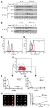
Figure 4. IL-12 and IFN-α differentially regulate the kinetics of STAT4 tyrosine phosphorylation in human CD4+ T cells
A and B, Human PBMCs were activated and expanded for two consecutive weeks with PHA in the presence of IL-2 and IL-12. On day 14, cells were rested in fresh medium for 30 minutes and stimulated with either IL-12 or IFN-αA for the indicated periods of time. Immunoprecipitation was performed using polyclonal antisera for human STAT4 or STAT1, and Western blotting was performed for phosphorylated STAT4 or STAT1, as indicated. Membranes were then stripped and re-probed for total STAT4 or STAT1. A, Time course of STAT4 and STAT1 phosphorylation in human Th1 PBMCs stimulated with IL-12 (lanes 2–7) or IFN-αA (lanes 8–13) for 0–24 hours. B, Time course of STAT4 phosphorylation in human Th1 PBMCs stimulated with IL-12 (lanes 2–7) or IFN-αA (lanes 8–13) for 0–6 hours. C through G, Freshly isolated PBMCs were activated with medium alone, IL-12, or IFN-α at various time points up to 24 hours. Cells were stained for CD4, CD45RA, and intracellular STAT4 and phosphotyrosine STAT4 (P-Y STAT4) as described in the Materials and Methods. C and D, Total STAT4 (C) and P-Y STAT4 (D) were measured from cells activated for 30 min by flow cytometry. Data are gated on live, CD4+, and CD45RA+ cells. Black line, unstimulated; green line, IL-12 stimulated; red line, IFN-α stimulated; gray shaded, non-immune rabbit Ig control. E, Gating scheme is shown for the analysis of CD4+, CD45RA- (R2) and CD45RA+ (R3) cells. F and G, CD45RA- (F) and CD45RA+ (G) gated cells are represented as a percentage of cells that display increased P-Y STAT4 staining over a 24 hour period. Open square, unstimulated; open triangle, IL-12 stimulated; open circle, IFN-α stimulated. H and I, Human PBMCs were activated and stained as above and analyzed on the ImageStream® imaging flow cytometer. Single cell images were gated on the live, CD4+, and CD45RA+ populations. H, Cells displaying positive staining for P-Y STAT4 were categorized for either low similarity (left panel) or high similarity (right panel) of co-localization of P-Y STAT4 within the nucleus based on nuclear dye staining (Draq-5). I, Cells displaying elevated P-Y STAT4 staining were quantified for co-localization of P-Y STAT4 within the nucleus of cells treated with IL-12 (closed square) or IFN-α (closed triangle).
Figure 5
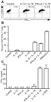
Figure 5. Cytokine-dependent IFN-γ secretion from fully polarized human Th1 cells
Purified human CD4+/CD45RA+ T cells were activated with plate-bound anti-CD3 and anti-CD28 in the presence of IL-2, anti-IL-4, anti-IFNAR2, and IL-12 for 3 days (Th1-inducing conditions). Cells were diluted into fresh medium and rested to day 7. A and B, Cells were restimulated for 4 hours in the presence of IL-12, IFN-αA, IL-18, or a combination of these cytokines. PMA + ionomycin was used as a positive control. Intracellular cytokine staining was performed with antibodies specific for hCD4 and hIFN-γ. Data were gated on live cell populations. A, Representative dot plots showing unstimulated cells and cells stimulated in the presence of IL-12 + IL-18 or IFN-αA + IL-18. B, Graphical representation of the proportion of CD4+ IFN-γ+ cells. C, Cells were restimulated for 24 hrs in the presence of the indicated cytokines or with plate-bound anti-CD3 antibody as described above. Cell culture supernatants were harvested and analyzed for the presence of IFN-γ protein by ELISA.
Figure 6
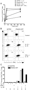
Figure 6. Ectopic IFNAR2 expression promotes sustained STAT4 phosphorylation and IFN-γ secretion in response to IFN-α
(A) Spleen and lymph node cells from DO11.10 mice were activated with OVA peptide under Th1-inducing conditions and transduced with retrovirus vectors expressing GFP alone (GFPRV) or the full-length mIFNAR2 subunit. Cells were sorted on day 7 based on GFP expression and restimulated with irradiated BALB/c splenocytes and OVA peptide. Following expansion for an additional 7 days, resting cells were activated with either IL-12 or IFN-α for the times indicated in the figure. Cells were then stained and analyzed for intracellular tyrosine-phosphorylated STAT4 as described in Fig. 5. (B and C) Day 14 transduced Th1 cells were activated with either IL-12 + IL-18, IFN-α + IL-18, or with the individual cytokines indicated in the figure for 24 hours. Brefelden A was added during the last 4 hours of stimulation. Cells were then stained for mCD4 and IFN-γ and analyzed by flow cytometry. Data were gated on live cells and GFP expression.
Figure 7
Figure 7. IFN-α does not promote stable T-bet expression in human CD4+ T cells
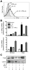
Purified human CD4+/CD45RA+ T cells were activated with plate-bound anti-CD3 + anti-CD28, IL-2, and anti-IL-4, and with either anti-IFNAR2 and anti-IL-12 (“Ctl”), anti-IFNAR2 and IL-12 (“IL-12”), IFN-αA and anti-IL-12 (“IFN-aA”), or with IFN-αA and IL-12 (“IL-12 + IFN-αA”). A, After 72 hours, cells were stained for surface expression of IL-12Rβ2: filled histogram, neutralizing antibodies alone; dashed line, IL-12 + anti-IFNAR2; dotted line, IFN-αA + αIL-12; solid line, IL-12 + IFN-αA. B, Total RNA was isolated from cells harvested 48 hours or 7 days after activation. Analysis of IL-12Rβ2 and T-bet transcript levels was performed by quantitative real-time PCR (qPCR) using the primers listed in Materials and Methods. Transcript levels for each condition were normalized to GAPDH, and the data were further normalized relative to cells activated under neutralizing conditions. C, Whole-cell lysates were prepared from day 7 activated cells and assessed for expression of T-bet protein by Western blotting (upper panel). Blots were stripped and re-probed for Lamin protein expression (lower panel).
Figure 8
Figure 8. Ectopic T-bet expression promotes Th1 development independent of IL-12 or IFN-α in naïve human CD4+ T cells
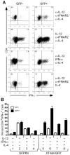
Purified naïve human CD4+ T cells were transduced with retrovirus constructs expressing GFP only (GFPRV) or with co-expression of human T-bet (hT-bet-GFP). During retroviral transduction, separate groups of cells were simultaneously activated in the presence or absence of cytokines or anti-cytokine antibodies as indicated in the figure. Cells were expanded on day 7 by restimulation on anti-CD3-coated plates. On day 14, resting cells were restimulated with PMA + ionomycin and analyzed for IFN-γ expression by intracellular cytokine staining. A, hT-bet-GFP-transduced cells were gated on live and either GFP negative (GFP-, left panels) or positive (GFP+, right panels) populations. The percentages of CD4+ and either IFN-γ- or IFN-γ+ populations are indicated within their respective quadrants. B, Triplicate cultures were analyzed for IFN-γ expression by intracellular cytokine staining. The percentage of CD4+/IFN-γ+ cells transduced with either the control GFPRV or hT-bet-GFP vectors are compared between the GFP- (open bars) and GFP+ (closed bars) populations. The error bars represent the standard deviation of the mean. These experiments were performed three times with similar results.

Similar articles

Cited by

References

    1. Takeda K, Kaisho T, Akira S. Toll-like receptors. Annu Rev Immunol. 2003;21:335–376. - PubMed
    1. Janeway CA, Jr, Medzhitov R. Innate immune recognition. Annu Rev Immunol. 2002;20:197–216. - PubMed
    1. Kadowaki N, Ho S, Antonenko S, Malefyt RW, Kastelein RA, Bazan F, Liu YJ. Subsets of human dendritic cell precursors express different toll-like receptors and respond to different microbial antigens. J Exp Med. 2001;194:863–869. - PMC - PubMed
    1. Boonstra A, Asselin-Paturel C, Gilliet M, Crain C, Trinchieri G, Liu YJ, O’Garra A. Flexibility of mouse classical and plasmacytoid-derived dendritic cells in directing T helper type 1 and 2 cell development: dependency on antigen dose and differential toll-like receptor ligation. J Exp Med. 2003;197:101–109. - PMC - PubMed
    1. Trinchieri G. Interleukin-12 and the regulation of innate resistance and adaptive immunity. Nat Rev Immunol. 2003;3:133–146. - PubMed

Publication types

MeSH terms