TRBO: a high-efficiency tobacco mosaic virus RNA-based overexpression vector
- PMID: 17720752
- PMCID: PMC2151719
- DOI: 10.1104/pp.107.106377
TRBO: a high-efficiency tobacco mosaic virus RNA-based overexpression vector
Abstract
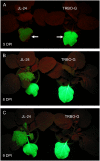
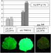
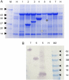
Transient expression is a rapid, useful approach for producing proteins of interest in plants. Tobacco mosaic virus (TMV)-based transient expression vectors can express very high levels of foreign proteins in plants. However, TMV vectors are, in general, not efficiently delivered to plant cells by agroinfection. It was determined that agroinfection was very efficient with a 35S promoter-driven TMV replicon that lacked the TMV coat protein gene sequence. This coat protein deletion vector had several useful features as a transient expression system, including improved ease of use, higher protein expression rates, and improved biocontainment. Using this TMV expression vector, some foreign proteins were expressed at levels of 3 to 5 mg/g fresh weight of plant tissue. It is proposed that this new transient expression vector will be a useful tool for expressing recombinant proteins in plants for either research or production purposes.
Figures


Similar articles
-
High-efficiency protein expression in plants from agroinfection-compatible Tobacco mosaic virus expression vectors.BMC Biotechnol. 2007 Aug 27;7:52. doi: 10.1186/1472-6750-7-52. BMC Biotechnol. 2007. PMID: 17723150 Free PMC article.
-
Removal of the selectable marker gene from transgenic tobacco plants by expression of Cre recombinase from a tobacco mosaic virus vector through agroinfection.Transgenic Res. 2006 Jun;15(3):375-84. doi: 10.1007/s11248-006-0011-6. Transgenic Res. 2006. PMID: 16779652
-
Assessment of the effectiveness of a nuclear-launched TMV-based replicon as a tool for foreign gene expression in plants in comparison to direct gene expression from a nuclear promoter.Transgenic Res. 2006 Feb;15(1):107-13. doi: 10.1007/s11248-005-2942-8. Transgenic Res. 2006. PMID: 16475015
-
Historical overview of research on the tobacco mosaic virus genome: genome organization, infectivity and gene manipulation.Philos Trans R Soc Lond B Biol Sci. 1999 Mar 29;354(1383):569-82. doi: 10.1098/rstb.1999.0408. Philos Trans R Soc Lond B Biol Sci. 1999. PMID: 10212936 Free PMC article. Review.
-
Plant virus expression vectors set the stage as production platforms for biopharmaceutical proteins.Virology. 2012 Nov 10;433(1):1-6. doi: 10.1016/j.virol.2012.06.012. Virology. 2012. PMID: 22979981 Review.
Cited by
-
Plant-Produced Chimeric Hepatitis E Virus-like Particles as Carriers for Antigen Presentation.Viruses. 2024 Jul 8;16(7):1093. doi: 10.3390/v16071093. Viruses. 2024. PMID: 39066255 Free PMC article.
-
TGBp3 triggers the unfolded protein response and SKP1-dependent programmed cell death.Mol Plant Pathol. 2013 Apr;14(3):241-55. doi: 10.1111/mpp.12000. Mol Plant Pathol. 2013. PMID: 23458484 Free PMC article.
-
Robust Agrobacterium-Mediated Transient Expression in Two Duckweed Species (Lemnaceae) Directed by Non-replicating, Replicating, and Cell-to-Cell Spreading Vectors.Front Bioeng Biotechnol. 2021 Nov 4;9:5. doi: 10.3389/fbioe.2021.761073. eCollection 2021. Front Bioeng Biotechnol. 2021. PMID: 34805101 Free PMC article.
-
Transmission of a New Polerovirus Infecting Pepper by the Whitefly Bemisia tabaci.J Virol. 2019 Jul 17;93(15):e00488-19. doi: 10.1128/JVI.00488-19. Print 2019 Aug 1. J Virol. 2019. PMID: 31092571 Free PMC article.
-
Extremely high-level and rapid transient protein production in plants without the use of viral replication.Plant Physiol. 2008 Nov;148(3):1212-8. doi: 10.1104/pp.108.126284. Epub 2008 Sep 5. Plant Physiol. 2008. PMID: 18775971 Free PMC article.
References
-
- Armstrong MR, Whisson SC, Pritchard L, Bos JI, Venter E, Avrova AO, Rehmany AP, Bohme U, Brooks K, Cherevach I, et al (2005) An ancestral oomycete locus contains late blight avirulence gene Avr3a, encoding a protein that is recognized in the host cytoplasm. Proc Natl Acad Sci USA 102 7766–7771 - PMC - PubMed
-
- Baron M, Norman DG, Campbell ID (1991) Protein modules. Trends Biochem Sci 16 13–17 - PubMed
-
- Beck DL, Dawson WO (1990) Deletion of repeated sequences from tobacco mosaic virus mutants with two coat protein genes. Virology 177 462–469 - PubMed
-
- Chalfie M (1995) Green fluorescent protein. Photochem Photobiol 62 651–656 - PubMed
Publication types
MeSH terms
Substances
LinkOut - more resources
Full Text Sources
Other Literature Sources
Research Materials

