. 2007 Aug 22;27(34):9155-68.
doi: 10.1523/JNEUROSCI.5492-06.2007.

The tau N279K exon 10 splicing mutation recapitulates frontotemporal dementia and parkinsonism linked to chromosome 17 tauopathy in a mouse model

Affiliations

The tau N279K exon 10 splicing mutation recapitulates frontotemporal dementia and parkinsonism linked to chromosome 17 tauopathy in a mouse model

Hana N Dawson et al. J Neurosci. .

Abstract

Intracellular tau deposits are characteristic of several neurodegenerative disorders called tauopathies. The tau protein regulates the stability and assembly of microtubules by binding to microtubules through three or four microtubule-binding repeats (3R and 4R). The number of microtubule-binding repeats is determined by the inclusion or exclusion of the second microtubule-binding repeat encoded by exon 10 of the TAU gene. TAU gene mutations that alter the inclusion of exon 10, and hence the 4R:3R ratio, are causal in the tauopathy frontotemporal dementia and parkinsonism linked to chromosome 17 (FTDP-17). A mutation located in exon 10 has been identified in several FTDP-17 families that present with increased exon 10 inclusion in both mRNA and protein, parkinsonism, movement disorders, and dementia. We have engineered a human tau minigene construct that was designed to allow alternative splicing of the tau exon 10. Here we demonstrate that transgenic mice expressing human tau protein with this mutation develop neurodegeneration as result of aberrant splicing. The mice recapitulate many of the disease hallmarks that are seen in patients with this mutation, including increased tau exon 10 inclusion in both mRNA and protein, motor and behavioral deficits, and tau protein accumulation in neurons and tufted astrocytes. Furthermore, these mice present with degeneration of the nigrostriatal dopaminergic pathway, suggesting a possible mechanism for parkinsonism in FTDP-17. Additionally, activated caspase-3 immunoreactivity in both neurons and astrocytes implicates the involvement of the apoptotic pathway in the pathology of these mice.


Figures

Figure 1.
Figure 1.
Schematic of tau constructs and splicing. A, The tau minigene construct. The first box on the left represents the putative TAU promoter region, and numbered boxes represent exons with the corresponding numbers. The hatched rectangles represent the locations of the four microtubule-binding domains. The thick black lines connecting the exons represent intronic sequences. The numbers above the lines indicate the number of intronic nucleotides included in the minigene. In the C-279 construct, the CMV promoter was inserted into the SalI/XhoI site instead of the TAU promoter. The position of the N279K mutation in the T-279 and the C-279 constructs is indicated below the construct. The last two boxes represent the SV40 polyadenylation (POLYA) and enhancer sequences, respectively. B, The tau cDNA construct is identical to the tau minigene constructs except it lacks the intronic sequences. The primers used for RT-PCR are indicated as arrows above each of the constructs in A and B, Primers 3 and 4 were used for RT-PCR of exon 10, whereas primers 1 and 2 were used for RT-PCR of exons 2 and 3. The figures are not drawn to scale. C, Representative data from RT-PCR of exons 2 and 3 and exon 10 using template mRNA from adult mouse and human brain homogenates. The lanes are marked and abbreviated as follows: markers (M), nontransgenic mouse (non-Tg), human control (human), TTg, C-279, T-279, and T-WT (see Table 1). Primers 3 and 4 are human specific and show the inclusion of exon 10 (top band) and exclusion of exon 10 (bottom band). RT-PCR of exons 2 and 3 shows the inclusion of both exons 2 and 3 (top band), inclusion of exon 2 only (middle band), and absence of both exons 2 and 3 (bottom band).
Figure 2.
Figure 2.
Expression of human tau protein in transgenic mice. A–F, The numbers above the lanes indicate the amount of total protein loaded in micrograms (A–D) and nanograms (E, F) for brain homogenate and in picograms for recombinant human tau (r-TAU). A1, A comparison of brain homogenates from 52-week-old T-279 mice and r-TAU protein, probed with the human-specific Tau13 antibody. A2, A comparison of brain homogenates from 23-week-old T-279 mice and r-TAU probed with the human-specific Tau13 antibody. For A1 and A2, 4 and 6 μg of brain homogenates from three animals and 50, 100, and 200 pg of r-TAU were loaded for comparison. A3, Same three 52-week-old mice as in A1 probed with human-specific HT7 antibody. A4, Three 1-d-old and three 5-d-old T-279 mice and one 52-week-old T-279 mouse probed with HT7 depicting the developmental shift in tau protein isotopes with (top band) and without (bottom band) exon 10. B, Brain homogenates from three 52-week-old WT nontransgenic mice (0.2 and 0.3 μg for each animal) and 400, 800, and 1600 pg of r-TAU were probed with the Tau5 antibody that recognizes both mouse and human tau protein. C, Brain homogenate from nontransgenic (mWT) and T-cDNA mice were loaded using equal amounts of total protein and probed with Tau 5. The TcDNA mouse was created on a tau knock-out background and thus only expresses human tau. The levels of expression can therefore be compared with the levels of mouse tau in nontransgenic mice. The blots were reprobed with an anti-GAPDH antibody to ensure equal loading. D, Human tau protein in brain homogenate from a T-cDNA mouse was compared with human tau protein from two T-279 mice probed with Tau13. Equal amounts (2, 4, and 6 μg) of total protein were loaded for each animal. The blot was probed with Tau13. The blots were reprobed with an anti-GAPDH antibody to ensure equal loading. E, F, A comparison of brain homogenates from three 52-week-old T-WT (E) and C-279 (F) mice (each two lanes labeled with 20 and 30 ng represent one animal) and r-TAU probed with the human-specific Tau13 antibody. M, Markers.
Figure 3.
Figure 3.
Comparison of 4R tau in adult and fetal tau from transgenic mice. A representative gel (bottom) and a graph form (top) of RT-PCR depict the inclusion of tau exon 10 (4R tau) in adult (A) and fetal (F) human tau using tau mRNA from T-WT, T-279, C-279, and TTg transgenic mice as a template. Human mRNA (Hu; bottom) was used as a control to indicate the expression of 4R (top band) or 3R (bottom band) tau. The leftmost column of the gel depicts markers. The expression of 4R tau was statistically decreased in fetal tau compared with adult tau in TTg and T-279 mice. The expression of 4R did not change between fetal and adult mice in T-WT and C-279 mice. A minimum of three experiments were averaged.
Figure 4.
Figure 4.
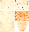
Tau pathology in CNS neurons of T-279 mice but not in T-WT or C-279 mice. A, A representative image (60×) of human Alzheimer's brain immunostained with the Tau13 human-specific antibody revealed neuronal and neuritic staining. B, H, I, Representative images of the frontal cortex of T-279, T-WT, and C-279 mice, respectively, immunostained with Tau13 (20×). C, Representative image of the frontal cortex corresponding to B, immunostained with AT8, a phosphorylation-dependent antibody (20×). D–F, Representative images of brain slices of the pons (D; 40×), olfactory bulb (E; 60×), and hypothalamus (F; 20×) from T-279 mice showing Tau13-immunostained neurons and neuronal processes. G, Representative Gallyas silver-stained brain slice from the hypothalamus of a T-279 mouse (60×). All animals represented in these images were 52 weeks of age.
Figure 5.
Figure 5.
Tau accumulation and degeneration of neuronal processes in T-279 mice. A, B, Tau13 immunostain (20×; A) and FluoroJade B histochemical stain (60×; B) of beaded and swollen neuronal processes in the midbrain. C, D, Tau13-immunopositive (C; 40×) and Gallyas silver-positive (60×; D) axons in the striatum. E, F, Representative image of Tau13-immunopositive staining of structures in the cerebellum (20×; E), which when examined at high power (60×; F) were identified as cerebellar rosettes. Arrows indicate beaded neuronal processes, and arrowheads indicate swollen neuronal processes. All animals represented in these images were 52 weeks of age.
Figure 6.
Figure 6.
Tau accumulation and neurodegeneration is age progressive. A, C, E, Images of Tau13-immunostained brain slices from 18-week-old T-279 mice. B, D, F, Images of Tau13-immunostained brain slices from 52-week-old T-279 mice. A, B, Lateral hypothalamic area. C, D, Lateral septal nucleus. E, F, Cerebellum. There was an age-dependent increase in tau accumulation in neurons and neuronal processes as well as an age-dependent degeneration of processes, as evidenced by beaded and disrupted neurites.
Figure 7.
Figure 7.
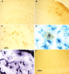
Tau protein accumulates in astrocytes. A–E, Images of brain slices from 52-week-old T-279 mice. A–C, Tau13-immunopositive tufted astrocytes in the hippocampus and cortex (A), frontal cortex (B), and amygdala (C). D, Tufted hippocampal astrocyte double immunostained with Tau13 (brown) and GFAP (blue). E, Gallyas silver-stained astrocytes in the hippocampus. F, Tau13 immunostaining of the hippocampus from a C-279 age-matched control. The animals in these images were selected because of their high numbers of tufted astrocytes.
Figure 8.
Figure 8.
Phosphorylated tau pathology in neurons and astrocytes of T-279 mice. A, A representative image of neurons and neuronal processes in the hypothalamus immunostained with AD2. B–D, Representative images of astrocytes in the hippocampus, immunostained with AD2, AT8, and AT100, respectively. The scale bar in D is representative of all four images. All animals represented in these images were 52 weeks of age.
Figure 9.
Figure 9.
TH-positive accumulations in varicosities and spheroids of neuronal processes present in T-279 mice. A, Darkly stained varicosities immunopositive with a tyrosine hydroxylase antibody were prominent in the substantia nigra compacta of T-279 mice (arrows). B, WT control mice showed only light staining of straight processes. C, Dark, TH-positive immunostaining of the nigrostriatal tract and neuronal processes of the striatum (inset) are present in the brains of T-279 mice. Small axonal swellings immunopositive with the TH antibody are interspersed throughout the nigrostriatal tracts and the striatum (arrows). D, WT mice show very light TH-positive immunostaining in the nigrostriatal tract. C, D, Images include a partial view of the anterior commissure (top of images; asterisks) for regional comparison between animals. The scale bar in A is valid for A–D.
Figure 10.
Figure 10.
Neurons and glia were immunopositive for activated caspase-3 in T-279 mice. A, Pyramidal neurons in layer 5 of the frontal cortex were immunopositive for activated caspase-3. B, Activated caspase-3 immunostaining was especially prominent in neurons and neuronal processes in the olfactory bulb. C, Tufted astrocytes immunopositive with the activated caspase-3 antibody were visible as a line (arrow) in layer 3 of the cortex. D, Higher magnification of C depicts the degenerated appearance of the astrocytes. Original images were taken at 20× magnification in A, B, and D and at 4× magnification in C.
Figure 11.
Figure 11.
T-279 mice show motor and cognitive deficits. A, T-279 mice with acute motor deficits lost the extension reflex (left) and eventually became completely paralyzed in their hind-limbs (right). B, Mice that did not appear affected by motor deficits at 23 and 52 weeks nevertheless showed a decreased latency on the rotarod task. C, D, Twenty-three-week-old (C) and 52-week-old (D) T-279 mice did not perform as well as their nontransgenic littermates on the radial arm water maze (RAWM) task.

Similar articles

Cited by

References

    1. Arai T, Ikeda K, Akiyama H, Shikamoto Y, Tsuchiya K, Yagishita S, Beach T, Rogers J, Schwab C, McGeer PL. Distinct isoforms of tau aggregated in neurons and glial cells in brains of patients with Pick's disease, corticobasal degeneration and progressive supranuclear palsy. Acta Neuropathol (Berl) 2001;101:167–173. - PubMed
    1. Arima K, Kowalska A, Hasegawa M, Mukoyama M, Watanabe R, Kawai M, Takahashi K, Iwatsubo T, Tabira T, Sunohara N. Two brothers with frontotemporal dementia and parkinsonism with an N279K mutation of the tau gene. Neurology. 2000;54:1787–1795. - PubMed
    1. Auboeuf D, Honig A, Berget SM, O'Malley BW. Coordinate regulation of transcription and splicing by steroid receptor coregulators. Science. 2002;298:416–419. - PubMed
    1. Baker M, Litvan I, Houlden H, Adamson J, Dickson D, Perez-Tur J, Hardy J, Lynch T, Bigio E, Hutton M. Association of an extended haplotype in the tau gene with progressive supranuclear palsy. Hum Mol Genet. 1999;8:711–715. - PubMed
    1. Berry RW, Abraha A, Lagalwar S, LaPointe N, Gamblin TC, Cryns VL, Binder LI. Inhibition of tau polymerization by its carboxy-terminal caspase cleavage fragment. Biochemistry. 2003;42:8325–8331. - PubMed

Publication types

MeSH terms