. 2007 Sep 6;50(18):4509-15.
doi: 10.1021/jm070482q. Epub 2007 Aug 16.

Potent new antiviral compound shows similar inhibition and structural interactions with drug resistant mutants and wild type HIV-1 protease

Affiliations

Potent new antiviral compound shows similar inhibition and structural interactions with drug resistant mutants and wild type HIV-1 protease

Yuan-Fang Wang et al. J Med Chem. .

Abstract

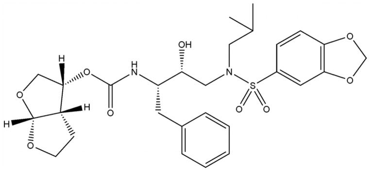
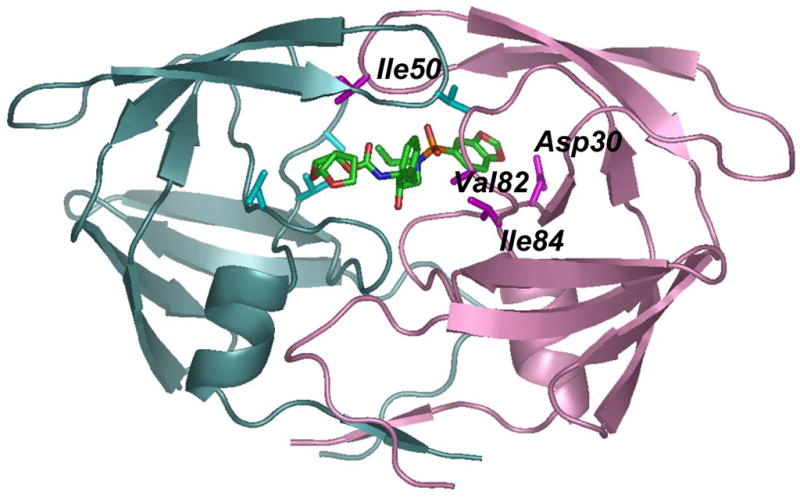
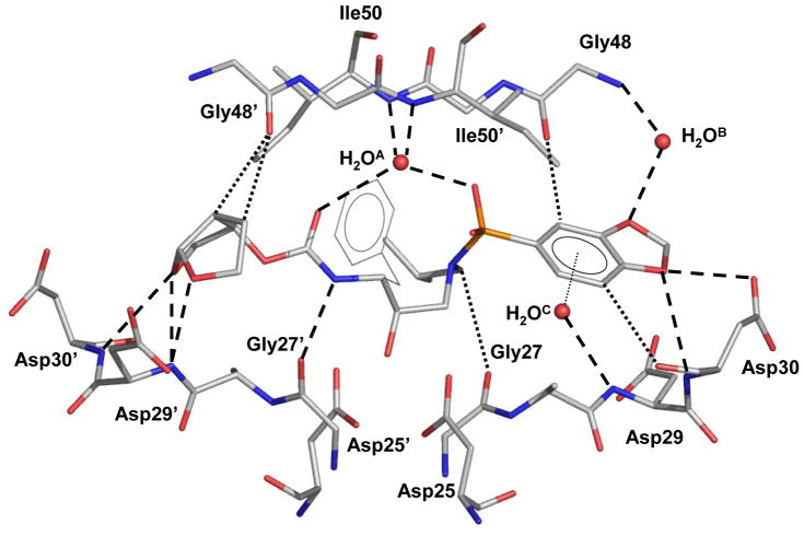
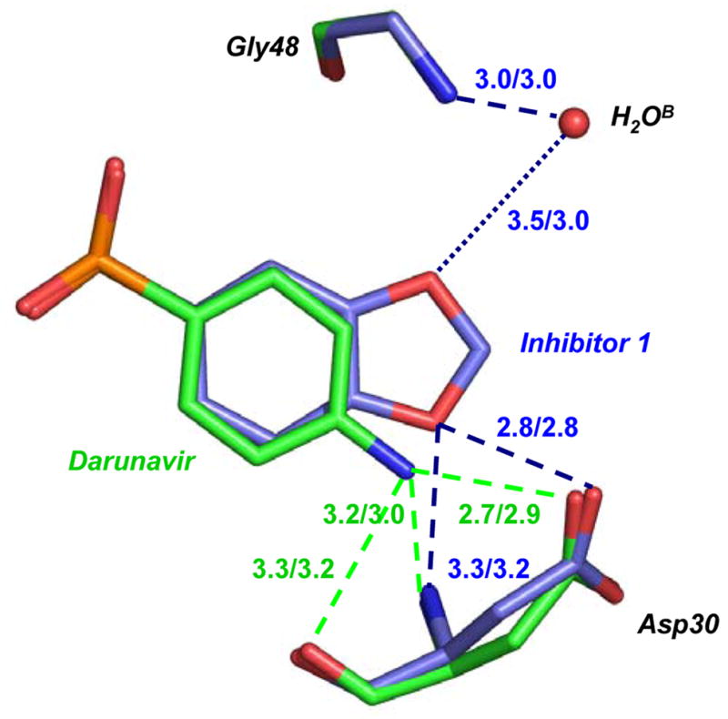
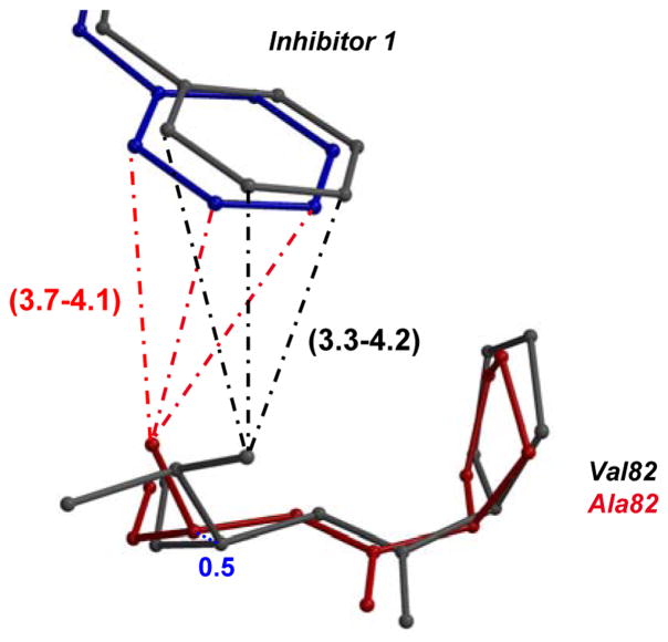
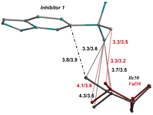
The potent new antiviral inhibitor GRL-98065 (1) of HIV-1 protease (PR) has been studied with PR variants containing the single mutations D30N, I50V, V82A, and I84V that provide resistance to the major clinical inhibitors. Compound 1 had inhibition constants of 17-fold, 8-fold, 3-fold, and 3-fold, respectively, for PR(D30N), PR(I50V), PR(V82A), and PR(I84V) relative to wild type PR. The chemically related darunavir had similar relative inhibition, except for PR(D30N), where inhibitor 1 was approximately 3-fold less potent. The high resolution (1.11-1.60 Angstrom) crystal structures of PR mutant complexes with inhibitor 1 showed small changes relative to the wild type enzyme. PR(D30N) and PR(V82A) showed compensating interactions with inhibitor 1 relative to those of PR, while reduced hydrophobic contacts were observed with PR(I50V) and PR(I84V). Importantly, inhibitor 1 complexes showed fewer changes relative to wild type enzyme than reported for darunavir complexes. Therefore, inhibitor 1 is a valuable addition to the antiviral inhibitors with high potency against resistant strains of HIV.


Figures

Figure 1
Figure 1. Structures of A) Inhibitor 1 (GRL-98065), B) Darunavir (TMC-114), and C) PR dimer indicating location of mutations
The backbone of one subunit is shown as pink ribbons with the sites of mutation (D30N, I50V, V82A and I84V) indicated by magenta side chains, while the other subunit is colored in light cyan with mutation sites in darker cyan. Only one subunit is labeled. Inhibitor 1 is colored by atom type.
Figure 2
Figure 2. Electron density map for inhibitor 1 in complex with PRV82A
The omit Fo-Fc map, using Fc calculated without inhibitor 1, is contoured at 8.5σ level. Inhibitor 1 had a single conformation.
Figure 3
Figure 3. Protease interactions with inhibitor 1 and darunavir
3A. Polar interactions of inhibitor 1 with PR The major conformation of inhibitor 1 is shown with interacting PR residues. Water is shown as red spheres. Hydrogen bond interactions are indicated by dotted lines, and CH…O interactions with main chain PR atoms by dashed lines. The H2OC O-H…π interaction with the P2′ aromatic ring is shown as a gray dotted line. The interactions of the alternate conformation of inhibitor are essentially the same. 3B. Comparison of PR interactions with inhibitor 1 and darunavir. Carbon atoms in PR/inhibitor 1 are colored in blue and those in PR/darunavir are green. The hydrogen bond interactions are indicated by the dashed lines, with distances in Å for the major/minor conformation of inhibitor, and colored blue for PR/inhibitor 1 and green for PR/darunavir. The dotted line indicates the water-mediated interaction with a weaker hydrogen bond for the major conformation.
Figure 4
Figure 4. Structural changes in mutants compared to wild type PR
The major and minor conformations of side chains in PR are shown in dark and light grey ball-and-stick, respectively. The alternate conformations in mutant complexes are shown in red and pink, and the alternate conformations of inhibitor are in blue and cyan. The polar interactions are indicated by the dashed lines, van der Waals interactions by the dotted lines, and C-H…π interactions in dot-and-dash line. The interatomic distances are shown in Å for both subunits. 4A. Interactions of P1′ of inhibitor 1 with residue 82′in PRV82A and PR. The minor conformation of inhibitor 1 and alternate conformation of Val82 in PR are omitted for clarity. The shift in position of Cα 82 between PR and mutant is shown in blue dotted lines. 4B. Van der Waals interactions of P1′ in inhibitor 1 with residue 82′ in PRV82A and PR. View and representation are similar to 4A. 4C. PRI84V and PR interactions with inhibitor 1. 4D. PRI50V and PR interactions with inhibitor 1. Only a single conformation is shown for clarity. 4E. PRD30N and PR interactions with inhibitor 1. 4F. PRD30N and PR interactions with darunavir.

Similar articles

Cited by

References

    1. Barbaro G, Lucchini A, Barbarini G. Highly active antiretroviral therapy in HIV-associated pulmonary hypertension. Minerva Cardioangiol. 2005;53:153–4. - PubMed
    1. Barlett JA, DeMasi R, Quinn J, Moxham C, Rousseau F. Overview of the Effectiveness of Triple Combination Therapy in Antiretroviral-Naïve HIV-1 Infected Adults. AIDS. 2001;15:1369–1377. - PubMed
    1. Palella FJ, Jr, Delaney KM, Moorman AC, Loveless MO, Fuhrer J, Satten GA, Aschman DJ, Holmberg SD. Declining morbidity and mortality among patients with advanced human immunodeficiency virus infection. HIV Outpatient Study Investigators. N Engl J Med. 1998;338:853–60. - PubMed
    1. Grabar S, Pradier C, Le Corfec E, Lancar R, Allavena C, Bentata M, Berlureau P, Dupont C, Fabbro-Peray P, Poizot-Martin I, Costagliola D. Factors associated with clinical and virological failure in patients receiving a triple therapy including a protease inhibitor. Aids. 2000;14:141–9. - PubMed
    1. Hertogs K, Bloor S, Kemp SD, Van den Eynde C, Alcorn TM, Pauwels R, Van Houtte M, Staszewski S, Miller V, Larder BA. Phenotypic and genotypic analysis of clinical HIV-1 isolates reveals extensive protease inhibitor cross-resistance: a survey of over 6000 samples. AIDS. 2000;14:1203–1210. - PubMed

Publication types

Substances