. 2007 Aug 22;26(16):3737-48.
doi: 10.1038/sj.emboj.7601813. Epub 2007 Aug 2.

Differential involvement of endocytic compartments in the biosynthetic traffic of apical proteins

Affiliations

Differential involvement of endocytic compartments in the biosynthetic traffic of apical proteins

Kerry O Cresawn et al. EMBO J. .

Abstract

Newly synthesized basolateral markers can traverse recycling endosomes en route to the surface of Madin-Darby canine kidney cells; however, the routes used by apical proteins are less clear. Here, we functionally inactivated subsets of endocytic compartments and examined the effect on surface delivery of the basolateral marker vesicular stomatitis virus glycoprotein (VSV-G), the raft-associated apical marker influenza hemagglutinin (HA), and the non-raft-associated protein endolyn. Inactivation of transferrin-positive endosomes after internalization of horseradish peroxidase (HRP)-containing conjugates inhibited VSV-G delivery, but did not disrupt apical delivery. In contrast, inhibition of protein export from apical recycling endosomes upon expression of dominant-negative constructs of myosin Vb or Sec15 selectively perturbed apical delivery of endolyn. Ablation of apical endocytic components accessible to HRP-conjugated wheat germ agglutinin (WGA) disrupted delivery of HA but not endolyn. However, delivery of glycosylphosphatidylinositol-anchored endolyn was inhibited by >50% under these conditions, suggesting that the biosynthetic itinerary of a protein is dependent on its targeting mechanism. Our studies demonstrate that apical and basolateral proteins traverse distinct endocytic intermediates en route to the cell surface, and that multiple routes exist for delivery of newly synthesized apical proteins.


Figures
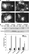
Figure 1
Figure 1
Inactivation of Tf-containing endosomes in nonpolarized and polarized cells inhibits surface delivery of tsVSV-G-YFP. (A) PTR9 cells grown on coverslips were infected with adenovirus-encoding tsVSV-G-YFP and incubated overnight at 40°C. The next day, the cells were incubated in serum-free media for 30 min before addition of HRP-Tf for 45 min at 40°C. Remaining surface HRP-Tf was stripped and Tf-containing endosomes were inactivated on ice as described in Materials and methods. As controls in this and subsequent experiments, cells were incubated either without HRP-Tf, DAB, or H2O2 (no add'n), with only HRP-Tf and DAB (no H2O2), or with only DAB and H2O2 (no HRP-Tf). Cells were then incubated at 32°C in the presence of cycloheximide for 1 h, washed with PBS, then fixed and imaged. Scale bar=5 μm (B) Polarized PTR9 cells grown on filters were infected with adenovirus encoding VSV-G and incubated overnight at 37°C. The following day, cells were starved in medium devoid of cysteine and methionine for 30 min and then radiolabeled for 15 min. HRP-Tf was included in the basolateral medium during the starve and radiolabeling period. After stripping residual surface HRP-Tf, cells were treated with or without DAB and H2O2 for 1 h on ice as in (A). After washing, cells were chased for 0 or 90 min at 37°C, and delivery to the basolateral surface was quantified using domain-selective biotinylation. Samples were immunoprecipitated and analyzed by SDS–PAGE and gels showing the total amount of radiolabeled protein in the cell and the amount at the basolateral surface in a representative experiment are shown (upper panel). The mean±s.e. of three experiments is plotted (lower panel). Surface delivery of VSV-G was significantly inhibited in the ‘complete' reaction samples compared with all three control conditions (*P<0.02 by Student's t-test).
Figure 2
Figure 2
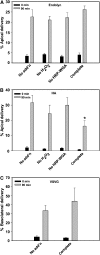
Delivery of endolyn and HA to the apical surface is not disrupted when Tf-containing endosomes are inactivated. Filter-grown PTR9 cells were infected with adenoviruses encoding endolyn (A) or HA (B) and incubated overnight at 37°C. Cells were radiolabeled, incubated with HRP-Tf at 37°C, and subjected to crosslinking as described for Figure 1B. Apical delivery was quantified by domain-selective biotinylation for endolyn (M=mature, I=immature) and by cell surface trypsinization for HA (resulting in two cleaved products marked HA1 and HA2). Uncleaved (intracellular) HA is marked as HA0. Samples were analyzed by SDS–PAGE and the gels from a representative experiment for each marker are shown (left panels). The percent apical delivery (mean±s.e.m.) for endolyn (n=3) and HA (n=3–6) are shown (right panels). Surface delivery of HA and endolyn in the ‘complete' reaction samples was statistically indistinguishable from all three control conditions.
Figure 3
Figure 3
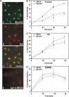
Biosynthetic apical delivery of endolyn, but not HA, is disrupted in cells expressing the myosin Vb tail. (A) IgA (200 μg/ml) was internalized from the basolateral surface of filter-grown MyoVbT or control MDCK cells for 10 min and chased for 60 min at 37°C. After rinsing, the cells were fixed, permeabilized, and incubated with Cy-5-conjugated anti-human IgA. Confocal sections taken just beneath the apical pole of the cell are shown; panel a: GFP-MyoVbT; panel b: IgA; panel c: overlay of a and b; panel d: IgA in control MDCK cells. Scale bar=10 μm. (BD) Filter-grown MyoVbT or control cells were infected with adenoviruses encoding endolyn (B), HA (C), or VSV-G (D). The following day, cells were starved, radiolabeled for 15 min, and chased for the indicated periods at 37°C before quantitation of surface delivery. The percent surface delivery is plotted as the mean±range of two experiments with duplicate samples for endolyn and HA and four experiments for VSV G. *P<0.001 compared with control cells.
Figure 4
Figure 4
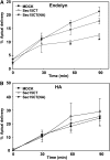
Expression of a dominant-negative inhibitor of Sec15 selectively disrupts apical delivery of endolyn. MDCK cells stably expressing Sec15 or Sec15(NA) and parental cells were co-infected with adenovirus-expressing transactivator and either endolyn (A) or HA (B). The following day, cells were radiolabeled for 15 min and chased from 0 to 90 min at 37°C before quantitation of apical delivery. The percent surface delivery is plotted as the mean±s.e. of three experiments for endolyn and two for HA. *P<0.05 compared with control cells.
Figure 5
Figure 5
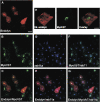
Newly synthesized endolyn colocalizes with MyoVbT and rab11a. Filter-grown MyoVbT cells were infected with adenovirus-expressing endolyn, incubated for 6 h at 37°C to initiate endolyn synthesis, and then moved to 19°C for 2 h to accumulate newly synthesized proteins in the TGN. Cells were then warmed to 37°C for 20 min to release staged proteins, then fixed and processed for detection of GFP-MyoVbT (green (B)), endolyn (red (A)), and rab11a (blue (D)). Individual confocal sections taken just beneath the apical pole of the cell are shown in each panel. Colocalization of GFP-MyoVbT with rab 11a is shown in (F); overlay of endolyn with GFP-MyoVbT, rab11a, and the triple overlay are shown in (C), (E), and (G), respectively. Cells in (H) were incubated with apically added monoclonal anti-endolyn antibody during the last hour at 19°C, then warmed to 37°C for 20 min to track the postendocytic itinerary of any endolyn that had reached the cell surface. Scale bar=20 μm, (A–G); 10 μm, (H).
Figure 6
Figure 6
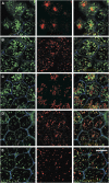
Characterization of WGA internalization conditions. WGA-FITC (25 μg/ml) was internalized from the apical surface of filter-grown PTR9 cells for 15 min at 37°C, and residual surface lectin was stripped with N-acetyl-D-glucosamine. After washing, cells were fixed, permeabilized, and processed for indirect immunofluorescence using antibodies against ZO-1 to visualize tight junctions (blue) and one of the following markers (red) (A) Rab11a, (B) EEA1, (C) furin, (D) mannose-6-phosphate receptor (M6PR), and (E) Lamp2. Single optical sections from apical-supranuclear levels of the cells are shown. Scale bar=10 μm.
Figure 7
Figure 7
Inactivation of apical WGA-containing endosomes disrupts delivery of HA, but not endolyn or VSV-G. Filter-grown PTR9 or control MDCK cells infected with adenoviruses encoding endolyn (A), HA (B), or VSV-G (C) were starved for 30 min and radiolabeled for 15 min. HRP-WGA was included in the apical medium during the 15 min pulse or afterwards for 1 h at 19°C. After internalization, remaining surface HRP-WGA was removed with 100 mM N-acetyl-D-glucosamine before endosome inactivation as described in Materials and methods. Subsequently, cells were chased for 0 or 90 min at 37°C and apical delivery was quantified. The percent surface delivery is plotted as the mean±s.e.m. for endolyn (n=3) and HA (n=6) and as mean±s.d. for VSV-G (n=1 experiment performed in sextuplicate). Ablation of apical WGA-containing endosomes reduced appearance of HA at the surface (*P<0.03 compared with the ‘no add'n' control).
Figure 8
Figure 8
Inactivation of WGA-containing compartments disrupts apical delivery of GPI-linked endolyn. Filter-grown MDCK cells stably expressing GPI-anchored endolyn were incubated with 2 mM butyrate overnight at 37°C to induce protein expression. The following day, cells were radiolabeled and incubated with HRP-WGA as described in the legend to Figure 7 before quantitation of GPI-endolyn apical delivery by domain selective biotinylation. The percent surface delivery of GPI-endolyn is plotted as the mean±s.e.m. of three experiments. *P<0.05 compared with the ‘no add'n' control.

Similar articles

Cited by

References

    1. Altschuler Y, Kinlough CL, Poland PA, Bruns JB, Apodaca G, Weisz OA, Hughey RP (2000) Clathrin-mediated endocytosis of MUC1 is modulated by its glycosylation state. Mol Biol Cell 11: 819–831 - PMC - PubMed
    1. Ang AL, Taguchi T, Francis S, Folsch H, Murrells LJ, Pypaert M, Warren G, Mellman I (2004) Recycling endosomes can serve as intermediates during transport from the Golgi to the plasma membrane of MDCK cells. J Cell Biol 167: 531–543 - PMC - PubMed
    1. Apodaca G, Katz LA, Mostov KE (1994) Receptor-mediated transcytosis of IgA in MDCK cells is via apical recycling endosomes. J Cell Biol 125: 67–86 - PMC - PubMed
    1. Barroso M, Sztul ES (1994) Basolateral to apical transcytosis in polarized cells is indirect and involves BFA and trimeric G protein sensitive passage through the apical endosome. J Cell Biol 124: 83–100 - PMC - PubMed
    1. Benting JH, Rietveld AG, Simons K (1999) N-Glycans mediate the apical sorting of a GPI-anchored, raft-associated protein in Madin–Darby canine kidney cells. J Cell Biol 146: 313–320 - PMC - PubMed

Publication types

MeSH terms

LinkOut - more resources