The conceptus increases secreted phosphoprotein 1 gene expression in the mouse uterus during the progression of decidualization mainly due to its effects on uterine natural killer cells
- PMID: 17636175
- PMCID: PMC2613481
- DOI: 10.1530/REP-07-0085
The conceptus increases secreted phosphoprotein 1 gene expression in the mouse uterus during the progression of decidualization mainly due to its effects on uterine natural killer cells
Abstract
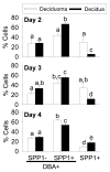
Within the mouse endometrium, secreted phosphoprotein 1 (SPP1) gene expression is mainly expressed in the luminal epithelium and some macrophages around the onset of implantation. However, during the progression of decidualization, it is expressed mainly in the mesometrial decidua. To date, the precise cell types responsible for the expression in the mesometrial decidua has not been absolutely identified. The goal of the present study was to assess the expression of SPP1 in uteri of pregnant mice (decidua) during the progression of decidualization and compared it with those undergoing artificially induced decidualization (deciduoma). Significantly (P<0.05) greater steady-state levels of SPP1 mRNA were seen in the decidua when compared with deciduoma. Further, in the decidua, the majority of the SPP1 protein was localized within a subpopulation of granulated uterine natural killer (uNK) cells but not co-localized to their granules. However, in addition to being localized to uNK cells, SPP1 protein was also detected in another cell type(s) that were not epidermal growth factor-like containing mucin-like hormone receptor-like sequence 1 protein-positive immune cells that are known to be present in the uterus at this time. Finally, decidual SPP1 expression dramatically decreased in uteri of interleukin-15-deficient mice that lack uNK cells. In conclusion, SPP1 expression is greater in the mouse decidua when compared with the deciduoma after the onset of implantation during the progression of decidualization. Finally, uNK cells were found to be the major source of SPP1 in the pregnant uterus during decidualization. SPP1 might play a key role in uNK killer cell functions in the uterus during decidualization.
Figures


Similar articles
-
Effect of the conceptus on uterine natural killer cell numbers and function in the mouse uterus during decidualization.Biol Reprod. 2007 Apr;76(4):579-88. doi: 10.1095/biolreprod.106.056630. Epub 2006 Dec 6. Biol Reprod. 2007. PMID: 17151350 Free PMC article.
-
Secreted phosphoprotein 1 (osteopontin) is expressed by stromal macrophages in cyclic and pregnant endometrium of mice, but is induced by estrogen in luminal epithelium during conceptus attachment for implantation.Reproduction. 2006 Dec;132(6):919-29. doi: 10.1530/REP-06-0068. Reproduction. 2006. PMID: 17127752
-
SPP1 expression in the mouse uterus and placenta: implications for implantation†.Biol Reprod. 2021 Oct 11;105(4):892-904. doi: 10.1093/biolre/ioab125. Biol Reprod. 2021. PMID: 34165144
-
Update on pathways regulating the activation of uterine Natural Killer cells, their interactions with decidual spiral arteries and homing of their precursors to the uterus.J Reprod Immunol. 2003 Aug;59(2):175-91. doi: 10.1016/s0165-0378(03)00046-9. J Reprod Immunol. 2003. PMID: 12896821 Review.
-
Functions of uterine natural killer cells are mediated by interferon gamma production during murine pregnancy.Semin Immunol. 2001 Aug;13(4):235-41. doi: 10.1006/smim.2000.0319. Semin Immunol. 2001. PMID: 11437631 Review.
Cited by
-
Angiopoietin-like gene expression in the mouse uterus during implantation and in response to steroids.Cell Tissue Res. 2012 Apr;348(1):199-211. doi: 10.1007/s00441-012-1337-4. Epub 2012 Feb 22. Cell Tissue Res. 2012. PMID: 22350948 Free PMC article.
-
Diet-induced obesity impairs endometrial stromal cell decidualization: a potential role for impaired autophagy.Hum Reprod. 2016 Jun;31(6):1315-26. doi: 10.1093/humrep/dew048. Epub 2016 Apr 6. Hum Reprod. 2016. PMID: 27052498 Free PMC article.
-
Analysis of uterine gene expression in interleukin-15 knockout mice reveals uterine natural killer cells do not play a major role in decidualization and associated angiogenesis.Reproduction. 2012 Mar;143(3):359-75. doi: 10.1530/REP-11-0325. Epub 2011 Dec 20. Reproduction. 2012. PMID: 22187674 Free PMC article.
-
Paracrine signals from the mouse conceptus are not required for the normal progression of decidualization.Endocrinology. 2009 Sep;150(9):4404-13. doi: 10.1210/en.2009-0036. Epub 2009 Jun 11. Endocrinology. 2009. PMID: 19520782 Free PMC article.
-
[The involvement of galectin-1 in implantation and pregnancy maintenance at the maternal-fetal interface].Zhejiang Da Xue Xue Bao Yi Xue Ban. 2017 May 25;46(3):321-327. doi: 10.3785/j.issn.1008-9292.2017.06.16. Zhejiang Da Xue Xue Bao Yi Xue Ban. 2017. PMID: 29039177 Free PMC article. Chinese.
References
-
- Apparao KB, Murray MJ, Fritz MA, Meyer WR, Chambers AF, Truong PR, Lessey BA. Osteopontin and its receptor alphavbeta(3) integrin are coexpressed in the human endometrium during the menstrual cycle but regulated differentially. J Clin Endocrinol Metab. 2001;86:4991–5000. - PubMed
-
- Ashkar AA, Black GP, Wei Q, He H, Liang L, Head JR, Croy BA. Assessment of requirements for IL-15 and IFN regulatory factors in uterine NK cell differentiation and function during pregnancy. J Immunol. 2003;171:2937–2944. - PubMed
-
- Austyn JM, Gordon S. F4/80, a monoclonal antibody directed specifically against the mouse macrophage. Eur J Immunol. 1981;11:805–815. - PubMed
-
- Bany BM, Cross JC. Post-implantation mouse conceptuses produce paracrine signals that regulate the uterine endometrium undergoing decidualization. Dev Biol. 2006;294:445–456. - PubMed
-
- Barber EM, Pollard JW. The uterine NK cell population requires IL-15 but these cells are not required for pregnancy nor the resolution of a Listeria monocytogenes infection. J Immunol. 2003;171:37–46. - PubMed
Publication types
MeSH terms
Substances
Grants and funding
LinkOut - more resources
Full Text Sources
Molecular Biology Databases
Research Materials
Miscellaneous

