. 2007 Sep;81(17):9142-51.
doi: 10.1128/JVI.02885-06. Epub 2007 Jul 3.

DCL4 targets Cucumber mosaic virus satellite RNA at novel secondary structures

Affiliations

DCL4 targets Cucumber mosaic virus satellite RNA at novel secondary structures

Quan-Sheng Du et al. J Virol. 2007 Sep.

Abstract

It has been reported that plant virus-derived small interfering RNAs (vsiRNAs) originated predominantly from structured single-stranded viral RNA of a positive single-stranded RNA virus replicating in the cytoplasm and from the nuclear stem-loop 35S leader RNA of a double-stranded DNA (dsDNA) virus. Increasing lines of evidence have also shown that hierarchical actions of plant Dicer-like (DCL) proteins are required in the biogenesis process of small RNAs, and DCL4 is the primary producer of vsiRNAs. However, the structures of such single-stranded viral RNA that can be recognized by DCLs remain unknown. In an attempt to determine these structures, we have cloned siRNAs derived from the satellite RNA (satRNA) of Cucumber mosaic virus (CMV-satRNA) and studied the relationship between satRNA-derived siRNAs (satsiRNAs) and satRNA secondary structure. satsiRNAs were confirmed to be derived from single-stranded satRNA and are primarily 21 (64.7%) or 22 (22%) nucleotides (nt) in length. The most frequently cloned positive-strand satsiRNAs were found to derive from novel hairpins that differ from the structure of known DCL substrates, miRNA and siRNA precursors, which are prevalent stem-loop-shaped or dsRNAs. DCL4 was shown to be the primary producer of satsiRNAs. In the absence of DCL4, only 22-nt satsiRNAs were detected. Our results suggest that DCL4 is capable of accessing flexibly structured single-stranded RNA substrates (preferably T-shaped hairpins) to produce satsiRNAs. This result reveals that viral RNA of diverse structures may stimulate antiviral DCL activities in plant cells.


Figures
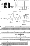
FIG. 1.
FIG. 1.
Origin and characterization of SD-satRNA-derived small RNAs (satsiRNAs). (A) Accumulation of satRNAs in SD-CMV-infected Arabidopsis. Total RNA extracted from wild-type (WT) and SD-CMV-infected (inf.) Arabidopsis plants was separated in agarose gel and stained with ethidium bromide (left), blotted, and hybridized with an α-32P-labeled probe of SD-satRNA sequence (right). Lane M, molecular size markers. (B) Size distribution of sequenced satsiRNAs. (C) Distribution of satsiRNA clones alongside both strands of SD-satRNA. Small RNAs were aligned with the SD-satRNA genome by sequence comparison. Short lines above the satRNA genome represent satsiRNAs derived from the positive strand, and those below the genome represent satsiRNAs derived from the negative strand. Clone frequencies were represented by the number of short lines at the same position. Small RNAs derived from SD-satRNA variants were also included. The identification serials of some satsiRNAs are indicated. Three pairs of satsiRNAs that were fully complementary in base pairing are boxed with dashes. Four small RNAs located at the 3′ end of satRNA (16 to 18 nt) were not counted as satsiRNAs and are indicated by dashed lines. (D) Alignment of D4-satRNA and SD-satRNA sequences, as well as two SD-satRNA variant sequences obtained from SD-CMV-infected Arabidopsis by reverse transcription-PCR and sequencing. Four major folding regions based on the D4-satRNA in vivo structure model are indicated. The C139 insertion in SD variants 1 and 2 and sequence heterogeneities are highlighted with dark gray. Some satsiRNA clones identical or perfectly complementary to the SD variants are shown.
FIG. 2.
FIG. 2.
Detection of small RNAs in SD-CMV-infected plants. (A) Northern blot analysis of representative satsiRNA. Thirty micrograms of small RNA extracted from wild-type (WT) A. thaliana Columbia or SD-CMV-infected Columbia was separated by PAGE and blotted. Membranes were hybridized with 32P-labeled probes of specific satsiRNA antisense (as) or sense (s) probes. Two signals detected with the satsiR-24 sense probe corresponding to RNAs larger than satsiRNAs are indicated by asterisks. (B) Accumulation of passenger strands of both miRNA* and tasiRNA* in SD-CMV-infected (inf.) plant. 32P-labeled probe-specific sequences for miR168, miR168*, tasiR255 and tasiR255*, and the U6 control are indicated.
FIG. 3.
FIG. 3.
SD-CMV infection symptoms and accumulation of satsiRNAs in wild-type strain Columbia and the dcl4-2 mutant. (A) One-month-old wild-type strain Columbia (Col.) and the dcl4-2 mutant (left two panels). The dcl4-2 mutant displays downward-curled leaf margins. Ten days post-SD-CMV-infection, wild-type strain Columbia and the dcl4-2 mutant (middle two panels) show similar developmental defect infection symptoms, which resulted in new leaves fasciated in the center of the plants (arrows). Influorescences of healthy and SD-CMV-infected Columbia strain and the infected snaky siliques (inset) are shown in the right panel. (B) Evidence for the contribution of DCL4 in the production of satsiRNAs. Thirty micrograms of RNA extracted from SD-CMV-infected wild-type strain Columbia and the dcl4-2 mutant was separated by PAGE and blotted. Membranes were hybridized with 32P-labeled probes specific for each satsiRNA, miR-159, and ta-siR255.
FIG. 4.
FIG. 4.
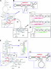
Localization of satsiRNAs in in vivo and in virion secondary structure models for CMV satRNA. (A and E) Structure model for D4-satRNA from total RNA extracted from infected cells (in vivo) (A) or from purified CMV particles (in virion) (E) taken from Fig. 2A and C in reference . Numbers I, II, III, and IV indicate the four major folding regions. (B) T-shaped hairpin for satsiR-2 (highlighted in red) twisted from the highly conserved 5′-end region I in panel A boxed in blue. (C) T-shaped hairpins for satsiR-18a/b/c (highlighted in purple) twisted from the highly conserved 3′-end region IV in panel A boxed with black lines. (D) Pair of potential imperfect duplex of satsiR-15 and satsiR-17 (indicated with green lines) positioned in the highly conserved 3′-end region IV in panel A boxed with black lines. (F) Pair of potential imperfect duplex of satsiR-3 and satsuR-11 (indicated with green lines) positioned in the nearly perfectly matched area in region II in panel E boxed in blue dashed lines. (G) T-shaped hairpin for satsiR-2 (highlighted in red) twisted from the highly conserved 5′-end region I in panel E boxed in blue lines. The four different residues in region III between D4-satR and SD-satR (SD) as well as mutations detected in the two variants (V1 and V2) are shown in red in panel A. The G269U base pair in D4- and SD-satRNA changed to the AU canonical base pair in the SD-satRNA variants 1 and 2 is shown in red in panel C. (Reprinted from Virology (28) with permission of the publisher.)
FIG. 5.
FIG. 5.
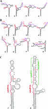
Predicted T-shaped hairpins for satsiR-5. Secondary structure prediction using the mFold software of RNA sequences including bases U79 to A190 of SD-satRNA (A) and variants with a C139 insertion (indicated in red) (B). (A) Four structures for the SD-satRNA sequence. Stem 8 in all of these structures was identical to that of the in virion model of D4-satRNA (see Fig. 4E). (B) Eight structures of SD-satRNA variant sequences. Structures 1 to 4 are similar to those shown for SD-satRNA in panel A. Structures 7 and 8 were twisted as illustrated in panel C to show the adequate T-shaped hairpins for satsiR-5 recognized to position at the upper part of the stem-loop region, and the long-stem-loop precursor for a potential imperfect duplex of satsiR-6 and satsiR-8a is indicated with a green line. Sequences of satsiR-5 and satsiR-8 are highlighted in red and purple, respectively. Stems 10, 11, and 14 formed in structure 8 are indicated.

Similar articles

Cited by

References

    1. Baulcombe, D. 2004. RNA silencing in plants. Nature 431:356-363. - PubMed
    1. Baumberger, N., and D. C. Baulcombe. 2005. Arabidopsis ARGONAUTE1 is an RNA Slicer that selectively recruits microRNAs and short interfering RNAs. Proc. Natl. Acad. Sci. USA 102:11928-11933. - PMC - PubMed
    1. Blevins, T., R. Rajeswaran, P. V. Shivaprasad, D. Beknazariants, A. S. Ammour, H. S. Park, F. Vazquez, D. Robertson, F. Meins, Jr., T. Hohn, and M. M. Pooggin. 2006. Four plant Dicers mediate viral small RNA biogenesis and DNA virus induced silencing. Nucleic Acids Res. 34:6233-6246. - PMC - PubMed
    1. Borsani, O., J. Zhu, P. E. Verslues, R. Sunkar, and J. K. Zhu. 2005. Endogenous siRNAs derived from a pair of natural cis-antisense transcripts regulate salt tolerance in Arabidopsis. Cell 123:1279-1291. - PMC - PubMed
    1. Bouche, N., D. Lauressergues, V. Gasciolli, and H. Vaucheret. 2006. An antagonistic function for Arabidopsis DCL2 in development and a new function for DCL4 in generating viral siRNAs. EMBO J. 25:3347-3356. - PMC - PubMed

Publication types

MeSH terms

LinkOut - more resources