

Antibodies to placental immunoregulatory ferritin with transfer of polyclonal lymphocytes arrest MCF-7 human breast cancer growth in a nude mouse model
- PMID: 17603631
- PMCID: PMC1899253
- DOI: 10.1593/neo.07259
Antibodies to placental immunoregulatory ferritin with transfer of polyclonal lymphocytes arrest MCF-7 human breast cancer growth in a nude mouse model
Abstract
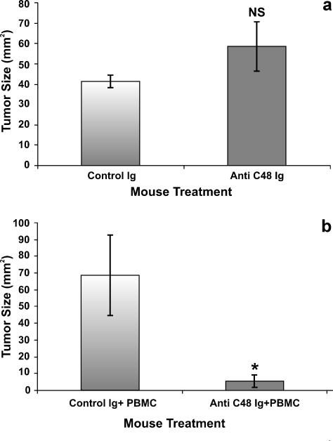
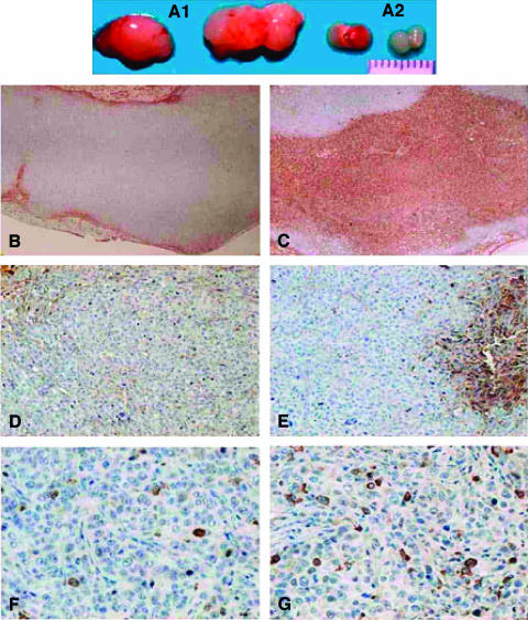
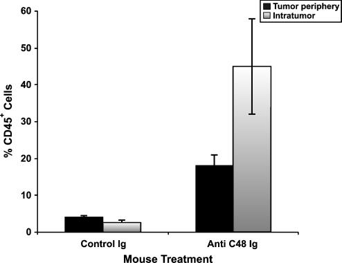
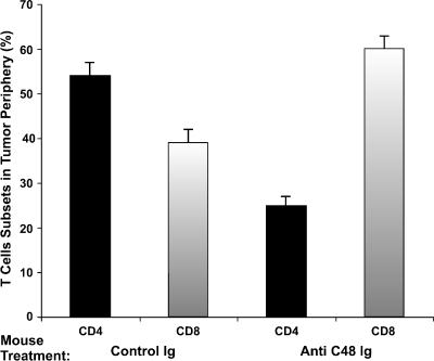
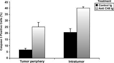
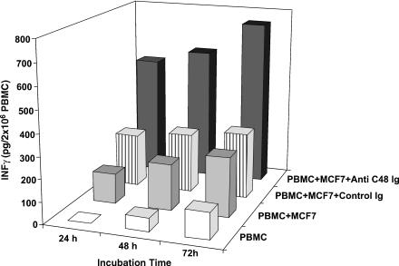
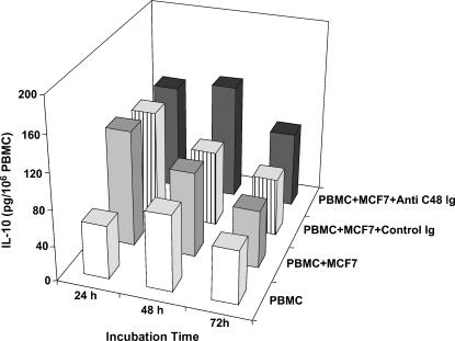
The recently cloned human gene named "placental immunoregulatory ferritin" (PLIF) is a pregnancy-related immunomodulator. Recombinant PLIF and its bioactive domain C48 are immune-suppressive and induce pronounced IL-10 production by immune cells. PLIF is expressed in the placenta and breast cancer cells. Blocking PLIF in pregnant mice by anti-C48 antibodies inhibited placental and fetal growth and modulated the cytokine network. It has been revealed that anti-C48 treatment inhibited MCF-7 tumor growth in nude mice. However, this significant effect was observed only in those transfused with human peripheral blood mononuclear cells. Blocking PLIF in tumor-engrafted human immune cell transfused mice resulted in massive infiltration of human CD45+ cells (mainly CD8+ T cells), both intratumorally and in the tumor periphery, and a significant number of caspase-3+ cells. In vitro, anti-C48 treatment of MCF-7 tumor cells cocultured with human lymphocytes induced a significant increase in interferon-gamma secretion. We conclude that blocking PLIF inhibits breast cancer growth, possibly by an effect on the cytokine network in immune cells and on breakdown of immunosuppression.
Figures

Similar articles
-
The novel C24D synthetic polypeptide inhibits binding of placenta immunosuppressive ferritin to human T cells and elicits anti-breast cancer immunity in vitro and in vivo.Neoplasia. 2014 Sep;16(9):741-50. doi: 10.1016/j.neo.2014.08.005. Neoplasia. 2014. PMID: 25246274 Free PMC article.
-
Blocking of the placental immune-modulatory ferritin activates Th1 type cytokines and affects placenta development, fetal growth and the pregnancy outcome.Hum Reprod. 2004 Mar;19(3):715-22. doi: 10.1093/humrep/deh099. Epub 2004 Jan 29. Hum Reprod. 2004. PMID: 14998975
-
Placental immunomodulator ferritin, a novel immunoregulator, suppresses experimental arthritis.Arthritis Rheum. 2003 Mar;48(3):846-53. doi: 10.1002/art.10850. Arthritis Rheum. 2003. PMID: 12632441
-
PLIF induces IL-10 production in monocytes: a calmodulin-p38 mitogen-activated protein kinase-dependent pathway.FASEB J. 2003 May;17(8):955-7. doi: 10.1096/fj.02-0960fje. Epub 2003 Mar 28. FASEB J. 2003. PMID: 12670872
-
Treatment of human bone marrow with recombinant placenta immunoregulator ferritin results in myelopoiesis and T-cell suppression through modulation of the cytokine-chemokine networks.Exp Hematol. 2006 Feb;34(2):159-66. doi: 10.1016/j.exphem.2005.10.006. Exp Hematol. 2006. PMID: 16459184
Cited by
-
Ferritin heavy chain in triple negative breast cancer: a favorable prognostic marker that relates to a cluster of differentiation 8 positive (CD8+) effector T-cell response.Mol Cell Proteomics. 2014 Jul;13(7):1814-27. doi: 10.1074/mcp.M113.037176. Epub 2014 Apr 17. Mol Cell Proteomics. 2014. PMID: 24742827 Free PMC article.
-
HLA-A2-restricted cytotoxic T lymphocyte epitopes from human heparanase as novel targets for broad-spectrum tumor immunotherapy.Neoplasia. 2008 Sep;10(9):977-86. doi: 10.1593/neo.08576. Neoplasia. 2008. PMID: 18714399 Free PMC article.
-
Epigenetic changes found in uterine decidual and placental tissues can also be found in the breast cancer microenvironment of the same unique patient: description and potential interpretations.Oncotarget. 2017 Dec 19;9(5):6028-6041. doi: 10.18632/oncotarget.23488. eCollection 2018 Jan 19. Oncotarget. 2017. PMID: 29464052 Free PMC article.
-
The novel C24D synthetic polypeptide inhibits binding of placenta immunosuppressive ferritin to human T cells and elicits anti-breast cancer immunity in vitro and in vivo.Neoplasia. 2014 Sep;16(9):741-50. doi: 10.1016/j.neo.2014.08.005. Neoplasia. 2014. PMID: 25246274 Free PMC article.
-
Neoplasia: the second decade.Neoplasia. 2008 Dec;10(12):1314-24. doi: 10.1593/neo.81372. Neoplasia. 2008. PMID: 19048110 Free PMC article.
References
-
- Emens LA, Reilly RT, Jaffee EM. Breast cancer vaccines: maximizing cancer treatment by tapping into host immunity. Endocr Relat Cancer. 2005;12:1–17. - PubMed
-
- Rouas-Freiss N, Paul P, Dausset J, Carosella ED. HLA-G promotes immune tolerance. J Biol Regul Homeost Agents. 2000;14:93–98. - PubMed
-
- Cavin LG, Venkatraman M, Factor VM, Kaur S, Schroeder I, Mercurio F, Beg AA, Thorgeirsson SS, Arsura M. Regulation of alpha-fetoprotein by nuclear factor-kappaB protects hepatocytes from tumor necrosis factor-alpha cytotoxicity during fetal liver development and hepatic oncogenesis. Cancer Res. 2004;64:7030–7038. - PubMed
-
- Moroz C, Traub L, Maymon R, Zahalka MA. PLIF, a novel human ferritin subunit from placenta with immunosuppressive activity. J Biol Chem. 2002;277:12901–12905. - PubMed
-
- Moroz C, Kupfer B, Twig S, Parhami-Seren B. Preparation and characterization of monoclonal antibodies specific to placenta ferritin. Clin Chim Acta. 1985;148:111–118. - PubMed
MeSH terms
Substances
LinkOut - more resources
Full Text Sources
Other Literature Sources
Medical
Research Materials
Miscellaneous