. 2007 Jul 25;26(14):3332-45.
doi: 10.1038/sj.emboj.7601776. Epub 2007 Jun 28.

Inhibition of c-Myc activity by ribosomal protein L11

Affiliations

Inhibition of c-Myc activity by ribosomal protein L11

Mu-Shui Dai et al. EMBO J. .

Erratum in

  • EMBO J. 2009 Apr 8;28(7):993

Abstract

The c-Myc oncoprotein promotes cell growth by enhancing ribosomal biogenesis through upregulation of RNA polymerases I-, II-, and III-dependent transcription. Overexpression of c-Myc and aberrant ribosomal biogenesis leads to deregulated cell growth and tumorigenesis. Hence, c-Myc activity and ribosomal biogenesis must be regulated in cells. Here, we show that ribosomal protein L11, a component of the large subunit of the ribosome, controls c-Myc function through a negative feedback mechanism. L11 is transcriptionally induced by c-Myc, and overexpression of L11 inhibits c-Myc-induced transcription and cell proliferation. Conversely, reduction of endogenous L11 by siRNA increases these c-Myc activities. Mechanistically, L11 binds to the Myc box II (MB II), inhibits the recruitment of the coactivator TRRAP, and reduces histone H4 acetylation at c-Myc target gene promoters. In response to serum stimulation or serum starvation, L11 and TRRAP display inverse promoter-binding profiles. In addition, L11 regulates c-Myc levels. These results identify L11 as a feedback inhibitor of c-Myc and suggest a novel role for L11 in regulating c-Myc-enhanced ribosomal biogenesis.


Figures

Figure 1
Figure 1
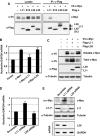
L11 inhibits c-Myc-dependent transactivation activity and reduces the percentage of cells in S phase. (A) L11 inhibits c-Myc-induced luciferase reporter expression driven by wild-type (WT), but not mutant, E box-containing E2F2 promoter. H1299 cells transfected with plasmids, as indicated, were subjected to luciferase assays to determine the reporter activity. *Indicates P<0.01 compared to control vector transfection; **indicates P<0.01 compared to transfection with c-Myc only. The protein expression of soluble c-Myc and L11 in this assay is shown in Supplementary Figure S1A. (B) Wild-type L11, but not its N-terminal deletion mutant (L1166−178), inhibited c-Myc-dependent luciferase reporter expression driven by WT, but not mutant, E box-containing E2F2 promoter. U2OS cells were transfected with plasmids as indicated and c-Myc driven luciferase activity were measured. (C) L11 inhibits c-Myc-dependent transcription of the endogenous c-Myc target genes. U2OS cells were infected with Ad-L11 and Ad-c-Myc individually or together and the expression of E2F2, nucleolin, and rRNA were measured by quantitative RT–PCR assays. The expression of total Ad-c-Myc and Ad-L11 proteins in this assay is shown in Figure 7H. (D, E) L11 reduces the percentage of cells in S phase determined by BrdU incorporation assay. U2OS cells infected with Ad-c-Myc and/or Ad-L11 were labeled with BrdU and subjected to anti-BrdU staining (red) and counter-stained with DAPI. Percentage of BrdU-positive cells is shown in (E).
Figure 2
Figure 2
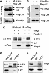
L11 interacts with c-Myc in cells. (A, B) Co-IP of ectopic L11 with ectopic c-Myc. H1299 cells were transfected with V5-c-Myc and Flag-L11 individually, or together and subjected to IP with anti-Flag (A) or anti-V5 (B), followed by IB with anti-Flag or anti-V5 antibody. (C) Overexpressed L11 interacts with untagged c-Myc. H1299 cells transfected with untagged c-Myc in the presence or absence of Flag-L11 were immunoprecipitated with anti-Flag antibody, followed by IB with indicated antibodies. (D, E) Endogenous L11 interacts with endogenous c-Myc. H1299 cell lysates were immunoprecipitated with monoclonal anti-c-Myc (C33) (D) or anti-L11 (E) antibody, followed by IB with anti-L11, polyclonal anti-c-Myc (N262) (D), or monoclonal anti-c-Myc (9E10) antibody (E).
Figure 3
Figure 3
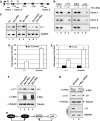
The N-terminal domain of L11 interacts with the MB II of c-Myc. (A) c-Myc interacts with the N-terminal domain of L11. H1299 cells transfected with plasmids encoding wild-type L11 or its deletion mutants together with the c-Myc plasmid were subjected to IP using anti-Flag antibodies, followed by IB with anti-V5 or anti-Flag antibodies. (B) Schematic diagram of L11 and its c-Myc-binding domains. (C) L11 binds to the MB II region of c-Myc. H1299 cells were transfected with the V5-tagged TAD of c-Myc (c-Myc1−144) and its deletion mutants and subjected to IP using anti-V5 antibodies, followed by IB using anti-Flag or anti-V5 antibodies. (D) The MB II region of c-Myc is required for c-Myc interaction with L11. H1299 cells were transfected with the V5-tagged WT c-Myc or MB II-deleted mutant of c-Myc (ΔMB II), together with Flag-L11, and subjected to IP using anti-Flag antibodies, followed by IB using antibodies as indicated. (E) Schematic diagram of c-Myc showing the L11 binding MB II of the TAD.
Figure 4
Figure 4
L11 co-resides with c-Myc in c-Myc target gene promoter. (A) Endogenous L11 binds to c-Myc target gene promoters. ChIP-PCR assays were conducted to detect the recruitment of endogenous c-Myc and L11 in c-Myc target gene promoters in H1299 cells, using anti-c-Myc (N262) or anti-L11 antibodies. (B) Overexpression of c-Myc enhances L11 binding to the nucleolin gene promoter. H1299 cells were transfected with or without V5-c-Myc. ChIP-PCR assays were conducted using anti-L11 antibodies to detect the recruitment of endogenous L11 at the nucleolin gene promoter. The expression of V5-c-Myc is shown in the bottom panels. (C) Knockdown of endogenous c-Myc decreases L11 binding to the nucleolin gene promoter. U2OS cells were transfected with scrambled or c-Myc siRNA. ChIP-PCR assays were conducted using anti-L11 antibodies to detect the recruitment of endogenous L11 at the nucleolin gene promoter. The expression of endogenous c-Myc is shown in the bottom panels. (D) L11 co-resides with c-Myc in its target gene promoters. H1299 cells were transfected with plasmids as indicated. ChIP-PCR assays were performed using anti-V5 or anti-Flag antibodies. Ten fold of initial lysates were used for anti-Flag IP compare to IP with anti-V5. The Flag-L11 protein–DNA complexes were eluted with Flag peptide competition and 10% of the elutes was used for DNA purification and PCR amplification (lane 7 and 8). The rest of elutes was subjected to a second IP using anti-V5 antibody, followed by PCR amplification (lanes 9 and 10). (E) Co-residence of L11 with c-Myc in the nucleolin gene promoter requires the MB II of c-Myc. H1299 cells were transfected with WT or ΔMB II mutant of c-Myc plasmid in the presence or absence of Flag-L11. Sequential ChIP-PCR assays were performed using anti-Flag antibody, followed by anti-V5 antibody as described in above (D). Real-time PCR was then performed to detect the immunoprecipitated nucleolin promoter DNA.
Figure 5
Figure 5
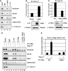
L11 reduces the recruitment of TRRAP to c-Myc target gene promoters and histone H4 acetylation at these promoters. (A) H1299 cell transfected with control or Flag-L11 plasmid were subjected to IB with anti-c-Myc or anti-Flag antibodies. (B) L11 reduces the recruitment of TRRAP to and histone H4 acetylation at c-Myc target gene promoters. The transfected cells in (A) were subjected to ChIP assays using goat anti-TRRAP, control goat IgG, rabbit polyclonal anti-acetyl histone H4, anti-c-Myc (N262), or control rabbit IgG followed by detection of the nucleolin gene promoter sequence using real-time PCR assays. (C, D) L11 competes the TRRAP for binding to the nucleolin gene promoter. H1299 cells transfected with Flag-TRRAP in the presence of increasing amounts of myc-tagged L11 were immunblotted with antibodies as indicated (C). The similar cells were also subjected to ChIP assays using anti-Flag antibody, followed by detection of the TRRAP binding to the nucleolin gene promoter using real-time PCR assays (D).
Figure 6
Figure 6
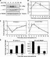
The dynamic binding of L11 to c-Myc target gene promoter inversely correlates with that of TRRAP in response to serum stimulation or serum starvation. (A) Dynamic expression of c-Myc and L11 in cells following serum-stimulation. U2OS cells cultured in 0.2% FCS containing medium for 48 h were stimulated with 20% FCS, and harvested at the indicated time points for IB assays using indicated antibodies. *Indicates nonspecific band. (B) The expression of the nucleolin in response to serum stimulation. Starved U2OS cells were stimulated with 20% FCS and harvested at the indicated time point as in (A) were assayed for expression of the nucleolin gene by real-time RT–PCR assays. (C) Dynamic association of L11 and TRRAP at the nucleolin gene promoter in response to serum stimulation. U2OS cells were serum starved and re-stimulated as in (A) and subjected to ChIP assays using anti-L11 or anti-TRRAP antibodies, followed by real-time PCR detection of the nucleolin promoter. (D) Dynamic association of L11 and TRRAP at the nucleolin gene promoter in response to serum starvation. Exponentially growing U2OS cells were starved in 0.2% FCS-containing medium and harvested at indicated time points. The cells were subjected to ChIP assays using anti-L11 or anti-TRRAP antibodies, followed by real-time PCR detection of the nucleolin promoter.
Figure 7
Figure 7
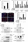
Endogenous L11 regulates c-Myc activity and levels. (A) Reduction of L11 by siRNA enhances c-Myc-induced luciferase reporter expression driven by WT, but not mutant, E box-containing E2F2 promoters in U2OS cells. (B) Knockdown of endogenous L11 enhances c-Myc-dependent transcription of the c-Myc target genes. U2OS cells were transfected with siRNAs as indicated and real-time RT–PCR assays were performed to determine the expression of E2F2, nucleolin, and rRNA genes. (C, D) Knockdown of endogenous L11 induces the percentage of cells in S phase. BrdU incorporation assay were performed in serum-starved U2OS cells transfected with siRNAs as indicated. Percentage of BrdU-positive cells is shown in (D). (E) The representative expression of endogenous c-Myc and L11 in above experiments (B), (C), and (D) as determined by IB with antibodies as indicated. (F) Knockdown of endogenous L11 by L11 siRNAs against two different sequences increases the protein levels of endogenous c-Myc in U2OS cells. *Indicates a nonspecific band. (G) Overexpression of L11 stabilizes ectopically expressed c-Myc. H1299 cells were transfected with plasmids as indicated. Whole-cell lysates were subjected to IB using antibodies as indicated. (H) Adenoviral-mediated overexpression of L11 stabilizes adenoviral encoded c-Myc. U2OS cells were infected with adenoviruses as indicated. Whole-cell lysates were subjected to IB using antibodies as indicated.
Figure 8
Figure 8
The inhibition of c-Myc activity by L11 is not a general effect of all individual ribosomal proteins. (A) L11, but not L29, L30, or S12, binds to c-Myc. H1299 cells were transfected with Flag-tagged L11, L29, L30, or S12 together with V5-c-Myc plasmids as indicated. Co-IPs were preformed using anti-Flag antibodies followed by IB with anti-V5 or anti-Flag antibodies. (B, C) Overexpression of L11, but not L29, inhibits the expression of the endogenous nucleolin gene. U2OS cells were transfected with plasmids as indicated. Half of the cells were used for IB assays to detect the protein expression of transfected c-Myc, L11, and L29 (C). The soluble c-Myc was determined from cleared lysates using NP40 lysis buffer, whereas the total level of c-Myc was determined using whole-cell lysates. The other half of the transfected cells were subjected to RT– real-time PCR to determine the expression of the nucleolin mRNA (B). (D, E) Knockdown of L11, but not L29, enhances the expression of the endogenous nucleolin gene. U2OS cells were transfected with siRNA as indicated. Half of the cells were used for IB assays to detect the protein expression of endogenous c-Myc and L11 (top panels) (E). The other half of the transfected cells were subjected to RNA extraction and RT, followed by real-time PCR to determine the relative expression of the nucleolin mRNA (D). The knockdown efficiency of L29 mRNA is shown in bottom panels of (E), as determined by semiquantitative RT–PCR.
Figure 9
Figure 9
L11 is a target gene of c-Myc. (A) Diagram of the l11 gene exon–intron regions. Bars indicate the three regions amplified by ChIP-PCR assays. Empty boxes indicate E boxes. Filled boxes indicate exons. Arrow indicates the transcription start. (B) c-Myc specifically binds to the E box-containing introns 1 and 2, but not intron 5, regions of the l11 gene in H1299 (lanes 1 to 4) and U2OS (lanes 5 to 8) cells determined by ChIP-PCR assays. (C) c-Myc-induced l11 mRNA expression in cells. H1299 (lanes 1 to 2), U2OS (lanes 3 to 4), and WI38 cells (lanes 5 to 7) were transfected or infected with c-Myc-expressing plasmid or adenovirus and the expression of l11 was determined within 48 h by a semiquantitative RT–PCR assay. (D, E) Comparison of c-Myc binding to the L11 and the nucleolin gene promoters. (D) U2OS cells were subjected to ChIP assays using anti-c-Myc (N262) antibodies or control rabbit IgG followed by real-time PCR assays to detect the L11 and the nucleolin promoter DNAs. (E) U2OS cells were serum starved for 48 h, followed by stimulation with 20% FCS for 6 h as in Figure 6A. The serum-starved and re-stimulated cells were subjected to ChIP assays using anti-c-Myc (N262) antibodies, followed by real-time PCR to detect the L11 and the nucleolin promoter DNAs. (F) c-Myc induces L11 protein expression in cells. U2OS (lanes 1 and 2) and H1299 (lanes 3 and 4) cells were infected with c-Myc expressing adenovirus or control virus. Immunoblot (IB) was performed using antibodies as indicated. (G) Knockdown of endogenous c-Myc reduces L11 mRNA and protein levels. U2OS cells were transfected with c-Myc or scrambled siRNA. IB was performed using antibody as indicated and l11 mRNA was examined by a semiquantitative RT–PCR assay. *Indicates nonspecific band recognized by anti-c-Myc (N262). (H) A schematic for the L11-c-Myc feedback regulation versus the L11-MDM2-p53 regulatory loop.

Similar articles

Cited by

References

    1. Adams JM, Harris AW, Pinkert CA, Corcoran LM, Alexander WS, Cory S, Palmiter RD, Brinster RL (1985) The c-myc oncogene driven by immunoglobulin enhancers induces lymphoid malignancy in transgenic mice. Nature 318: 533–538 - PubMed
    1. Adhikary S, Eilers M (2005) Transcriptional regulation and transformation by Myc proteins. Nat Rev Mol Cell Biol 6: 635–645 - PubMed
    1. Adhikary S, Marinoni F, Hock A, Hulleman E, Popov N, Beier R, Bernard S, Quarto M, Capra M, Goettig S, Kogel U, Scheffner M, Helin K, Eilers M (2005) The ubiquitin ligase HectH9 regulates transcriptional activation by Myc and is essential for tumor cell proliferation. Cell 123: 409–421 - PubMed
    1. Amati B, Dalton S, Brooks MW, Littlewood TD, Evan GI, Land H (1992) Transcriptional activation by the human c-Myc oncoprotein in yeast requires interaction with Max. Nature 359: 423–426 - PubMed
    1. Arabi A, Wu S, Ridderstrale K, Bierhoff H, Shiue C, Fatyol K, Fahlen S, Hydbring P, Soderberg O, Grummt I, Larsson LG, Wright AP (2005) c-Myc associates with ribosomal DNA and activates RNA polymerase I transcription. Nat Cell Biol 7: 303–310 - PubMed

Publication types

MeSH terms