. 2007 Jun 13:8:50.
doi: 10.1186/1471-2199-8-50.

Zfra affects TNF-mediated cell death by interacting with death domain protein TRADD and negatively regulates the activation of NF-kappaB, JNK1, p53 and WOX1 during stress response

Affiliations

Zfra affects TNF-mediated cell death by interacting with death domain protein TRADD and negatively regulates the activation of NF-kappaB, JNK1, p53 and WOX1 during stress response

Qunying Hong et al. BMC Mol Biol. .

Abstract

Background: Zfra is a 31-amino-acid zinc finger-like protein, which is known to regulate cell death by tumor necrosis factor (TNF) and overexpressed TNF receptor- or Fas-associated death domain proteins (TRADD and FADD). In addition, Zfra undergoes self-association and interacts with c-Jun N-terminal kinase 1 (JNK1) in response to stress stimuli. To further delineate the functional properties of Zfra, here we investigated Zfra regulation of the activation of p53, WOX1 (WWOX or FOR), NF-kappaB, and JNK1 under apoptotic stress.

Results: Transiently overexpressed Zfra caused growth suppression and apoptotic death of many but not all types of cells. Zfra either enhanced or blocked cell death caused by TRADD, FADD, or receptor-interacting protein (RIP) in a dose-related manner. This modulation is related with Zfra binding with TRADD, NF-kappaB, JNK1 and WOX1, as determined by GST pull-down analysis, co-immunoprecipitation, and mapping by yeast two-hybrid analysis. Functionally, transiently overexpressed Zfra sequestered NF-kappaB (p65), WOX1, p53 and phospho-ERK (extracellular signal-activated kinase) in the cytoplasm, and TNF or UV light could not effectively induce nuclear translocation of these proteins. Zfra counteracted the apoptotic functions of Tyr33-phosphorylated WOX1 and Ser46-phosphorylated p53. Alteration of Ser8 to Gly abolished the apoptotic function of Zfra and its regulation of WOX1 and p53.

Conclusion: In response to TNF, Zfra is upregulated and modulates TNF-mediated cell death via interacting with TRADD, FADD and RIP (death-inducing signaling complex) at the receptor level, and downstream effectors NF-kappaB, p53, WOX1, and JNK1.


Figures

Figure 1
Figure 1
Zfra induces apoptotic cell death. (A) Transiently overexpressed Zfra induced death of human ovarian ME180, embryonic kidney HEK-293, neuroblastoma SK-N-SH, breast MDA-MB-231 and MCF7 cells, Molt4 T lymphocytes and murine L929 fibroblasts, but not human prostate DU145 and mink lung epithelial Mv1Lu cells. These cells were transfected with an EGFP (vector) or EGFP-Zfra expression vector (150 ng/well in 96-well microtiter plates) or medium (control) by CaPO4, followed by culturing for 48 hr and then staining with crystal violet for determining the extent of death (n = 8; mean ± standard deviation; p < 0.001, EGFP vector-transfected cells versus EGFP-Zfra-transfected cells except DU145 and Mv1Lu; Student's t tests). Molt4 T cells were transfected with the DNA constructs by electroporation. Data shown in the buffer and vector (EGFP) controls are from testing L929 cells. Similar data were obtained using other cells. The numbers of cells with positive EGFP or EGFP-Zfra protein expression were ~60–75%, as determined by fluorescence microscopy. EGFP-Zfra was present ubiquitously in cell compartments in majority of the tested cells (see representative L929 cells; 200× magnification). Interestingly, EGFP-Zfra was mainly present in the cytoplasm of MDA-MB-231 cells (400× magnification). (B) In adherence- or anchorage-independent growth assay on agarose, Zfra was shown to block colony formation of L929 cells, as opposed to empty vector controls (n = 3, p < 0.001; Student's t test). Live colonies were stained with a soluble tetrazolium-based MTS proliferation reagent. (C) Molt4 T cells were electroporated with the EGFP-Zfra expression vector, followed by culturing for 48 hr and analyzing the extent of apoptosis using Annexin V assay.
Figure 2
Figure 2
Ser8 is involved in Zfra-induced apoptosis. Ser8 is a conserved phosphorylation site in Zfra [10]. Ser8 was altered to Gly (S8G). L929 cells were electroporated with EGFP-Zfra, S8G, or EGFP, followed by culturing for 48 hr and staining with crystal violet. Transiently overexpressed Zfra induced death of L929 cells (34.8 ± 8.6%; n = 8), whereas S8G mutant had a significantly reduced activity in causing cell death (17.1 ± 5.6%; n = 8; p < 0.005, Student's t test; see *). The extent of protein expression is shown.
Figure 3
Figure 3
Effect of Zfra on TRADD, FADD, or RIP-mediated cell death. ME180, HEK-293 and DU145 cells were transfected with cytotoxic doses of TRADD, FADD, or RIP cDNA (200 ng/well), in the presence or absence of EGFP-Zfra (200 ng/well) by CaPO4. These cells were cultured for 48 hr and then stained with crystal violet. In Zfra-sensitive ME180 and HEK-293 cells, Zfra and TRADD increased cell death in an additive manner (n = 8; p < 0.001, TRADD alone versus TRADD/Zfra in combination; Student's t tests), whereas no additive effect was shown for Zfra with FADD or RIP (n = 8; p > 0.05, FADD or RIP alone versus Zfra/FADD or Zfra/RIP; Student's t tests). In Zfra-resistant DU145 cells, Zfra alone did not induce death, but enhanced the death by ectopic TRADD, FADD and RIP by ~100–150% increases (n = 8; p < 0.001; Student's t tests). EGFP-Zfra was expressed in approximately 60–70% of total cells, as visualized by fluorescence microscopy.
Figure 4
Figure 4
Synergistic enhancement of cell death by non-cytotoxic levels of Zfra and TRADD, FADD or RIP. Zfra-sensitive ovarian ME-180 cells were transfected with non-cytotoxic doses of TRADD (FADD or RIP) (20, 40, 80 ng/well) and/or EGFP-Zfra by CaPO4. Zfra and TRADD, FADD, or RIP synergistically induced ME180 cell death in 48 hr (n = 16; mean ± standard deviation; p < 0.001; Zfra, TRADD, FADD, or RIP alone versus Zfra/TRADD, Zfra/FADD, or Zfra/RIP; Student's t tests). Cells were stained with crystal violet. EGFP-Zfra was expressed in approximately 60–70% of total cells, as visualized by fluorescence microscopy.
Figure 5
Figure 5
Zfra regulates cell growth in a biphasic manner. (A) L929 cells were transfected with various amounts of EGFP-Zfra and/or a non-toxic dose of RIP (100 ng/well) by CaPO4. Zfra alone enhanced cell growth at low doses, but induced death at higher doses (n = 8; mean ± standard deviation). RIP blocked the Zfra-mediated cell growth. (B) Similarly, L929 cells were transfected with various amounts of RIP and/or a non-toxic dose of Zfra (60 ng/well). At low levels, RIP had little or no effect on cell growth. When in combination, both Zfra and RIP effectively increased the cell death (n = 8; mean ± standard deviation). Cells were stained with crystal violet. EGFP-Zfra was expressed in approximately 60–70% of total cells, as visualized by fluorescence microscopy.
Figure 6
Figure 6
Zfra physically binds TRADD and the N-terminal first WW domain and C-terminal SDR domain in WOX1. Binding of Zfra (as target) with WOX1 (as bait) was mapped by Ras rescue-based yeast two-hybrid analysis (see Materials and Methods). Positive binding is evidenced by the growth of mutant yeast at 37°C in a selective medium. Zfra bound to the N-terminal first WW domain and the C-terminal SRD domain of WOX1. Alteration of the known phosphorylation site Tyr33 to Arg33 (Y33R) in WOX1 abrogated the binding. Zfra did not interact with the mitochondria-targeting region in the SDR domain (Mito-SDR). No yeast cell growth at 37°C was shown when empty vector versus empty vector was tested as a negative control. In a positive control, the self-binding of MafB is shown. In addition, Zfra was shown to bind the full-length TRADD, but not RIP. WOX1 also bound TRADD. Neg cont: negative control; Pos cont: positive control.
Figure 7
Figure 7
TNF and UV light increase the binding of Zfra with WOX1, JNK1 and NF-κB, but weakly with p53 in SK-N-SH cells. (A) Exposure of SK-N-SH cells to TNF (50 ng/ml) for 40 min resulted in upregulation of Zfra and degradation of IκBα. (B) In GST pull-down analysis using SK-N-SH cells, TNF increased the binding of GST-Zfra with NF-κB (p65) and JNK1 (greater than 50%), and weakly with p53 but not IκBα. GST-Zfra physically interacted with endogenous Zfra and WOX1, and TNF limitedly increased the binding (less than 30%). In negative controls, GST alone could not bind the above-indicated proteins. The relative amounts of GST and GST-Zfra used in the pull down are shown. One-twentieth amounts of protein input for endogenous Zfra and other indicated proteins are shown in Western blotting. (C) Similarly, during a 10-min treatment, TNF increased the binding of GST-Zfra with p-WOX1, but not with FADD, RIP, IκBα and Fas. (D) SK-N-SH cells were exposed to UV light (120 mJoule/cm2) and then cultured for 1 hr, followed by processing GST pull-down analysis. UV light increased the binding of endogenous Zfra with p-WOX1 and JNK1. (E) By co-immunoprecipitation, UV light (120 mJoule/cm2) increased the binding of endogenous Zfra with itself and p-JNK1 (at Thr183/Tyr185) (greater than 50% increase), but limitedly with WOX1 (less than 20% increase), in SK-N-SH cells.
Figure 8
Figure 8
Transiently overexpressed Zfra sequesters NF-κB, WOX1, p53 and ERK in the cytoplasm. Breast MCF7 and neuroblastoma SK-N-SH cells were electroporated with an EGFP-Zfra construct or EGFP only, cultured overnight, and exposed to TNF for 40 min. Expression of GFP and GFP-Zfra is shown (A, B). (A) Without stimulation, EGFP-Zfra alone inhibited nuclear localization of NF-κB (p65), WOX1, p53 and p-ERK (Y204 phosphorylated) in SK-N-SH cells, whereas it had little or no effect on JNK1/2. TNF (50 ng/ml) could not induce nuclear translocation of endogenous JNK1/2, NF-κB (p65), p-ERK, p53 and WOX1 in Zfra-expressing cells during treatment for 40 min. (B) Similarly, in MCF7 cells, EGFP-Zfra suppressed nuclear localization of NF-κB and WOX1, and TNF could not induce nuclear translocation of these proteins. (C) EGFP-Zfra did not have a significant effect on the nuclear localization of WOX1 in Molt4 T cells; however, it blocked UV light-induced nuclear translocation of WOX1. Ectopic expression of EGFP and EGFP-Zfra in the above cells is shown in each panel. The levels of cytosolic α-tubulin are regarded as loading controls.
Figure 9
Figure 9
Zfra antagonizes the apoptotic function of WOX1 and JNK1. (A) L929 cells were transfected with a cytotoxic dose of EGFP-WOX1 in the presence or absence of low doses of EGFP-Zfra by CaPO4 (n = 8). The cells were cultured 24 and 48 hrs and stained with crystal violet. Zfra blocked WOX1-mediated growth suppression and death in 24 hr (data not shown for 48 hr). Expression of EGFP-tagged Zfra and WOX1 is shown, as determined by fluorescence microscopy (400× magnification). (B) Similarly, L929 cells were electroporated with WOX1 and/or Zfra, cultured 48 hr, and then processed for DNA fragmentation. An apoptosis-inducing amount of WOX1 and a non-apoptosis-inducing dose of Zfra were used. In a positive control, cells were treated with staurosporine (stauro; 500 nM) for 16 hr. In negative controls, cells were electroporated with medium or GFP vector only. ep, electroporation. (C) L929 cells were electroporated with an apoptosis-inducing amount of WOX1 and/or Zfra, or JNK1 (or medium only), and cultured for 24 and 48 hrs, respectively. Apoptosis occurred in 24 hr, as evidenced by increased percentages of cells in the subG1 phase from cell cycle analysis (data not shown for 48 hr). WOX1, JNK1, or Zfra alone suppressed cell growth, as indicated by reduced cell populations at the G1. However, both Zfra and WOX1 or JNK1 could not increase apoptosis in a synergistic manner. X-axis: DNA content; Y-axis: Cell numbers.
Figure 10
Figure 10
Phosphorylation of WOX1 at Tyr33 is essential for its apoptotic function and for counteracting Zfra-mediated cell death. (A) Transiently overexpressed EGFP-Zfra and EGFP-WOX1 induced death of COS7 fibroblasts (n = 8, mean ± standard deviation). Zfra counteracted with WOX1 in causing cell death. Alteration of Tyr33 to Arg33 (Y33R) abolished the apoptosis-inducing activity of WOX1, and this mutant did not block Zfra-mediated cell death. Expression of EGFP-tagged Zfra, WOX1 and WOX1(Y33R) is shown in fluorescent micrographs (400× magnification). WOX1 is shown in the perinuclear area, whereas the Y33R mutant tends to aggregate in the nuclei. Zfra is expressed ubiquitously in cells. (B) Similar experiments were carried out in breast MDA-MB-231 cells. Again, Zfra and WOX1 did not increase cell death in a synergistic manner (n = 8, mean ± standard deviation). The Y33R mutant lost its apoptotic function and had no apparent effect on Zfra-mediated cell death. (C) L929 cells were transfected with the SDR domain plus a C-terminal tail (SDR/C) or the SDR domain alone (tagged with ECFP at the N-terminus). These cells were cultured for 24 hr in the presence or absence of a synthetic full-length Zfra peptide (10 μM), and the extent of cell death was determined by crystal violet staining. Zfra enhanced cell death caused by SDR/C (n = 8, mean ± standard deviation). SDR alone could not cause cell death, and Zfra did not increase the cell death. Protein expression of ECFP-SDR/C and ECFP-SDR is shown. (D) Constructs made for the above experiments are shown.
Figure 11
Figure 11
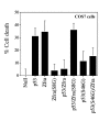
Phosphorylation of p53 at serine 46 (S46) is essential for counteracting Zfra-mediated cell death. COS7 cells were co-transfected with both p53 and/or Zfra by electroporation, followed by culturing for 24 hr and then determining the extent of cell death by staining with crystal violet. In addition, the cells were also introduced with mutants of p53 (S46) and/or Zfra(S8G). The results show that p53 and Zfra nullified each other's activity in causing cell death (n = 8, mean ± standard deviation). The apoptotic function of both Zfra(S8G) and p53(S46G) was significantly reduced. S8G mutant had no effect in reducing p53-mediated cell death.
Figure 12
Figure 12
A schematic model of Zfra/WOX1 involvement in TNF signaling. TNF is able to initiate two counteractive pathways – one apoptotic and the other protective. (A) In the death pathway, binding of TNF to the cognate p55-TNF receptor (TNFR) results in recruitment of death domain proteins TRADD, FADD and RIP, thus generating the so called death-inducing signaling complex (DISC). Caspase 8 and downstream effectors are then activated to induce cell death at the mitochondrial and nuclear levels [16,17]. In this study, we discovered that Zfra physically interacts with TRADD and WOX1, and that WOX1 binds TRADD. Thus, Zfra and WOX1 are likely to be recruited to the DISC. WOX1 enhances TNF cytotoxicity [3], and Zfra counteracts the WOX1 function. Zfra either enhances or inhibits the function of death domain proteins (open arrow). Thus, the ying and yang of cell death depends upon the strength of DISC formation and the counteractive or enhancing force of Zfra. (B) In the protective pathway, p55-TNF recruits TRADD, TRAF2 and RIP, followed by activating several downstream adaptors and finally JNK1 and NF-κB. We determined that overexpressed Zfra sequesters p53, WOX1, and NF-κB in the cytoplasm. Thus, Zfra is likely to bind and block the function of these proteins during TNF signaling (see each step marked by a number). In Step 1, at the membrane level, Zfra binds TRADD in the presence of TRADD, TRAF2, RIP and WOX1. In Step 2, a trimolecular complex of Zfra/JNK1/WOX1 may form when JNK1 is activated by the upstream activated MEK. Zfra binds and counteracts the apoptotic function of JNK1. Also, JNK1 counteracts the apoptotic function of WOX1 [6]. In Step 3, MEK activates ERK, and that Zfra may bind and sequester ERK to the cytoplasm. In Step 4, phosphorylation of IκBα by IKK causes degradation of IκBα and release of NF-κB for nuclear translocation. Again, Zfra is able to bind and sequester NF-κB in the cytoplasm. In Step 5 and 6, TNF induces NF-κB activation, and then NF-κB activates p53 [22]. The non-ankyrin C-terminus of IκBα physically interacts with cytosolic p53 [25]. p53 is functionally associated with WOX1, and both proteins may induce apoptosis synergistically [3,4,6,8]. Thus, an in vivo complex of Zfra with IκBα/p53/WOX1 or p53/WOX1 is likely (Chang et al., submitted).

Similar articles

Cited by

References

    1. Bednarek AK, Laflin KJ, Daniel RL, Liao Q, Hawkins KA, Aldaz CM. WWOX, a novel WW domain-containing protein mapping to human chromosome 16q23.3-24.1, a region frequently affected in breast cancer. Cancer Res. 2000;60:2140–2145. - PubMed
    1. Ried K, Finnis M, Hobson L, Mangelsdorf M, Dayan S, Nancarrow JK, Woollatt E, Kremmidiotis G, Gardner A, Venter D, Baker E, Richards RI. Common chromosomal fragile site FRAD16D sequence: identification of the FOR gene spanning FRAD16D and homozygous deletions and translcoation breakpoints in cancer cells. Hum Mol Genet. 2000;9:1651–1663. doi: 10.1093/hmg/9.11.1651. - DOI - PubMed
    1. Chang N-S, Pratt N, Heath J, Schultz L, Sleve D, Carey GB, Zevotek N. Hyaluronidase induction of a WW domain-containing oxidoreductase that enhances tumor necrosis factor cytotoxicity. J Biol Chem. 2001;276:3361–3370. doi: 10.1074/jbc.M007140200. - DOI - PubMed
    1. Chang N-S, Hsu LJ, Lin YS, Lai FJ, Sheu HM. WW domain-containing oxidoreductase: a candidate tumor suppressor. Trends Mol Med. 2007;13:12–22. doi: 10.1016/j.molmed.2006.11.006. - DOI - PubMed
    1. Smith DI, McAvoy S, Zhu Y, Perez DS. Large common fragile site genes and cancer. Semin Cancer Biol. 2007;17:31–41. doi: 10.1016/j.semcancer.2006.10.003. - DOI - PubMed

Publication types

MeSH terms