. 2007 Sep;81(17):9490-501.
doi: 10.1128/JVI.00364-07. Epub 2007 Jun 13.

Identification of a novel virulence factor in recombinant pneumonia virus of mice

Affiliations

Identification of a novel virulence factor in recombinant pneumonia virus of mice

Christine D Krempl et al. J Virol. 2007 Sep.

Abstract

Pneumonia virus of mice (PVM) is a murine relative of human respiratory syncytial virus (HRSV). Here we developed a reverse genetics system for PVM based on a consensus sequence for virulent strain 15. Recombinant PVM and a version engineered to express green fluorescent protein replicated as efficiently as the biological parent in vitro but were 4- and 12.5-fold attenuated in vivo, respectively. The G proteins of HRSV and PVM have been suggested to contribute to viral pathogenesis, but this had not been possible to study in a defined manner in a fully permissive host. As a first step, we evaluated recombinant mutants bearing a deletion of the entire G gene (Delta G) or expressing a G protein lacking its cytoplasmic tail (Gt). Both G mutants replicated as efficiently in vitro as their recombinant parent, but both were nonpathogenic in mice at doses that would otherwise be lethal. We could not detect replication of the Delta G mutant in mice, indicating that its attenuation is based on a severe reduction in the virus load. In contrast, the Gt mutant appeared to replicate as efficiently in mice as its recombinant parent. Thus, the reduction in virulence associated with the Gt mutant could not be accounted for by a reduction in viral replication. These results identified the cytoplasmic tail of G as a virulence factor whose effect is not mediated solely by the viral load. In addition to its intrinsic interest, a recombinant virus that replicates with wild-type-like efficiency but does not cause disease defines optimal properties for vaccine development.


Figures

FIG. 1.
FIG. 1.
Construction of plasmids expressing the antigenomes of rPVM and rPVM-GFP7. (A) Map of the antigenome of rPVM showing the locations of the viral genes and, below, the five overlapping cDNA fragments (numbered 1 to 5) used to construct its cDNA. PT7, T7 RNA polymerase promoter; δ, hepatitis delta virus ribozyme; TT7, T7 RNA polymerase terminator. Unique restriction enzyme sites used to construct the cDNA are given, with their nucleotide positions in parentheses; the AgeI and BstBI sites that were created during construction are underlined. (B) Nucleotide changes introduced into rPVM to create AgeI (upper panel) and BstBI (lower panel) markers in the SH-G and M2-L intergenic regions, respectively. Predicted transcription GS and GE signals are boldfaced, intergenic sequences are roman, restriction enzyme sites are underlined, and nucleotide substitutions and additions in rPMV are italicized, while deletions are indicated by gaps. (C) Insertion of a transcription cassette encoding enhanced GFP into the AgeI site of rPVM, placing GFP as the seventh gene in the 3′-to-5′ order. The GFP ORF (open rectangle, not drawn to scale) was modified by PCR to add a GS sequence derived from the N gene (which also is identical in the P gene) on the upstream side of the ORF and a GE sequence derived from the F gene on the downstream side (each underlined). The fragment was flanked by AgeI restriction sites whose positions in the final cDNA are indicated.
FIG. 2.
FIG. 2.
In vitro analysis of recovered rPVM and rPVM-GFP7. (A) RT-PCR and restriction analysis of viral RNA from BHK-21 cells infected with the biological wild-type virus PVM15 (lanes 1 and 2), the wild-type virus rPVM (lanes 3 and 4), or rPVM-GFP7 (lanes 5 and 6). The RT-PCR products were made using primers that hybridized within the N and F genes, spanning PVM positions 3312 to 5981. The amplified fragments were digested with AgeI (lanes marked +) or left untreated (lanes marked −) and electrophoresed on a 1% agarose gel. The predicted sizes of the fragments are given on the left. The last lane represents a 1-kbp DNA ladder. (B) Multistep growth kinetics of biological and recombinant PVMs in vitro. Replicate monolayers of BHK-21 cells were infected with the biological parental virus PVM15, rPVM, or rPVM-GFP7 at an input MOI of 0.01 PFU per cell. At the indicated time points, the cells and medium were harvested, freeze-thawed, clarified, and flash frozen. Virus titers were determined by a plaque assay. Error bars, standard errors of the means. (C) Cells from the experiment for which results are shown in panel B were examined for GFP expression on days 1 to 4 using an inverted fluorescence microscope.
FIG. 3.
FIG. 3.
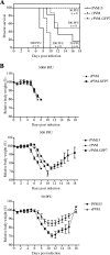
Virulence of wild-type and recombinant PVM. Six- to eight-week-old BALB/c mice were infected intranasally with the indicated doses of PVM15, rPVM, and rPVM-GFP7 in an inoculum volume of 80 μl. Mice were observed closely and weighed daily. Mice that met the end point regulations of the German animal protection board were euthanized by cervical dislocation. The results of two independent experiments, each including at least four animals per group, were combined. (A) Survival after infection with 500 PFU (PVM15, rPVM, and rPVM-GFP7) or 50 PFU (PVM15 and rPVM) of the indicated viruses. The total numbers of animals are given beneath the doses. (B) Body weight (relative to that on day 0, taken as 100%) following infection with the indicated viruses at the indicated doses.
FIG. 4.
FIG. 4.
Replication of rPVM and rPVM-GFP7 in the upper and lower respiratory tracts of BALB/c mice. Mice were inoculated intranasally with 500 PFU of PVM15, rPVM, or rPVM-GFP7. Mice were sacrificed on day 3 (A and C) or day 6 (B and D) after infection, lungs (A and B) and nasal turbinates (NT) (C and D) were removed and homogenized, and viral titers were determined by a plaque assay. The mean virus titers and the percentages of animals with detectable virus are given below the corresponding graphs. Neighboring groups (indicated with brackets) were compared using the Mann-Whitney test. Asterisks indicate statistically significant differences (P < 0.05). The results of two independent experiments were combined in each graph.
FIG. 5.
FIG. 5.
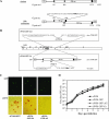
rPVM-GFP7 viruses with mutations in the G gene. (A) Schematic diagrams (not drawn to scale) of the G genes of the biologically derived virulent and nonpathogenic isolates of PVM strain 15 (23, 34). Open rectangles, ORFs. The amino acid length of the encoded protein is given in each rectangle. Translational start (filled triangles pointing right) or stop (filled vertical bars) codons are identified by their nucleotide positions in the respective G gene. The GS and GE signals are indicated, and nontranslated gene regions are shown as thin horizontal lines. The nucleotide sequence (negative sense) of positions 166 to 190 of the G gene is given, and the inserted U residue that results in a frameshift is shown in parentheses. The complement of the translational stop codon (nucleotides 188 to 190) that is accessed by the frameshift is italicized, and the complement to the alternative start codon at nucleotides 183 to 185 (Met-34) is boldfaced. (B) Schematic representations (not drawn to scale) of rPVM-GFP-ΔG and rPVM-GFP-Gt. The diagram in the middle represents the rPVM-GFP7 genome (filled box, GFP gene). The box above the diagram illustrates the deletion of the entire G gene to create rPVM-GFP-ΔG. The nucleotide sequence (negative sense) shows the GFP/F gene junction and the AgeI site (italicized) left following the 1,350-nt deletion. The sequence is numbered according to the sequence of rPVM-GFP7, and the GFP GE and F GS signals are underlined. The box below the diagram illustrates the deletion of the cytoplasmic tail of G to create rPVM-GFP-Gt. The unmodified G gene is depicted as in panel A, with the nucleotide positions of its ends numbered according to the complete rPVM-GFP7 sequence. The two potential translational start codons at positions 83 (Met-1) and 182 (Met-34) relative to the G gene sequence are indicated. The nucleotide sequence (negative sense) shows the G GS signal (underlined), the 167-nt deletion that deletes the cytoplasmic tail, and the UAC triplet at positions 182 to 184 that is the complement of the new translational initiation site (Met-34 in the original G ORF). (C) Characterization of rPVM-GFP7, rPVM-GFP-Gt, and rPVM-GFP-ΔG with respect to the expression of G protein. Vero cell monolayers in six-well plates were infected with serial dilutions of the indicated viruses and incubated at 32°C under a methylcellulose overlay for 5 days. (Upper panels) GFP-expressing foci were photographed without further treatment by using an inverted fluorescence microscope. (Lower panels) This was followed by fixation and staining using a G-specific antiserum as described for the plaque assay procedure. Each pair of upper and lower panels depicts approximately the same field of view. (D) Multistep growth kinetics of rPVM, rPVM-GFP7, rPVM-GFP-ΔG, and rPVM-GFP-Gt in BHK-21 cells. Duplicate monolayers were infected at an input MOI of 0.01 PFU per cell. At the indicated days postinfection, the medium supernatants were harvested and flash frozen, and fresh medium was added. Viral titers were determined by a plaque assay. Means from two independent experiments were used to generate the diagram. Error bars, standard errors of the means.
FIG. 6.
FIG. 6.
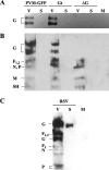
(A and B) Western blot analysis of virus-associated and soluble secreted viral proteins. BHK-21 cells were infected with rPVM-GFP7, rPVM-GFP-Gt, or rPVM-GFP-ΔG. Virus particles (V) and soluble proteins in the medium overlying the infected cells (S) were collected separately, subjected to SDS-PAGE under nonreducing conditions, and transferred to nitrocellulose membranes. PVM proteins were detected using a G-specific rabbit antiserum (A) or serum from a convalescent mouse (B). (C) As a positive control, HEp-2 cells were infected with HRSV strain A2. Virus particles and soluble proteins were separated in parallel to those of PVM, and proteins were detected using a commercial polyclonal HRSV-specific antibody. V, virus particles; S, supernatant; M, supernatant from mock-infected cells.
FIG. 7.
FIG. 7.
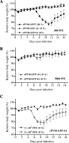
Virulence of rPVM-GFP-ΔG and rPVM-GFP-Gt in mice, measured by changes in body weight. Six- to eight-week-old BALB/c mice were infected intranasally with 500 PFU of rPVM-GFP7 (eight mice), rPVM-GFP-ΔG (nine mice), or rPVM-GFP-Gt (nine mice) (A), 5,000 PFU of rPVM-GFP-ΔG or rPVM-GFP-Gt (five mice per group) (B), or 5,000 or 50,000 PFU of rPVM-GFP-Gt (five mice per group), as indicated (C). Mice were observed closely and weighed daily. Weights are expressed relative to the weight at day 0, taken as 100%. When necessary, mice that met the end point regulations of the German animal protection board were euthanized by cervical dislocation. The percentage of dead animals per group is given in parentheses in each graph. For panel A, two independent experiments involving four and five animals per group, respectively, were combined.

Similar articles

Cited by

References

    1. Alff, P. J., I. N. Gavrilovskaya, E. Gorbunova, K. Endriss, Y. Chong, E. Geimonen, N. Sen, N. C. Reich, and E. R. Mackow. 2006. The pathogenic NY-1 hantavirus G1 cytoplasmic tail inhibits RIG-I- and TBK-1-directed interferon responses. J. Virol. 80:9676-9686. - PMC - PubMed
    1. Barr, J., P. Chambers, P. Harriott, C. R. Pringle, and A. J. Easton. 1994. Sequence of the phosphoprotein gene of pneumonia virus of mice: expression of multiple proteins from two overlapping reading frames. J. Virol. 68:5330-5334. - PMC - PubMed
    1. Bermingham, A., and P. L. Collins. 1999. The M2-2 protein of human respiratory syncytial virus is a regulatory factor involved in the balance between RNA replication and transcription. Proc. Natl. Acad. Sci. USA 96:11259-11264. - PMC - PubMed
    1. Biacchesi, S., Q. N. Pham, M. H. Skiadopoulos, B. R. Murphy, P. L. Collins, and U. J. Buchholz. 2005. Infection of nonhuman primates with recombinant human metapneumovirus lacking the SH, G, or M2-2 protein categorizes each as a nonessential accessory protein and identifies vaccine candidates. J. Virol. 79:12608-12613. - PMC - PubMed
    1. Biacchesi, S., M. H. Skiadopoulos, K. C. Tran, B. R. Murphy, P. L. Collins, and U. J. Buchholz. 2004. Recovery of human metapneumovirus from cDNA: optimization of growth in vitro and expression of additional genes. Virology 321:247-259. - PubMed

Publication types

MeSH terms