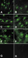
Expression of ciliary neurotrophic factor (CNTF) and its tripartite receptor complex by cells of the human optic nerve head
- PMID: 17563726
- PMCID: PMC2768760
Expression of ciliary neurotrophic factor (CNTF) and its tripartite receptor complex by cells of the human optic nerve head
Abstract
Purpose: Ciliary neurotrophic factor (CNTF) promotes gene expression, cell survival and differentiation in various types of peripheral and central neurons, glia and nonneural cells. The level of CNTF rises rapidly upon injury to neural tissue, suggesting that CNTF exerts its cytoprotective effects after release from cells via mechanisms induced by cell injury. The purpose of this study was to determine if cells in the optic nerve head express CNTF and its tripartite receptor complex.
Methods: Well-established optic nerve head astrocytes (ONHA) and lamina cribrosa (LC) cell cultures were derived from normal human donors. Total RNA was reverse transcribed and polymerase chain reaction (PCR) amplified for mRNA detection. Cytoplasmic protein expression was determined by immunocytochemistry and Western blot analysis of the cellular lysates. Serum free medium was concentrated and used for detecting extracellular proteins. CNTF complexed with CNTFR-alpha was assayed by immunoprecipitation.
Results: Cells isolated from the human optic nerve head express CNTF and its tripartite receptor complex members (CNTFR-alpha, gp130, LIFR-beta).
Conclusions: Taken together, these data suggest a possible neuroprotective role of CNTF in the optic nerve head.
Figures


Similar articles
-
Expression of biologically active mouse ciliary neutrophic factor (CNTF) and soluble CNTFRalpha in Escherichia coli and characterization of their functional specificities.Eur Cytokine Netw. 2004 Jul-Sep;15(3):255-62. Eur Cytokine Netw. 2004. PMID: 15542451
-
Ciliary neurotrophic factor and its receptors are differentially expressed in the optic nerve transected adult rat retina.Brain Res. 2004 Jul 9;1013(2):152-8. doi: 10.1016/j.brainres.2004.03.030. Brain Res. 2004. PMID: 15193523
-
Leukemia inhibitory factor and neurotrophins support overlapping populations of rat nodose sensory neurons in culture.Dev Biol. 1994 Feb;161(2):338-44. doi: 10.1006/dbio.1994.1035. Dev Biol. 1994. PMID: 8313987
-
The tripartite CNTF receptor complex: activation and signaling involves components shared with other cytokines.J Neurobiol. 1994 Nov;25(11):1454-66. doi: 10.1002/neu.480251111. J Neurobiol. 1994. PMID: 7852997 Review.
-
The ciliary neurotrophic factor and its receptor, CNTFR alpha.Pharm Acta Helv. 2000 Mar;74(2-3):265-72. doi: 10.1016/s0031-6865(99)00050-3. Pharm Acta Helv. 2000. PMID: 10812968 Review.
Cited by
-
Assessment of ability of human adipose derived stem cells for long term overexpression of IL-11 and IL-13 as therapeutic cytokines.Cytotechnology. 2020 Oct;72(5):773-784. doi: 10.1007/s10616-020-00421-8. Epub 2020 Sep 15. Cytotechnology. 2020. PMID: 32935166 Free PMC article.
-
Hesperidin ameliorates hypobaric hypoxia-induced retinal impairment through activation of Nrf2/HO-1 pathway and inhibition of apoptosis.Sci Rep. 2020 Nov 10;10(1):19426. doi: 10.1038/s41598-020-76156-5. Sci Rep. 2020. PMID: 33173100 Free PMC article.
-
The Role of Endogenous Neuroprotective Mechanisms in the Prevention of Retinal Ganglion Cells Degeneration.Front Neurosci. 2018 Nov 15;12:834. doi: 10.3389/fnins.2018.00834. eCollection 2018. Front Neurosci. 2018. PMID: 30524222 Free PMC article. Review.
-
Cell proliferation and interleukin-6-type cytokine signaling are implicated by gene expression responses in early optic nerve head injury in rat glaucoma.Invest Ophthalmol Vis Sci. 2011 Jan 25;52(1):504-18. doi: 10.1167/iovs.10-5317. Print 2011 Jan. Invest Ophthalmol Vis Sci. 2011. PMID: 20847120 Free PMC article.
-
Effect of CNTF on retinal ganglion cell survival in experimental glaucoma.Invest Ophthalmol Vis Sci. 2009 May;50(5):2194-200. doi: 10.1167/iovs.08-3013. Epub 2008 Dec 5. Invest Ophthalmol Vis Sci. 2009. PMID: 19060281 Free PMC article.
References
-
- Lambert W, Agarwal R, Howe W, Clark AF, Wordinger RJ. Neurotrophin and neurotrophin receptor expression by cells of the human lamina cribrosa. Invest Ophthalmol Vis Sci. 2001;42:2315–23. - PubMed
-
- Hudgins SN, Levison SW. Ciliary neurotrophic factor stimulates astroglial hypertrophy in vivo and in vitro. Exp Neurol. 1998;150:171–82. - PubMed
-
- Ji JZ, Elyaman W, Yip HK, Lee VW, Yick LW, Hugon J, So KF. CNTF promotes survival of retinal ganglion cells after induction of ocular hypertension in rats: the possible involvement of STAT3 pathway. Eur J Neurosci. 2004;19:265–72. - PubMed
-
- Hibi M, Nakajima K, Hirano T. IL-6 cytokine family and signal transduction: a model of the cytokine system. J Mol Med. 1996;74:1–12. - PubMed
MeSH terms
Substances
LinkOut - more resources
Full Text Sources
Miscellaneous
