. 2007 Jun 18;177(6):981-93.
doi: 10.1083/jcb.200702145. Epub 2007 Jun 11.

Cdc14-regulated midzone assembly controls anaphase B

Affiliations

Cdc14-regulated midzone assembly controls anaphase B

Anton Khmelinskii et al. J Cell Biol. .

Abstract

Spindle elongation in anaphase of mitosis is a cell cycle-regulated process that requires coordination between polymerization, cross-linking, and sliding of microtubules (MTs). Proteins that assemble at the spindle midzone may be important for this process. In this study, we show that Ase1 and the separase-Slk19 complex drive midzone assembly in yeast. Whereas the conserved MT-bundling protein Ase1 establishes a midzone, separase-Slk19 is required to focus and center midzone components. An important step leading to spindle midzone assembly is the dephosphorylation of Ase1 by the protein phosphatase Cdc14 at the beginning of anaphase. Failure to dephosphorylate Ase1 delocalizes midzone proteins and delays the second, slower phase of anaphase B. In contrast, in cells expressing nonphosphorylated Ase1, anaphase spindle extension is faster, and spindles frequently break. Cdc14 also controls the separase-Slk19 complex indirectly via the Aurora B kinase. Thus, Cdc14 regulates spindle midzone assembly and function directly through Ase1 and indirectly via the separase-Slk19 complex.


Figures

Figure 1.
Figure 1.
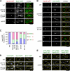
Spindle binding of midzone components. (A and B) ASE1-GFP SLK19-tdTomato (A) and ASE1-GFP BIM1-eqFP (B) cells were analyzed by time-lapse microscopy. Selected frames (maximum z-series projections) are shown. Time after the beginning of image acquisition is indicated in minutes. The inset in A is a magnification of the extending anaphase spindle of the ASE1-GFP SLK19-tdTomato cell. The arrow points toward Ase1 at the spindle center. (B) Asterisks mark spindle poles. (C) Analysis of α-factor synchronized ESP1-GFP SLK19-tdTomato cells. Early (i), mid- (ii), and late (iii–iv) anaphase cells. Arrowheads point to spindle poles. (D) Quantification of C. Approximate cell morphologies are indicated by yellow outlines drawn onto the overlaid images. Bars, 5 μm.
Figure 2.
Figure 2.
Spindle midzone localization of Slk19 and Ase1 is interdependent. (A and B) Slk19-GFP localization in SLK19-GFP mCherry-TUB1 cells with and without ASE1 synchronized with α factor (G1; t = 0). (B) Cells were analyzed at each time point for large buds (dotted lines), spindle length, and Slk19-GFP localization at the spindle midzone and poles (red dots) or only at the spindle poles (green dots). The mean spindle length (blue lines) with SD (error bars; black bars) is indicated for each time point. n > 50 per time point. (C and D) Ase1-GFP localization in ASE1-GFP mCherry-TUB1 SPC42-eqFP cells with and without SLK19. (C) The percentage of anaphase cells with centered or mislocalized Ase1-GFP spindle signal is shown on the right side of the figure. (D) Quantification of C. Mean value of the Ase1-GFP zone length with SD is shown for each spindle class. n > 30 per spindle class. Approximate cell morphologies are indicated by yellow outlines drawn onto the overlaid images. Bars, 5 μm.
Figure 3.
Figure 3.
Regulation of Ase1 localization by Esp1 is independent of Cdc14. (A) Ase1-GFP localization in esp1-1 ASE1-GFP SPC42-eqFP cells with and without the expression of pGal1-CDC14. Cells grown in raffinose medium were synchronized with α factor at 23°C in G1 and released into a new cell cycle after removing α actor in the presence of galactose at 37°C. (B) Levels of Cdc14 before (−Gal) and 1 h after induction of the pGal1 promoter with galactose (+Gal) as determined by immunoblotting with anti-Cdc14 antibodies. The asterisk indicates a protein band that cross reacts with the anti-Cdc14 antibodies. (C and D) Quantification of A. (C) Mean value of the Ase1-GFP zone length with SD (error bars) is shown for each spindle class. (D) Anaphase cells with mislocalized (not centered) Ase1-GFP spindle signal were counted for each spindle class. n > 30 per spindle class. Approximate cell morphologies are indicated by yellow outlines drawn onto the overlaid images. Bar, 5 μm.
Figure 4.
Figure 4.
Multiple roles of Cdc14 in the regulation of anaphase spindle assembly. (A–E) Localization of spindle midzone proteins in WT, cdc14-2, cdc14-2 SLI156A, and sli15-3 cells. SPC42-GFP or SPC42-eqFP was used as a marker for SPBs. Cells were synchronized with α factor at 23°C and released into a new cell cycle at 37°C. (A) Localization of Esp1-4GFP in CDC14 SLI15 (WT), cdc14-2 SLI15, cdc14-2 SLI156A, and CDC14 sli15-3 cells. (B) Ase1-4GFP localization in CDC14 SLI15 (WT), cdc14-2 SLI15 (i–iii), cdc14-2 SLI156A (iv–vi), and CDC14 sli15-3 cells (vii–viii). (C) Quantification of Ase1-4GFP localization in anaphase cells of the indicated types. n > 200 for each strain. (D) Localization of 4GFP-tagged Cin8, Stu1, and Bim1 in CDC14 SLI15 (WT), cdc14-2 SLI15, and cdc14-2 SLI156A cells. (E) Localization of 4GFP-tagged Cin8, Stu1, and Bim1 in SLI15 (WT) and sli15-3 cells. Approximate cell morphologies are indicated by yellow outlines drawn onto the overlaid images. Bars, 5 μm.
Figure 5.
Figure 5.
Cdc14 directly dephosphorylates Ase1. (A) Ase1 and Cdc14-N interact in the yeast two-hybrid system. Mean values from three independent experiments are shown with SD (error bars). (B) CDC14 ASE1-6HA and td-cdc14 ASE1-6HA cells were synchronized with α factor in G1 phase (t = 0) at 23°C and released from the block at 37°C (see Materials and methods). Samples were analyzed with the indicated antibodies. (C) cdc26Δ pMet3-CDC20 cells with CDC14 (lane 1), pGal1-CDC14 (lane 3), and pGal1-CDC14C283A (lane 5) were arrested in metaphase through Cdc20 depletion (t = 0). Galactose was then added, and the strains were incubated for 1 h at 37°C (lanes 2, 4, and 6). The percentage of metaphase cells was determined for each time point (DAPI staining; n > 100). Samples were analyzed with the indicated antibodies. (D) Cdc14 levels of cells in C (lanes 4 and 6) were similar. (E) Immunoprecipitated Ase1-6HA was incubated with Cdc14, Cdc14C283A, or AP as indicated.
Figure 6.
Figure 6.
Dephosphorylation of Ase1 is essential for spindle midzone organization. (A) Distribution of the seven Cdk1 consensus sites in Ase1. CC, coiled-coil region; MB, MT-binding domain. (B) td-cdc14 cells expressing ASE1-GFP, ASE17A-GFP, or ASE17D-GFP were synchronized with α factor at 23°C and released into a new cell cycle at 37°C. After 3 h of incubation at 37°C, cells were collected and analyzed by immunoblotting with anti-GFP antibodies. (C) Spindle localization of GFP-tagged Ase1, Ase17A, and Ase17D in CDC14 cells. Spc42-eqFP was used as a pole marker. (D) CDC14 and td-cdc14 cells with ASE1-GFP, ASE17A-GFP, and ASE17D-GFP were grown and synchronized as described in Materials and methods. Spindle localization of Ase1-GFP was quantified as outlined in the figure. n > 100 anaphase cells per CDC14 strain; n > 500 anaphase cells per td-cdc14 strain. (E and F) Localization of Cin8-, Stu1-, and Slk19-GFP in ASE1, ASE17A, and ASE17D cells. Cells were grown in YPD at 30°C to mid-log phase. Cells with a single centered GFP domain between the spindle poles were counted as having normal midzone localization. n > 100 anaphase cells per strain. Approximate cell morphologies are indicated by yellow outlines drawn onto the overlaid images. Bars, 5 μm.
Figure 7.
Figure 7.
Dephosphorylation of Ase1 is required for continuous extension of the anaphase spindle. (A) Kinetics of anaphase spindle extension of ASE1 WT, ase1Δ, ASE17A, and ASE17D cells with GFP-TUB1 by time-lapse microscopy. Pole to pole distance was measured in three dimensions. Insets depict the different phases of extension: 1, 2, and S (stalled). (B) The spindle breaks during anaphase in ASE17A cells. Selected frames (maximum z-series projections) are shown. Time is indicated in minutes. (C) cdc15-1 GFP-TUB1 cells with WT ASE1, ase1Δ, ASE17A, or ASE17D were released from α-factor block and incubated at 37°C (the restrictive temperature). Broken spindles (two half-spindles without continuous GFP-Tub1 signal in between) were scored over time. n > 100 spindles per time point. Bar, 5 μm.

Similar articles

Cited by

References

    1. Araki, H., K. Awane, N. Ogawa, and Y. Oshima. 1992. The CDC26 gene of Saccharomyces cerevisiae is required for cell growth only at high temperature. Mol. Gen. Genet. 231:329–331. - PubMed
    1. Balasubramanian, M.K., E. Bi, and M. Glotzer. 2004. Comparative analysis of cytokinesis in budding yeast, fission yeast and animal cells. Curr. Biol. 14:R806–R818. - PubMed
    1. Belmont, L.D., A.A. Hyman, K.E. Sawin, and T.J. Mitchison. 1990. Real-time visualization of cell cycle-dependent changes in microtubule dynamics in cytoplasmic extracts. Cell. 62:579–589. - PubMed
    1. Berlin, V., C.A. Styles, and G.R. Fink. 1990. BIK1, a protein required for microtubule function during mating and mitosis in Saccharomyces cerevisiae, colocalizes with tubulin. J. Cell Biol. 111:2573–2586. - PMC - PubMed
    1. Bouck, D.C., and K.S. Bloom. 2005. The kinetochore protein Ndc10p is required for spindle stability and cytokinesis in yeast. Proc. Natl. Acad. Sci. USA. 102:5408–5413. - PMC - PubMed

Publication types

MeSH terms