. 2007 Jul;10(7):870-9.
doi: 10.1038/nn1916. Epub 2007 Jun 10.

Cannabinoids mediate analgesia largely via peripheral type 1 cannabinoid receptors in nociceptors

Affiliations

Cannabinoids mediate analgesia largely via peripheral type 1 cannabinoid receptors in nociceptors

Nitin Agarwal et al. Nat Neurosci. 2007 Jul.

Abstract

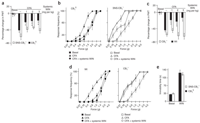
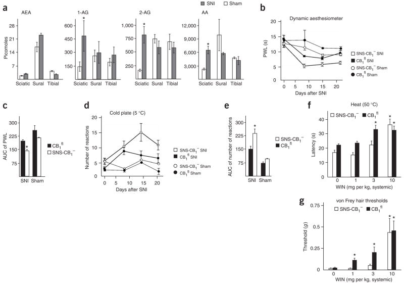
Although endocannabinoids constitute one of the first lines of defense against pain, the anatomical locus and the precise receptor mechanisms underlying cannabinergic modulation of pain are uncertain. Clinical exploitation of the system is severely hindered by the cognitive deficits, memory impairment, motor disturbances and psychotropic effects resulting from the central actions of cannabinoids. We deleted the type 1 cannabinoid receptor (CB1) specifically in nociceptive neurons localized in the peripheral nervous system of mice, preserving its expression in the CNS, and analyzed these genetically modified mice in preclinical models of inflammatory and neuropathic pain. The nociceptor-specific loss of CB1 substantially reduced the analgesia produced by local and systemic, but not intrathecal, delivery of cannabinoids. We conclude that the contribution of CB1-type receptors expressed on the peripheral terminals of nociceptors to cannabinoid-induced analgesia is paramount, which should enable the development of peripherally acting CB1 analgesic agonists without any central side effects.


Figures

Figure 1
Figure 1
Demonstration of conditional deletion of CB1 specifically in nociceptive neurons of the DRG in sensory neuron–specific CB1 knockout mice (SNS-CB1). (a) mRNA in situ hybridization for expression of CB1 or GABAB(1) (control) on DRG sections from SNS-CB1 mice and control littermates (CB1fl). (b) Quantitative size analysis of DRG neurons expressing CB1 mRNA showed that small-diameter neurons lost and large-diameter neurons maintained CB1 expression in SNS-CB1 mice. (c) A goat anti-CB1 used throughout this study yielded specific labeling of DRG neurons that was entirely lost in the DRG of globally CB1 mice. (d) Typical examples of anti-CB1 immunoreactivity in subpopulations of DRG neurons labeled using binding to IB4 or using antibodies to TRPV1, Nav1.8 and neurofilament 200 (NF200) in wild-type, CB1fl and SNS-CB1 mice. In SNS-CB1 mice, CB1 immunoreactivity was nearly abrogated from nociceptors (IB4-, substance P– or Nav1.8-positive neurons) and reduced in a large fraction of TRPV1-positive C- and A-δ neurons, but entirely preserved in NF200-positive large-diameter neurons. (e) Quantitative summary of DRG cell populations expressing CB1 protein in wild-type (Wt), CB1fl mice and SNS-CB1 mice from experiments represented in d (mean ± s.e.m.; n = 10–15 DRG sections each). *P < 0.001, ANOVA, post hoc Fisher’s test. Scale bars, 40 μm in (a,c,d).
Figure 2
Figure 2
Expression of CB1 mRNA and CB1 protein is similar in the brain and spinal cord of CB1fl mice and SNS-CB1 mice. (a,b) Antisense mRNA riboprobes revealed comparable expression of CB1 in hippocampal interneurons (arrows, a) and spinal neurons (b) of SNS-CB1 mice and their CB1fl/fl littermates, but a loss of signal in global CB1 mice or on usage of the sense probes. (c,d) Immunostaining with a goat anti-CB1 revealed comparable expression of CB1 in the brain (hippocampus shown in c) and in the spinal cord (d) of SNS-CB1 mice and their CB1fl littermates, but a loss of signal in global CB1 mice or in staining controls. (e) Autoradiography with a synthetic cannabinoid 3H-CP-55940 revealed similar levels of binding (mean ± s.e.m.) in various brain regions of SNS- CB1 as compared to CB1fl mice. (f) The pattern of termination of primary nociceptive afferents in the spinal dorsal horn was similar in CB1fl and SNS-CB1 mice, as shown via binding to TRITC-labeled isolectin-B4 (IB4) and immunoreactivity for substance P. Scale bars, 150 (ad) and 100 μm (f).
Figure 3
Figure 3
Nociceptive responses, locomotive performance and nociceptive activity–induced expression of proteins in SNS-CB1 mice and their CB1fl littermates. (a) SNS-CB1 mice (n = 12) showed significant reductions in paw withdrawal latency (PWL; P = 0.001) in response to radiant heat and in paw withdrawal threshold (PWT; P = 0.003) in response to punctuate pressure in comparison with CB1fl mice (n = 12). (b) SNS-CB1 mice (n = 8) showed a significant reduction in the duration of acute nocifensive responses to intraplantar paw injection of capsaicin (P = 0.002) or formalin (phase I; P = 0.049), as compared with CB1fl mice (n = 8). (c) Latency to fall from a rotating rod was similar in SNS-CB1 mice (n = 6) and CB1fl mice (n = 6; P = 0.203). (d,e) Quantitative analysis of neurons immunoreactive for either Fos or phosphorylated ERK1/2 per section of DRG or spinal dorsal horn in the basal state (naive) or 1 h after intraplantar hindpaw injection of formalin in SNS-CB1 mice (n = 6) and CB1fl mice (n = 6). *P < 0.05, ANOVA, post hoc Fisher’s test. All data points represent mean ± s.e.m.
Figure 4
Figure 4
Analysis of endocannabinoid levels in the paws and spinal segments (L4–L6) of SNS- CB1 mice, CB1fl mice, global CB1 mice and their wild-type controls (n = 6 each) in the basal state (naive) or in wild-type mice after injection of CFA into the hindpaw. (a) Levels of AEA, 1-AG, 2-AG and arachidonic acid (AA) rose in the paw skin after inflammation, as compared with naive state (P < 0.05; n = 8 paws in each group), whereas oleoylethanolamide (OEA) levels remained unchanged (P = 0.9). (b) Levels of endocannabinoids did not change significantly in the L4–L6 spinal cord after paw inflammation over the naive state (P > 0.05; n = 6 mice in each group). (c,d) Levels of endocannabinoids in the paw or in the spinal cord were not significantly different across SNS-CB1, CB1fl, global CB1 and wild-type mice. *P < 0.05, ANOVA, post hoc Fisher’s test. All data points represent mean ± s.e.m.
Figure 5
Figure 5
Behavioral and electrophysiological analysis of SNS-CB1 mice in models of inflammatory pain. (a) Comparison of response frequency to von Frey hairs in SNS-CB1 mice (n = 12), CB1fl mice (n = 12), global CB1 knockout mice (CB1; n = 6) and their wild-type littermates (Wt; n = 6) before and 27 h after intraplantar injection of CFA. Note that SNS-CB1 mice and CB1 mice demonstrated comparable deviations from their respective control littermates. (b) Summary of response thresholds (defined as a force eliciting a response frequency of at least 40%) before and at 6–7 h, 14 h, 27 h or 52 h after intraplantar injection of CFA to SNS-CB1, CB1fl, global CB1 and Wt mice. (c) Response frequency to abdominal application of von Frey filaments after induction of acute pancreatitis was significantly greater in SNS-CB1 mice (n = 7) than in CB1 mice (n = 8) (P < 0.01, ANOVA, post hoc Fisher’s test). (d) Electrophysiological recordings from C-mechanoreceptors in the skin-nerve preparation derived from the paw showed that the frequency of responsive C-fibers was significantly greater at 1–2 mN force in SNS-CB1mice (n = 29 fibers) than in CB1fl mice (n = 31 fibers) (P < 0.05, chi square analysis). y axes in ac indicate force exerted by individual von Frey filaments. All data points represent mean ± s.e.m.
Figure 6
Figure 6
Effects of a systemically applied CB1/CB2-agonist, WIN, on inflammation-induced mechanical hypersensitivity and immobilization behavior. (ad) Paw inflammation was induced by unilateral intraplantar injection of CFA and mechanical hypersensitivity was derived as the percentage change in paw withdrawal threshold (PWT) over uninjected paw using an automated dynamic aesthesiometer (a,c) or by recording stimulus force-response frequency curves upon manual application of von Frey filaments (b,d) on the same cohort of animals. Systemically applied WIN (1 or 3 mg per kg) reduced CFA-induced mechanical hypersensitivity to a greater extent in CB1fl mice (n = 5 or 6 mice for each dose) than it did in SNS-CB1 mice (n = 5 or 6 for each dose) (a,b). Systemically applied WIN (1 or 3 mg per kg) reduced CFA-induced mechanical hypersensitivity in wild-type mice (Wt; n = 5 or 6 for each dose), but not in classical CB1 knockout mice (CB1; n = 5 or 6 for each dose) (c,d). *P < 0.05 as compared with CFA-induced mechanical hyperalgesia in (a,c), ANOVA, post hoc Fisher’s test. (e) Intraperitoneal injection of WIN induced immobilization responses in the ring catalepsy test in both CBfl mice and SNS-CB1 mice. *P < 0.05 over basal state, ANOVA, post hoc Fisher’s test. All data points represent mean ± s.e.m.
Figure 7
Figure 7
Effects of WIN. (ad) WIN was applied via intrathecal (a,b) or intraplantar (c,d) routes of administration on inflammation-induced mechanical hypersensitivity. Intrathecally applied WIN reduced CFA-induced mechanical hypersensitivity in both CB1fl mice (n = 6) and in SNS-CB1 mice (n = 6). Intraplantar application of WIN (10–30 μg) significantly reduced CFA-induced mechanical hypersensitivity in CB1fl mice (n = 5 or 6 for each dose), but not in SNS-CB1 mice (n = 5 or 6 for each dose). *P < 0.05 as compared with CFA-induced mechanical hyperalgesia in a and c, ANOVA, Fisher’s test. All data points represent mean ± s.e.m.
Figure 8
Figure 8
Endocannabinoid levels, pain behavior and analgesic effects of WIN in SNS-CB1mice and CB1fl mice in the SNI model for neuropathic pain. (a) Levels of endocannabinoids in innervation territories of the ‘sural’ and ’saphenous/tibial’ branches of the sciatic nerve or in the sciatic nerve just proximal to the site of ligation. *P < 0.02, ANOVA, Fisher’s test; n = 3 or 4 samples in each group. (b,c) Latency of PWL in response to mechanical stimuli (represented as integrated area under the curve, AUC, in c) in SNS-CB1 mice and CB1fl mice (n = 7 each for SNI and 3 each for sham). SNS-CB1 mice showed an exaggerated drop in PWL as compared with controls after SNI (*P < 0.05, ANOVA, Fisher’s test). (d,e) Number of reactions to a cold stimulus (5 °C) (represented as integrated AUC in panel e) in SNS-CB1 mice and CB1fl mice (n = 6 each). *P < 0.05, ANOVA, Fisher’s test. (f,g) Effects of intraperitoneal injections of WIN (1, 3 or 10 mg per kg) on latency of paw withdrawal to heat at 50 °C (f) or plantar response threshold to von Frey hairs (g) in SNS-CB1 mice (n = 7) and CB1fl mice (n = 9). *P < 0.05 as compared with values before WIN application (0) in the respective group, ANOVA, Fisher’s test. All data points represent mean ± s.e.m.

Similar articles

Cited by

References

    1. Walker JM, Hohmann AG. Cannabinoid mechanisms of pain suppression. Handb Exp Pharmacol. 2005;168:509–554. - PubMed
    1. Pacher P, Batkai S, Kunos G. The endocannabinoid system as an emerging target of pharmacotherapy. Pharmacol Rev. 2006;58:389–462. - PMC - PubMed
    1. Freund TF, Katona I, Piomelli D. Role of endogenous cannabinoids in synaptic signaling. Physiol Rev. 2003;83:1017–1066. - PubMed
    1. Piomelli D. The endocannabinoid system: a drug discovery perspective. Curr Opin Investig Drugs. 2005;6:672–679. - PubMed
    1. Patwardhan AM, et al. The cannabinoid WIN55,212-2 inhibits transient receptor potential vanilloid 1 (TRPV1) and evokes peripheral antihyperalgesia via calcineurin. Proc Natl Acad Sci USA. 2006;103:11393–11398. - PMC - PubMed

Publication types

MeSH terms