

Review
doi: 10.1083/jcb.200701146.
Epub 2007 May 29.
Building blood vessels--stem cell models in vascular biology
Affiliations
- PMID: 17535968
- PMCID: PMC2064276
- DOI: 10.1083/jcb.200701146
Item in Clipboard
Review
Building blood vessels--stem cell models in vascular biology
J Cell Biol.
.
Abstract
Spheroids of differentiating embryonic stem cells, denoted embryoid bodies, constitute a high-quality model for vascular development, particularly well suited for loss-of-function analysis of genes required for early embryogenesis. This review examines vasculogenesis and angiogenesis in murine embryoid bodies and discusses the promise of stem cell-based models for the study of human vascular development.
Figures
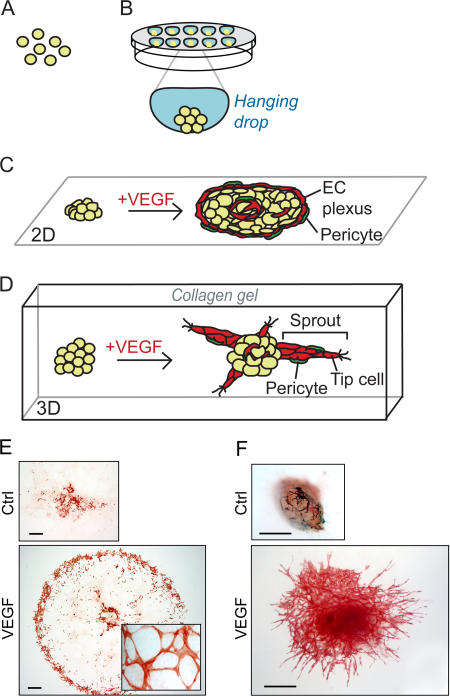
Outline of 2D and 3D EB models for vasculogenesis and angiogenesis. Stem cells are trypsinized (d 0), (A) and aggregated to create EBs, in drops hanging from the lid of a Petri dish (B). Aggregation can also occur spontaneously by seeding ESCs in suspension in a nonadhesive Petri dish, resulting in EBs of variable size. After 4 d, EBs are seeded on a tissue culture slide (2D), (C) or alternatively embedded in a 3D collagen gel (D). Addition of VEGF induces formation of a peripheral vascular plexus in 2D (C and E) and endothelial cell sprouts (“angiogenesis”) in 3D (D and F) (bottom panel in F adopted from Magnusson et al., [2005]). Whole-mount stainings for CD31 of 2D (E) or 3D (F) EBs at d 10 of differentiation, untreated (Ctrl, top) or induced with VEGF (bottom). Bars, 500 μm.
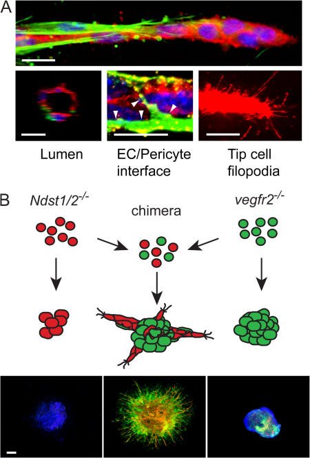
Angiogenic sprouts invade the surrounding matrix. (A) Features of blood vessel sprouts formed in 3D collagen matrix in response to VEGF. Expression is shown of the endothelial cell marker CD31/platelet-endothelial cell adhesion molecule (PECAM; red), the pericyte markers αSMA (green, top), and NG2 (green, bottom middle). Hoechst 33342 was used to indicate nuclei (blue). Lumen formation is evident in larger vessels (left, cross section [z-stack] of a sprout [d 18] generated by confocal microscopy). The tip cell at the front of growing sprouts send out filopodia to sense growth factor gradients. Occasional filopodia are also detected on stalk cells that lack pericyte coverage. Bars, 10 μm. (B) Knockout EBs and the assembly of chimeric EBs. Cells deficient in production of HS (Ndst1/2
−/−) or lacking VEGFR-2 (vegfr2
−/−) do not form vascular sprouts in the EB model. However, chimeric EBs generated by mixing of the two ESC lines before EB formation respond to VEGF and form sprouts. In the chimeras, the endothelial cells (CD31; red) are derived from Ndst1/2
−/− cells expressing VEGFR-2, whereas functional HS is provided by pericytes (αSMA; green) lacking VEGFR-2 (Jakobsson et al., 2006). Bar, 300 μm.
Similar articles
-
In vitro differentiation of mouse embryonic stem cells into primitive blood vessels.Methods Enzymol. 2008;443:103-17. doi: 10.1016/S0076-6879(08)02006-5. Methods Enzymol. 2008. PMID: 18772013
-
No evidence for vasculogenesis regulation by angiostatin during mouse embryonic stem cell differentiation.J Cell Physiol. 2007 Oct;213(1):27-35. doi: 10.1002/jcp.21084. J Cell Physiol. 2007. PMID: 17450519
-
Identification of RSK and TTK as Modulators of Blood Vessel Morphogenesis Using an Embryonic Stem Cell-Based Vascular Differentiation Assay.Stem Cell Reports. 2016 Oct 11;7(4):787-801. doi: 10.1016/j.stemcr.2016.08.004. Epub 2016 Sep 8. Stem Cell Reports. 2016. PMID: 27618721 Free PMC article.
-
Vascular engineering using human embryonic stem cells.Biotechnol Prog. 2009 Jan-Feb;25(1):2-9. doi: 10.1002/btpr.129. Biotechnol Prog. 2009. PMID: 19197982 Review.
-
Embryonic stem cell models in vascular biology.J Thromb Haemost. 2009 Jul;7 Suppl 1:53-6. doi: 10.1111/j.1538-7836.2009.03427.x. J Thromb Haemost. 2009. PMID: 19630768 Review.
Cited by
-
Blood and lymphatic vessel formation.Cold Spring Harb Perspect Biol. 2015 Mar 2;7(3):a008268. doi: 10.1101/cshperspect.a008268. Cold Spring Harb Perspect Biol. 2015. PMID: 25731762 Free PMC article. Review.
-
Endothelial cells dynamically compete for the tip cell position during angiogenic sprouting.Nat Cell Biol. 2010 Oct;12(10):943-53. doi: 10.1038/ncb2103. Epub 2010 Sep 26. Nat Cell Biol. 2010. PMID: 20871601
-
Characterization of tubular liquid crystal structure in embryonic stem cell derived embryoid bodies.Cell Biosci. 2017 Jan 3;7:3. doi: 10.1186/s13578-016-0130-6. eCollection 2017. Cell Biosci. 2017. PMID: 28066542 Free PMC article.
-
Bone Morphogenetic Protein 9 Regulates Early Lymphatic-Specified Endothelial Cell Expansion during Mouse Embryonic Stem Cell Differentiation.Stem Cell Reports. 2019 Jan 8;12(1):98-111. doi: 10.1016/j.stemcr.2018.11.024. Epub 2018 Dec 27. Stem Cell Reports. 2019. PMID: 30595547 Free PMC article.
-
Formation of composite endothelial cell-mesenchymal stem cell islets: a novel approach to promote islet revascularization.Diabetes. 2008 Sep;57(9):2393-401. doi: 10.2337/db07-0981. Epub 2008 Jun 2. Diabetes. 2008. PMID: 18519803 Free PMC article.
References
-
- Bautch, V.L., S.D. Redick, A. Scalia, M. Harmaty, P. Carmeliet, and R. Rapoport. 2000. Characterization of the vasculogenic block in the absence of vascular endothelial growth factor-A. Blood. 95:1979–1987. - PubMed
-
- Carmeliet, P., V. Ferreira, G. Breier, S. Pollefeyt, L. Kieckens, M. Gertsenstein, M. Fahrig, A. Vandenhoeck, K. Harpal, C. Eberhardt, et al. 1996. Abnormal blood vessel development and lethality in embryos lacking a single VEGF allele. Nature. 380:435–439. - PubMed
-
- Carmeliet, P., M.G. Lampugnani, L. Moons, F. Breviario, V. Compernolle, F. Bono, G. Balconi, R. Spagnuolo, B. Oostuyse, M. Dewerchin, et al. 1999. Targeted deficiency or cytosolic truncation of the VE-cadherin gene in mice impairs VEGF-mediated endothelial survival and angiogenesis. Cell. 98:147–157. - PubMed
-
- Choi, K., M. Kennedy, A. Kazarov, J.C. Papadimitriou, and G. Keller. 1998. A common precursor for hematopoietic and endothelial cells. Development. 125:725–732. - PubMed
