Review
. 2007:23:519-47.
doi: 10.1146/annurev.cellbio.23.090506.123319.

Biogenesis and function of multivesicular bodies

Affiliations
Review

Biogenesis and function of multivesicular bodies

Robert C Piper et al. Annu Rev Cell Dev Biol. 2007.

Abstract

The two major cellular sites for membrane protein degradation are the proteasome and the lysosome. Ubiquitin attachment is a sorting signal for both degradation routes. For lysosomal degradation, ubiquitination triggers the sorting of cargo proteins into the lumen of late endosomal multivesicular bodies (MVBs)/endosomes. MVB formation occurs when a portion of the limiting membrane of an endosome invaginates and buds into its own lumen. Intralumenal vesicles are degraded when MVBs fuse to lysosomes. The proper delivery of proteins to the MVB interior relies on specific ubiquitination of cargo, recognition and sorting of ubiquitinated cargo to endosomal subdomains, and the formation and scission of cargo-filled intralumenal vesicles. Over the past five years, a number of proteins that may directly participate in these aspects of MVB function and biogenesis have been identified. However, major questions remain as to exactly what these proteins do at the molecular level and how they may accomplish these tasks.


Figures

Figure 1
Figure 1
Ubiquitination and endosomal ubiquitin-sorting receptors. The transfer of ubiquitin (Ub) to substrate proteins involves the sequential action of three classes of enzymes: an activating enzyme (E1), a conjugation enzyme (E2), and a ligase (E3) (Hershko & Ciechanover 1998). Most eukaryotic cells have a single E1 enzyme, which uses ATP to form a high-energy thiol-ester bond with the C-terminal glycine (G76) of Ub E1 and then transfers Ub to one of a handful of E2 enzymes, which also forms a thiol-ester bond with Ub G76. E2s can bind RING-finger-type E3 ligases that bridge the substrate protein with E2, thus facilitating the transfer of Ub from E2 to the substrate. E2 can also transfer Ub to HECT-type E3 ligases, which also form a thiol-ester bond and directly transfer Ub onto substrate proteins. Ub is typically attached to lysine side chains (K) of substrate proteins and forms an isopeptide bond. E3s can sometimes interact directly with their substrate or use other adaptor proteins (Ap) to target their substrates. Ub is removed from proteins by Ub-specific proteases or deubiquitinating enzymes (DUbs). The major DUb families are grouped on the basis of the similarities of their active site (Ubp, Ub peptidase; Uch, Ub C-terminal hydrolase; OTU, otubain; JAMM, Jab1/MPN domain metalloenzyme; JD, Josephin domain).
Figure 2
Figure 2
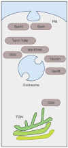
Ub-binding proteins involved in protein trafficking throughout the post-Golgi/endocytic system. Proteins such as Eps15, Epsin, Tom1, Tollip, GGA, Hrs, STAM, TSG101, and Vps36/Eap45 (see text) may work as receptors for Ub-cargo and catalyze a number of distinct transport steps that convey cargo toward the lysosome for degradation. These transport steps include internalization from the plasma membrane (PM), vesicle formation at the trans-Golgi network (TGN), and lumenal vesicle formation at the endosome.
Figure 3
Figure 3
Recognition of ubiquitin. (a) Ub is depicted as a cartoon in two orientations. The flexible C terminus containing G75 and G76 is oriented at the bottom and colored blue. Some, but not all, of the lysines of Ub are labeled in red, and the hydrophobic patch on the surface of Ub composed of L8, I44, and V70 is shown in yellow. (b) The corresponding orientations of Ub interfaced with the indicated Ub-binding domains. On the left are the domains found in the endosomal sorting complexes required for transport (ESCRT)-I/II supercomplex; on the right are Ub-binding motifs found in peripheral Ub receptors. The GAT domain contains two separable Ub-binding sites (S1 and S2), which are shown in the side orientation. For the face-on view of the GAT domain and the GRAM-like Ub binding on Eap45 (GLUE) domain, part of the domain is removed to highlight the interface with Ub. All Ub-binding motifs interact to some degree with the I44 hydrophobic patch, and none interact with the alpha-helix region on the back of Ub. The red, highlighted residue in the Ub-E2-like variant domain (UEV) structure corresponds to M85.
Figure 4
Figure 4
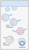
MVB anatomy. Schematic of the endocytic pathway indicating the progression from early endosomes to multivesicular late endosomes and finally to multilamellar lysosomes. Internal vesicle formation occurs during endosome maturation. Pictured are two types of internal membranes: phosphatidylinositol 3-phosphate [PI(3)P] positive (red ) and LBPA positive ( yellow).
Figure 5
Figure 5
Electron and fluorescence micrographs of MVBs and late endosomes. (a) An early endosome with a tubular extension, which serves as a carrier for recycling proteins such as a transferrin receptor. An electron-dense protein coat can be seen on the surface of endosomes. This subdomain is highlighted by arrowheads and is labeled with colloidal gold for clathrin [from M. Sacshe and J. Klumpermann (Sachse et al. 2002), with permission]. The electron micrographs in the lower panels show late endosomes from BHK cells labeled for phosphatidylinositol 3-phosphate [PI(3)P] singly (left) or double labeled (right) for PI(3)P (red arrows) and for LBPA ( yellow arrows). Provided by Rob Parton, University of Brisbane. (b) Fluorescence micrographs of wild-type and class E vps mutant cells expressing GFP-tagged MVB cargo and labeled with the lipophilic dye FM4-64 (red ), which marks the limiting membrane (provided by A. Oestreich, Mayo Clinic). MVB cargo is localized within the limiting membrane of the vacuole in wild-type cells. The mutant class E vps cells mislocalize the MVB cargo to the limiting membrane of the vacuole and the aberrant class E compartment.
Figure 6
Figure 6
Map of key MVB biogenesis machinery. Schematic representation of protein-protein interactions among components involved in MVB sorting. ESCRT-I, -II, and -III are highlighted in blue, green, and orange, respectively. The Vps27-Hse1 complex is shown in pink. No distinction between yeast and mammalian proteins is shown in an attempt to highlight some of the conserved interactions.
Figure 7
Figure 7
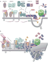
MVB core machinery. (a) Schematic for how class E Vps proteins may control MVB sorting. Pictured is the sequential recruitment of ESCRT complexes that are nucleated by the association of ESCRT-0 (STAM/Hrs) with endosomal membranes by binding phosphatidylinositol 3-phosphate [PI(3)P]. Vps27/Hrs recruits clathrin to form an endosomal subdomain and also binds and activates ESCRT-I, which in turn binds and recruits the ESCRT-II complex to endosomes. ESCRT-II then recruits ESCRT-III, which forms a polymer. This network is then disassembled at the last step of ILV formation. Ub-cargo is sorted in a sequential manner as well: It is first recognized by ESCRT-0 and then passed first to ESCRT-I and then to ESCRT-II before being incorporated into ILVs. At some point, Ub-cargo is deubiquitinated so that Ub is recycled. (b) An alternative model, wherein the Hrs complex is one of many peripheral Ub-sorting receptors that gather Ub-cargoes on endosomes and concentrate them in clathrin-rich subdomains. These receptors then transfer cargo to the ESCRT-I/II supercomplex, which has a variety of Ub-binding sites that can recognize a host Ub-cargo. ESCRT-I/II sits enmeshed in a specialized subdomain enriched in tetraspanins, sphingolipids, and other components that will be incorporated into ILVs. This meshwork is organized by ESCRT-III, which forms a polymer (represented as a flat checkerboard lattice) that can incarcerate cargo and hold ESCRT-I/II. This coated subdomain houses DUbs like Doa4, which can remove Ub without allowing cargo to escape. The ESCRT-III polymer is contained within endosomal subdomains by the Vps4 ATPase. ILV formation is catalyzed by the aggregation of cargo and tetraspanins that form large oligomeric structures. ILV formation is also facilitated by sorting of lipids conducive to membrane curvature.

Similar articles

Cited by

References

    1. Abrami L, Lindsay M, Parton RG, Leppla SH, Van Der Goot FG. Membrane insertion of anthrax protective antigen and cytoplasmic delivery of lethal factor occur at different stages of the endocytic pathway. J Cell Biol. 2004;166:645–51. - PMC - PubMed
    1. Alam SL, Langelier C, Whitby FG, Koirala S, Robinson H, et al. Structural basis for ubiquitin recognition by the human ESCRT-II EAP45 GLUE domain. Nat Struct Mol Biol. 2006;13:1029–30. - PubMed
    1. Alam SL, Sun J, Payne M, Welch BD, Blake BK, et al. Ubiquitin interactions of NZF zinc fingers. EMBO J. 2004;23:1411–21. - PMC - PubMed
    1. Almeida CG, Takahashi RH, Gouras GK. Beta-amyloid accumulation impairs multivesicular body sorting by inhibiting the ubiquitin-proteasome system. J Neurosci. 2006;26:4277–88. - PMC - PubMed
    1. Amerik AY, Nowak J, Swaminathan S, Hochstrasser M. The Doa4 deubiquitinating enzyme is functionally linked to the vacuolar protein-sorting and endocytic pathways. Mol Biol Cell. 2000;11:3365–80. - PMC - PubMed

Publication types

LinkOut - more resources