Assessing functional connectivity in the human brain by fMRI
- PMID: 17499467
- PMCID: PMC2169499
- DOI: 10.1016/j.mri.2007.03.007
Assessing functional connectivity in the human brain by fMRI
Abstract
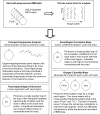
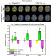
Functional magnetic resonance imaging (fMRI) is widely used to detect and delineate regions of the brain that change their level of activation in response to specific stimuli and tasks. Simple activation maps depict only the average level of engagement of different regions within distributed systems. FMRI potentially can reveal additional information about the degree to which components of large-scale neural systems are functionally coupled together to achieve specific tasks. In order to better understand how brain regions contribute to functionally connected circuits, it is necessary to record activation maps either as a function of different conditions, at different times or in different subjects. Data obtained under different conditions may then be analyzed by a variety of techniques to infer correlations and couplings between nodes in networks. Several multivariate statistical methods have been adapted and applied to analyze variations within such data. An approach of particular interest that is suited to studies of connectivity within single subjects makes use of acquisitions of runs of MRI images obtained while the brain is in a so-called steady state, either at rest (i.e., without any specific stimulus or task) or in a condition of continuous activation. Interregional correlations between fluctuations of MRI signal potentially reveal functional connectivity. Recent studies have established that interregional correlations between different components of circuits in each of the visual, language, motor and working memory systems can be detected in the resting state. Correlations at baseline are changed during the performance of a continuous task. In this review, various methods available for assessing connectivity are described and evaluated.
Figures


Comment in
-
Comment on "Assessing functional connectivity in the human brain by fMRI".Magn Reson Imaging. 2008 Jan;26(1):146. doi: 10.1016/j.mri.2007.06.002. Epub 2007 Jul 24. Magn Reson Imaging. 2008. PMID: 17651935 No abstract available.
Similar articles
-
Comment on "Assessing functional connectivity in the human brain by fMRI".Magn Reson Imaging. 2008 Jan;26(1):146. doi: 10.1016/j.mri.2007.06.002. Epub 2007 Jul 24. Magn Reson Imaging. 2008. PMID: 17651935 No abstract available.
-
Functional connectivity in fMRI: A modeling approach for estimation and for relating to local circuits.Neuroimage. 2007 Feb 1;34(3):1093-107. doi: 10.1016/j.neuroimage.2006.10.008. Epub 2006 Nov 28. Neuroimage. 2007. PMID: 17134917 Free PMC article.
-
Probabilistic framework for brain connectivity from functional MR images.IEEE Trans Med Imaging. 2008 Jun;27(6):825-33. doi: 10.1109/TMI.2008.915672. IEEE Trans Med Imaging. 2008. PMID: 18541489
-
Biophysical and neural basis of resting state functional connectivity: Evidence from non-human primates.Magn Reson Imaging. 2017 Jun;39:71-81. doi: 10.1016/j.mri.2017.01.020. Epub 2017 Feb 2. Magn Reson Imaging. 2017. PMID: 28161319 Free PMC article. Review.
-
Principles of magnetic resonance assessment of brain function.J Magn Reson Imaging. 2006 Jun;23(6):794-807. doi: 10.1002/jmri.20587. J Magn Reson Imaging. 2006. PMID: 16649206 Review.
Cited by
-
Tinnitus Neural Mechanisms and Structural Changes in the Brain: The Contribution of Neuroimaging Research.Int Arch Otorhinolaryngol. 2015 Jul;19(3):259-65. doi: 10.1055/s-0035-1548671. Epub 2015 Mar 30. Int Arch Otorhinolaryngol. 2015. PMID: 26157502 Free PMC article. Review.
-
Increasing structural atrophy and functional isolation of the temporal lobe with duration of disease in temporal lobe epilepsy.Epilepsy Res. 2015 Feb;110:171-8. doi: 10.1016/j.eplepsyres.2014.12.006. Epub 2014 Dec 13. Epilepsy Res. 2015. PMID: 25616470 Free PMC article.
-
Reward networks in the brain as captured by connectivity measures.Front Neurosci. 2009 Dec 15;3(3):350-62. doi: 10.3389/neuro.01.034.2009. eCollection 2009. Front Neurosci. 2009. PMID: 20198152 Free PMC article.
-
Multi-dynamic modelling reveals strongly time-varying resting fMRI correlations.Med Image Anal. 2022 Apr;77:102366. doi: 10.1016/j.media.2022.102366. Epub 2022 Jan 29. Med Image Anal. 2022. PMID: 35131700 Free PMC article.
-
Presurgical temporal lobe epilepsy connectome fingerprint for seizure outcome prediction.Brain Commun. 2022 May 17;4(3):fcac128. doi: 10.1093/braincomms/fcac128. eCollection 2022. Brain Commun. 2022. PMID: 35774185 Free PMC article.
References
-
- Horwitz B. The elusive concept of brain connectivity. Neuroimage. 2003;19(2 Pt 1):466–70. - PubMed
-
- Goebel R, Roebroeck A, Kim DS, Formisano E. Investigating directed cortical interactions in time-resolved fMRI data using vector autoregressive modeling and Granger causality mapping. Magn Reson Imaging. 2003;21(10):1251–61. - PubMed
-
- Friston KJ, Buechel C, Fink GR, Morris J, Rolls E, Dolan RJ. Psychophysiological and modulatory interactions in neuroimaging. Neuroimage. 1997;6(3):218–29. - PubMed
-
- Gonzalez-Lima F, McIntosh AR. Structural equation modeling and its application to network analysis in functinoal brain imaging. Human Brain Mapping. 1994;2:2–22.
Publication types
MeSH terms
Grants and funding
LinkOut - more resources
Full Text Sources
Other Literature Sources
Medical

