Comparative Study
. 2007 Jul 1;582(Pt 1):177-94.
doi: 10.1113/jphysiol.2007.133330. Epub 2007 May 3.

Mechano- and chemosensitivity of rat nodose neurones--selective excitatory effects of prostacyclin

Affiliations
Comparative Study

Mechano- and chemosensitivity of rat nodose neurones--selective excitatory effects of prostacyclin

Vladislav Snitsarev et al. J Physiol. .

Abstract

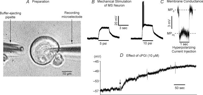
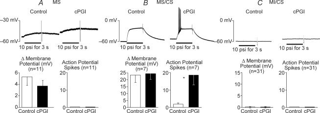
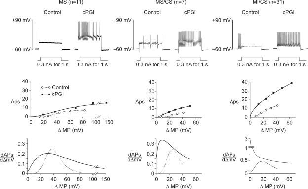
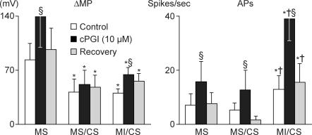
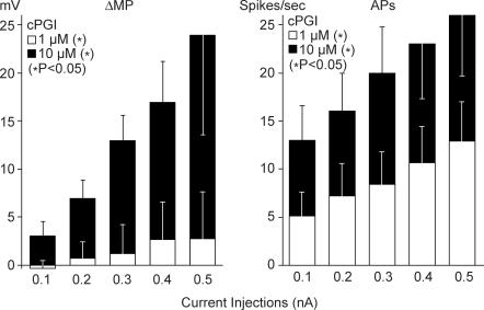
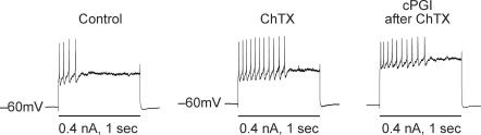
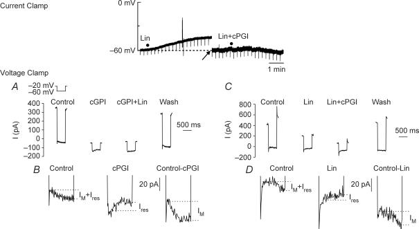
Nodose ganglion sensory neurones exert a significant reflex autonomic influence. We contrasted their mechanosensitivity, excitability and chemosensitivity in response to the stable prostacyclin (PGI2) analogue carbacyclin (cPGI) in culture. Under current clamp conditions we measured changes in membrane potential (DeltamV) and action potential (AP) responses to mechanically induced depolarizations and depolarizing current injections before and after superfusion of cPGI (1 microM and 10 microM). Chemosensitivity was indicated by augmentation of AP firing frequency and increased maximum gain of AP frequency (max. dAP/dDeltamV), during superfusion with cPGI. Results indicate that two groups of neurones, A and B, are mechanosensitive (MS) and one group, C, is mechanoinsensitive (MI). Group A shows modest depolarization without AP generation during mechanical stimulation, and no increase in max. dAP/dDeltamV, despite a marked increase in electrical depolarization with cPGI. Group B shows pronounced mechanical depolarization accompanied by enhanced AP discharge with cPGI, and an increase in max. dAP/dDeltamV. Group C remains MI after cPGI but is more excitable and markedly chemosensitive (CS) with a pronounced enhancement of max. dAP/dDeltamV with cPGI. The effect of cPGI on ionic conductances indicates that it does not sensitize the mechanically gated depolarizing degenerin/epithelial Na+ channels (DEG/ENaC), but it inhibits two voltage-gated K+ currents, Maxi-K and M-current, causing enhanced AP firing frequency and depolarization, respectively. We conclude that MS nodose neurones may be unimodal MS or bimodal MS/CS, and that MI neurones are unimodal CS, and much more CS to cPGI than MS/CS neurones. We suggest that the known excitatory effect of PGI2 on baroreceptor and vagal afferent fibres is mediated by inhibition of voltage-gated K+ channels (Maxi-K and M-current) and not by an effect on mechanically gated DEG/ENaC channels.


Figures

Figure 1
Figure 1. Experimental preparation and responses to mechanical stimulation and to superfusion with cPG1
A, the experimental set-up shows a nodose neurone with a sharp microelectrode and a buffer ejecting pipette for mechanical stimulation. B, mechanical stimulation (horizontal bars) with a jet of extracellular buffer ejected under pressure of 5 psi depolarized this neurone. With a stronger stimulation of 10 psi, the depolarization was greater and triggered transient action potentials (APs). Membrane potential (MP) returned gradually to the baseline after the end of mechanical stimulation. This neurone belonged to the mechanosensitive (MS) and chemosensitive (CS) group. C, membrane conductance (MG) was calculated from the change in MP from MPo to MPinj (ΔMP) during 1 s current injection of a hyperpolarizing current of 0.1 nA. D, the beginning of carbacyclin (cPGI) superfusion is indicated by the arrow. The maximal depolarizations caused by cPGI (10 μm) occurred over a period of approximately 5 min and averaged 6.0 ± 2.4 mV in MS neurones (n= 18) and 7.8 ± 2.0 mV in mechanoinsensitive (MI) neurones (n= 31) and were not statistically different in these two groups. The response to cPGI was sustained and a wash-out period of 15 min reversed it and allowed studies in the recovery period. A dose of 1 μm of cPGI did not cause depolarization (not shown).
Figure 2
Figure 2. Responses to mechanical stimulation (10 psi for 3 s) before and after cPGI (10 μm)
Tracings represent depolarizations (ΔmV) and action potential (AP) spikes of 3 individual neurones in response to mechanical stimulation from their resting membrane potential. Bar graphs show means ±s.e.m. of changes in membrane potential (ΔmV), and the number of AP spikes generated during mechanical stimulation before (Control – open bars) and after cPGI (filled bars). Three groups of neurones are represented. Two groups, A and B, were mechanosensitive (MS) and one group, C, was mechanoinsensitive (MI). Group A (MS) neurones (n= 11) depolarized less than Group B and did not generate AP spikes before or after cPGI. Group B (MS/CS) (n= 7) depolarized significantly more and generated AP spikes. cPGI did not increase the magnitude of mechanically induced depolarizations in either A or B but enhanced the AP generation in B. *Significant difference (P < 0.05). Because of their enhanced APs after cPGI, group B neurones were considered also chemosensitive (CS). Group C (MI/CS) neurones were mechanoinsensitive (MI) before cPGI and remained MI after cPGI. cPGI sensitized their AP responses to electrical depolarization hence their label as CS (see Fig. 3).
Figure 3
Figure 3. Current-induced depolarizations and corresponding AP generations in three groups of nodose neurones: MS, MS/CS and MI/CS
(Refer to group data in Table 2.) The upper tracings from 3 representative neurones show depolarizations and AP firings in response to current injections of 0.3 nA for 1 s before (control) and after cPGI. Resting MPs were maintained at −60 mV. The increase in APs after cPGI in an MS neurone (left tracings) was induced by a pronounced increase in depolarization. In the MS/CS neurone (middle tracings), the increase in APs after cPGI occurred without a significant increase in depolarization. In MI/CS neurone (right tracings) the increase in APs was significantly greater than in the other two neurones before cPGI and much greater after cPGI. Analyses of group data shown in Table 2 are presented here in two graphs under each of the 3 groups: MS, MS/CS and MI/CS. The upper graphs portray the sigmoid curves calculated from the AP spikes during the depolarizations induced by current injections. The circles and dashed lines represent the control mean values of ΔMP (mV) and the corresponding mean values of AP obtained during each of the 5 current injections (0.1, 0.2, 0.3, 0.4 and 0.5 nA) before cPGI (see Table 2 for s.e.m. of each value). The squares and continuous lines represent the values obtained with the same interventions after cPGI (10 μm). The lower graphs portray the derivatives dAPs/dΔMP (number s−1 mV−1), obtained in each group before (dashed lines) and after (continous line) cPGI. The following significant differences with respect to the effect of cPGI were determined by analyses of logistical equations and ANOVA (see also Table 2). In the MS group, ‘control’ depolarizations were greater than in both other groups (P < 0.05) and cPGI caused a significantly greater increase in the magnitude of depolarization without an increase in the maximal gain in APs. In the MS/CS group, cPGI did not increase the magnitude of depolarization but it caused a significant increase and a shift to the left of the maximal gain. In the MI/CS group, the control gain was higher than the other 2 groups (P < 0.05) and cPGI caused a most pronounced enhancement of the maximal gain of APs with a significant shift to the left.
Figure 4
Figure 4. Recovery from the enhanced responses to current injections (0.5 nA) caused by cPGI
Bars indicate means ±s.e.m. of ΔMP (mV) and APs in response to 0.5 nA current injections in MS (n= 11), MS/CS (n= 7) and MI/CS (n= 31) neurones that were obtained before cPGI (open bars), during cPGI (filled bars), and during recovery following the washout period (shaded bars). Although we show responses to only 0.5 nA for simplicity, differentially enhanced responses to 0.1, 0.2, 0.3 and 0.4 nA by cPGI were qualitatively similar (as shown in Table 2). *Responses in MS/CS and MI/CS neurones were significantly different (lesser for ΔMP and greater for APs) from those seen in MS neurones; †greater AP responses in MI/CS neurones than MS/CS neurones; §significant increases with cPGI compared to corresponding controls in each group which were reversed back to control values after washout periods of 15 min. Absence of symbols indicates lack of significant differences.
Figure 5
Figure 5. Dose-related enhancement of responses to electrical depolarization by 1 μmversus 10 μm cPGI
Bars indicate means ±s.e.m. of the increases in ΔMP (mV) and in APs in response to incremental current injections that were provoked by 1 μm cPGI (open bars) and by 10 μm cPGI (black bars) over corresponding responses seen in the absence of cPGI. The small dose of cPGI (1 μm) enhanced the AP responses (*P < 0.05) without causing a significant increase in ΔMP (NS) over the changes observed in the absence of cPGI. The larger dose (10 μm) caused a more pronounced enhancement of APs (*P < 0.05) with a significant increase in ΔMP (*P < 0.05) over control values without cPGI.
Figure 6
Figure 6. Effect of ChTX and cPGI on the ΔMP (mV) and APs responses to current injections
The tracings show representative responses to 3 current injections of 0.4 nA over 1 s in the same neurone from a constant MP of −60 mV. ChTX (100 nm) increased AP generation from ‘control’ of 4 spikes to 11 spikes without an increase in current-induced depolarization. After the additional superfusion of cPGI (10 μm), current-induced depolarizations increased but without a further increment in APs (10 spikes). Table 3 includes responses in 8 neurones to graded current injections that indicate that ChTX enhances APs and abrogates further increases in APs by cPGI despite the enhanced effect on depolarization (ΔMP).
Figure 7
Figure 7. Effects of cPGI and linopirdine on MP and MG under current clamp and on M-current under voltage clamp conditions
Current clamp: linopirdine (Lin) depolarized the neurone by 20.1 mV and decreased MG by 7.7 nS. When MP was adjusted back to −60 mV by a hyperpolarizing current injection (−0.58 nA) (see arrow), the addition of cPGI in the presence of Lin had no further effect on MP and MG. During Lin applications, occasional spontaneous APs could be observed. Negative deflections are responses to −0.1 nA current injections of 0.1 s used to calculate MG, which was decreased by Lin. Voltage clamp (M-current): A, tracings from one neurone show current excursions produced by a series of four, 500 ms hyperpolarizing voltage steps from a holding potential of −20 mV to −60 mV obtained before (Control), after the addition of cPGI (10 μm), after further addition of linopirdine (30 μm) (cPGI + Lin), and after the washout period (Wash). B, tracings show a tenfold magnification of the bottom part of the first and second current excursions in A while the MP was held at −60 mV (during the 500 ms duration of the hyperpolarizing voltage step) in Control (IM+Ires), and after cPGI (Ires). An electronically derived subtraction of the current after cPGI from the control current (Control–cPGI) is also shown (IM). C and D are similar to tracings A and B except that they were acquired from a different neurone to which Lin was added first followed by cPGI and the washout period. At the holding potential of −20 mV before the hyperpolarizing step, the M-current was partly activated contributing to a steady outward current (A and C, Control), which was reduced dramatically by cPGI (A, cPGI) or by linopirdine (C, Lin). During the 500 ms hyperpolarizing step at −60 mV, the deactivation of the M-current resulted in a net slow inward current relaxation (IM+Ires). After cPGI (10 μm) (A and B, cPGI) there was a reversal of the slow inward current relaxation during the hyperpolarizing step giving rise instead to a gradual outward current (Ires). The addition of linopirdine in the presence of cPGI had no significant additional effect on the current. Subtraction of the gradual outward current (Ires) from the slow net inward current relaxation in control (IM+Ires) resulted in a total inward current (IM) of over 30 pA. When linopirdine was applied first (n= 6) (C and D) the inward current relaxation in control (IM+Ires) was reversed (C and D Lin), and was not affected by further addition of cPGI in the presence of linopirdine (C, Lin + cPGI). The washout period restored both the steady outward current and the slow inward current relaxation (A and C, Wash).

Similar articles

Cited by

References

    1. Abysique A, Lucchini S, Orsoni P, Mei N, Bouvier M. Effects of alverine citrate on cat intestinal mechanoreceptor responses to chemical and mechanical stimuli. Aliment Pharmacol Ther. 1999;13:561–566. - PubMed
    1. Adaikan PG, Karim SM, Lau LC. Platelet and other effects of carboprostacyclin – a stable prostacyclin analogue. Prostaglandins Med. 1980;5:307–320. - PubMed
    1. Aiken JW, Shebuski RJ. Comparison in anesthetized dogs of the anti-aggregatory and hemodynamic effects of prostacyclin and a chemically stable prostacyclin analog, 6a-carba-PGI2 (carbacyclin) Prostaglandins. 1980;19:629–643. - PubMed
    1. Aiken SP, Zaczek R, Brown BS. Pharmacology of the neurotransmitter release enhancer linopirdine (DuP 996), and insights into its mechanism of action. Adv Pharmacol. 1996;35:349–384. - PubMed
    1. Armour JA, Huang MH, Pelleg A, Sylven C. Responsiveness of in situ canine nodose ganglion afferent neurones to epicardial mechanical or chemical stimuli. Cardiovasc Res. 1994;28:1218–1225. - PubMed

Publication types

MeSH terms

Substances

LinkOut - more resources