

Human adenovirus type 37 and the BALB/c mouse: progress toward a restricted adenovirus keratitis model (an American Ophthalmological Society thesis)
- PMID: 17471351
- PMCID: PMC1809897
Human adenovirus type 37 and the BALB/c mouse: progress toward a restricted adenovirus keratitis model (an American Ophthalmological Society thesis)
Abstract
Purpose: To establish a mouse model of adenovirus keratitis in order to study innate immune mechanisms in the adenovirus-infected cornea.
Methods: Balb/c 3T3 fibroblasts were inoculated with human adenovirus (HAdV) serotypes 8, 19, or 37 and observed for cytopathic effect. Viral growth titers were performed, and apoptosis was measured by TUNEL assay. Viral and host cytokine gene expression was assessed by RT-PCR in cultured Balb/c 3T3 fibroblasts and in the corneas of virus-injected Balb/c mice. Western blot analysis was performed to detect cell signaling in the virus-infected cornea.
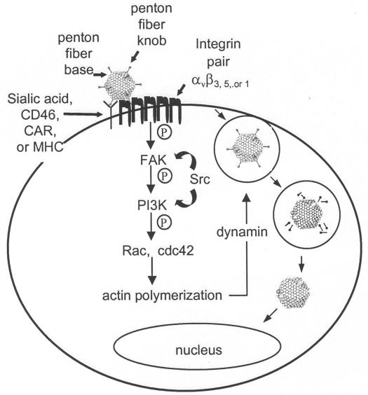
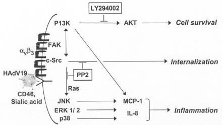
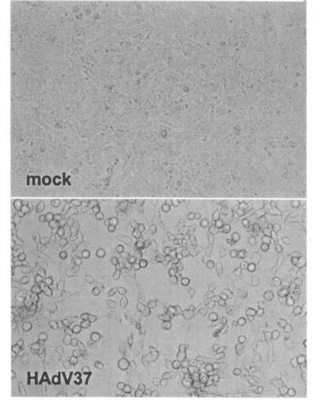
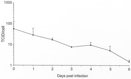
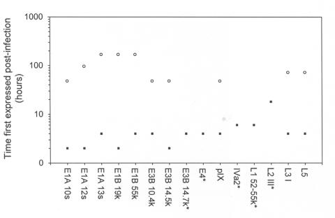
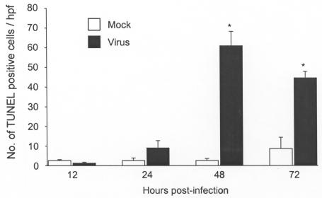
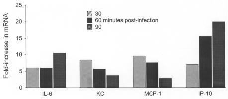
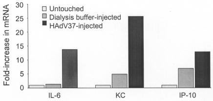
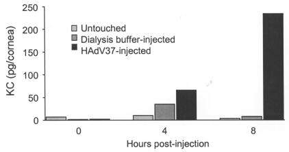
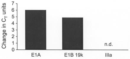
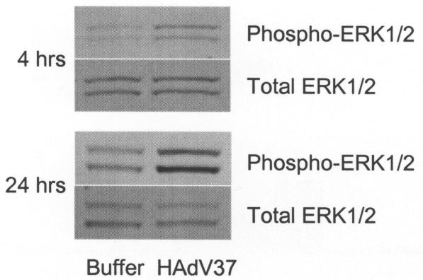
Results: Only HAdV37 induced cytopathic effect in mouse cells. Viral gene expression was limited, and viral replication was not detected. Apoptotic cell death in HAdV37-infected Balb/c cells was evident 48 and 72 hours postinfection (P < .01). MCP-1, IL-6, KC, and IP-10 mRNA levels were increased maximally by 8.4, 9.6, 10.5, and 20.0-fold, respectively, at 30 to 90 minutes after HAdV37 infection. Similar cytokine elevations were observed in the corneas of Balb/c mice 4 hours after stromal injection of HAdV37, when viral gene expression for the viral capsid protein IIIa was not detected. Western blot showed increased phosphorylation of ERK1/2 at 4 and 24 hours after corneal infection.
Conclusions: Despite limited viral gene expression, HAdV37 infection of Balb/c 3T3 fibroblasts results in increased proinflammatory gene expression. A similar pattern of cytokine expression in the corneas of HAdV37-infected Balb/c mice suggests the mouse adenoviral keratitis model may be useful for the study of early innate immune responses in the adenovirus-infected corneal stroma.
Figures
Similar articles
-
Adenovirus and the Cornea: More Than Meets the Eye.Viruses. 2021 Feb 13;13(2):293. doi: 10.3390/v13020293. Viruses. 2021. PMID: 33668417 Free PMC article. Review.
-
Adenovirus type 37 keratitis in the C57BL/6J mouse.Invest Ophthalmol Vis Sci. 2007 Feb;48(2):781-8. doi: 10.1167/iovs.06-1036. Invest Ophthalmol Vis Sci. 2007. PMID: 17251478
-
Adenovirus keratitis: a role for interleukin-8.Invest Ophthalmol Vis Sci. 2000 Mar;41(3):783-9. Invest Ophthalmol Vis Sci. 2000. PMID: 10711694
-
Chemokine CXCL1/KC and its receptor CXCR2 are responsible for neutrophil chemotaxis in adenoviral keratitis.J Interferon Cytokine Res. 2009 Oct;29(10):657-66. doi: 10.1089/jir.2009.0006. J Interferon Cytokine Res. 2009. PMID: 19642907 Free PMC article.
-
Human Adenovirus Species D Interactions with Corneal Stromal Cells.Viruses. 2021 Dec 14;13(12):2505. doi: 10.3390/v13122505. Viruses. 2021. PMID: 34960773 Free PMC article. Review.
Cited by
-
Adenovirus and the Cornea: More Than Meets the Eye.Viruses. 2021 Feb 13;13(2):293. doi: 10.3390/v13020293. Viruses. 2021. PMID: 33668417 Free PMC article. Review.
-
Resident corneal c-fms(+) macrophages and dendritic cells mediate early cellular infiltration in adenovirus keratitis.Exp Eye Res. 2016 Jun;147:144-147. doi: 10.1016/j.exer.2016.05.016. Epub 2016 May 13. Exp Eye Res. 2016. PMID: 27185163 Free PMC article.
-
Inhibition of angiogenic factor production from murine mast cells by an antiallergic agent (epinastine hydrochloride) in vitro.Mediators Inflamm. 2008;2008:265095. doi: 10.1155/2008/265095. Mediators Inflamm. 2008. PMID: 18725994 Free PMC article.
-
The 5'UTR in human adenoviruses: leader diversity in late gene expression.Sci Rep. 2017 Apr 4;7(1):618. doi: 10.1038/s41598-017-00747-y. Sci Rep. 2017. PMID: 28377580 Free PMC article.
-
Ocular tropism of respiratory viruses.Microbiol Mol Biol Rev. 2013 Mar;77(1):144-56. doi: 10.1128/MMBR.00058-12. Microbiol Mol Biol Rev. 2013. PMID: 23471620 Free PMC article. Review.
References
-
- Gordon YJ, Aoki K, Kinchington PR. Adenovirus keratoconjunctivitis. In: Pepose JS, Holland GN, Wilhelmus KR, eds. Ocular Infection & Immunity. St Louis: Mosby; 1996; 877–894.
-
- Chodosh J, Astley RA, Butler MG, Kennedy RC. Adenovirus keratitis: a role for interleukin-8. Invest Ophthalmol Vis Sci. 2000;41:783–789. - PubMed
-
- Natarajan K, Ghalayini AJ, Sterling RS, Holbrook RM, et al. Activation of focal adhesion kinase in adenovirus-infected human corneal fibroblasts. Invest Ophthalmol Vis Sci. 2002;43:2685–2690. - PubMed
-
- Natajaran K, Rajala M, Chodosh J. c-Src activation induces early IL-8 expression in adenovirus-infected human corneal fibroblasts. J Immunol. 2003;170:6234–6243. - PubMed
-
- Xiao J, Chodosh J. JNK regulates MCP-1 expression by adenovirus type 19 infected human corneal fibroblasts. Invest Ophthalmol Vis Sci. 2005;46:3777–3782. - PubMed
Publication types
MeSH terms
Substances
Grants and funding
LinkOut - more resources
Full Text Sources
Miscellaneous
