. 2007 Apr 25;27(17):4562-71.
doi: 10.1523/JNEUROSCI.5110-06.2007.

Phosphatidylinositol 3-Akt-kinase-dependent phosphorylation of p21(Waf1/Cip1) as a novel mechanism of neuroprotection by glucocorticoids

Affiliations

Phosphatidylinositol 3-Akt-kinase-dependent phosphorylation of p21(Waf1/Cip1) as a novel mechanism of neuroprotection by glucocorticoids

Christoph Harms et al. J Neurosci. .

Abstract

The role of glucocorticoids in the regulation of apoptosis remains incongruous. Here, we demonstrate that corticosterone protects neurons from apoptosis by a mechanism involving the cyclin-dependent kinase inhibitor p21(Waf1/Cip1). In primary cortical neurons, corticosterone leads to a dose- and Akt-kinase-dependent upregulation with enhanced phosphorylation and cytoplasmic appearance of p21(Waf1/Cip1) at Thr 145. Exposure of neurons to the neurotoxin ethylcholine aziridinium (AF64A) results in activation of caspase-3 and a dramatic loss of p21(Waf1/Cip1) preceding apoptosis in neurons. These effects of AF64A are reversed by pretreatment with corticosterone. Corticosterone-mediated upregulation of p21(Waf1/Cip1) and neuroprotection are completely abolished by glucocorticoid and mineralocorticoid receptor antagonists as well as inhibitors of PI3- and Akt-kinase. Both germline and somatically induced p21(Waf1/Cip1) deficiency abrogate the neuroprotection by corticosterone, whereas overexpression of p21(Waf1/Cip1) suffices to protect neurons from apoptosis. We identify p21(Waf1/Cip1) as a novel antiapoptotic factor for postmitotic neurons and implicate p21(Waf1/Cip1) as the molecular target of neuroprotection by high-dose glucocorticoids.


Figures
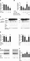
Figure 1.
Figure 1.
Corticosterone protects neurons from apoptosis via activation of the PI3-Akt-kinase pathway. A, Cortical neurons were pretreated (20 h + 1 h) with corticosterone (Cort.), before addition of AF64A (40 μm). LDH release into the medium was used as a marker of cellular disintegrity. Data are presented as increase in LDH release above the baseline (108.7 ± 2.3 at 48 h; mean values ± SEM; n = 24–88 wells; data pooled from 2–6 experiments). Treatment of naive cells with Cort. did not significantly change basal LDH release (124.5 ± 2.2 U/ml medium); *p < 0.001 versus AF64A; one-way ANOVA followed by Tukey's post hoc test. B, Receptor antagonists reverse the neuroprotective effect of Cort. Cortical neurons were pretreated with the glucocorticoid receptor antagonist mifepristone (10 μm) 30 min before corticosterone (10 μm) or the mineralocorticoid receptor antagonist spironolactone (50 μm) 30 min before corticosterone (50 μm). In naive cells, the basal LDH release at 72 h (41.4 ± 1.4) was not affected by treatment with mifepristone or Cort. plus mifepristone (35.7 ± 2.6 and 48.7 ± 3.0). Basal release of LDH (35.5 ± 2.7) also did not change in the presence of spironolactone or Cort. plus spironolactone (34.8 ± 0.6 and 46.7 ± 2.5 U/ml medium); *p < 0.05 versus AF64A; #p < 0.001 versus corresponding AF64A plus Cort. (10 or 50 μm); one-way ANOVA followed by Tukey's post hoc test (n = 16). C, Cortical neurons were pretreated with the Akt-specific inhibitor SH6 (5 μm) 21 h and Cort. (50 μm) 20 h before addition of AF64A. Cells were harvested at the indicated time points after AF64A treatment and subjected to Western blot analysis. Membranes were probed with antibodies raised against total caspase-3 and performed in duplicate. D, Cortical neurons were treated with the PI3-kinase inhibitor LY294002 (5 μm) or the Akt-kinase inhibitor SH6 (5 μm) 21 h and/or Cort. (50 μm) 20 h before AF64A (40 μm) was added. LDH release is presented as increase in LDH release above basal levels (71.3 ± 4.8) at 48 h after addition of AF64A. Treatment of naive cells with Cort. or Cort. plus LY294002 did not change basal LDH release (64.9 ± 5.8 and 67.0 ± 7.8, respectively), nor did SH6 or SH6 plus Cort. (65.8 ± 3.3 and 69.4 ± 5.1 U/ml medium); n = 12–23 wells pooled from two representative independent experiments. *p < 0.001 versus control cultures; #p < 0.001 versus AF64A plus Cort.; one-way ANOVA followed by Tukey's post hoc test. E, Cortical neurons were treated with Cort. (50 μm) or Cort. (50 μm) plus SH6 (5 μm; 1 h pretreatment). Cells were harvested at the indicated time points after Cort., and samples were analyzed by immunoblot procedure. Membranes were incubated with antibodies raised against Pi-Akt [Ser 473] and total Akt. A representative blot of three independent experiments is shown. F, Cortical neurons were pretreated with Cort. (50 μm; 20 h) before AF64A (40 μm) and harvested at 24 h after AF64A. Caspase-3 activity was measured as described previously (Harms et al., 2004). n = 5–6 wells pooled from two independent experiments. *p = 0.039 versus control cultures; #p = 0.046 versus AF64A; one-way ANOVA followed by Tukey's post hoc test. AMC, 7-Amino-4-methylcoumarin.
Figure 2.
Figure 2.
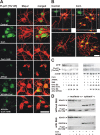
Cytoplasmic appearance of p21Waf1/Cip1 and Pi-p21 [Thr 145] phosphorylation after corticosterone is inhibited by Akt-kinase inhibition with SH6. A, Cortical neurons immobilized on microscope glass coverslips were pretreated with the Akt inhibitor SH6 (21 h) and/or 50 μm corticosterone (Cort.; 20 h) before AF64A was added. Cells were fixed 24 h after addition of AF64A, and immunocytochemistry with antibodies raised against the phosphorylated form of p21Waf1/Cip1 (Pi-p21 [Thr 145]; green) or neuronal Map-2 expression (red) was performed. Images were taken with a confocal microscope. B, The two top images represent confocal Z-series along the y–z-axis (right narrow panel) and the x–z-axis (bottom narrow panels) through the somata of Map-2-positive cells. Control cultures (left) revealed nuclear localization of Pi-p21 [Thr 145], whereas after treatment with Cort. (right), cytoplasmic localization was observed (respective green insets at higher magnification). This pattern of localization of Pi-p21 [Thr 145] is also visible in the three-dimensional reconstructions (bottom). Scale bars: A, B, 30 μm. Inset in B, twofold magnification. C, Cortical neurons were pretreated with SH6 and/or 50 μm Cort. before AF64A was added. Cells were harvested 1 h after treatment with corticosterone or 24 h after treatment with AF64A and subjected to SDS-PAGE and immunoblotting. Membranes were probed with a rabbit polyclonal antibody against p21Waf1/Cip1, Pi-p21 [Thr 145], and actin to demonstrate equal loading. Three to six independent experiments were performed. Densitometric quantification of the blots revealed an increase in the p21/actin ratio from 0.345 ± 0.051 in control cultures to 0.582 ± 0.078 in Cort.-treated cells (n = 6; p < 0.05). Similarly, the Pi-p21/actin ratio significantly increased from 0.150 ± 0.015 in control cultures to 0.373 ± 0.090 44 h after Cort. (n = 3; p < 0.05). The Cort.-induced increase in both proteins was withheld by cotreatment with SH6 (0.353 ± 0.039 and 0.083 ± 0.020, respectively). D, Cortical neurons were pretreated with SH6 (1 h before Cort.) and/or 50 μm Cort. (20 h) before AF64A was added. Cells were harvested 44 h after treatment with Cort. or 24 h after treatment with AF64A and subjected to cellular fractionation. To confirm successful separation of the compartments, HDAC1 and GAPDH were used as indicators for the nuclear and cytoplasmic fraction. Pi-p21 [Thr 145] signal was detected in cultures treated with or without the Akt kinase inhibitor SH6. Exposure times were kept constant during visualization. Two independent experiments were performed.
Figure 3.
Figure 3.
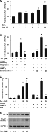
Transcriptional activation of p21Waf1/Cip1 expression is blocked by glucocorticoid receptor antagonists and the Akt-kinase inhibitor SH6. A, Cortical neurons were treated with 50 μm Cort., and cells were harvested at the indicated time points. Real time-PCR for p21Waf1/Cip1 mRNA was performed from two independent samples in duplicate and compared with actin as a housekeeping protein. Data are shown as fold induction ± SEM; *p < 0.001 versus control cultures; one-way ANOVA followed by Tukey's post hoc test. B, Receptor antagonists reverse the transcriptional activation of p21Waf1/Cip1. Cortical neurons were pretreated with the glucocorticoid receptor antagonist mifepristone (10 μm) 30 min before corticosterone (10 μm) or the mineralocorticoid receptor antagonist spironolactone (50 μm) 30 min before corticosterone (50 μm), and cells were harvested 24 h after application of corticosterone. Real time-PCR for p21Waf1/Cip1 mRNA was performed from three independent samples in duplicate and compared with actin as a housekeeping protein. Data are shown as fold induction ± SEM; *p < 0.001 versus control cultures; #p < 0.001 versus corresponding Cort. dose plus vehicle without receptor antagonist; *one-way ANOVA followed by Tukey's post hoc test. C, Cortical neurons were treated with the indicated concentration of Cort. or 5 μm SH6, and cells were harvested after 24 h. Real time-PCR for p21Waf1/Cip1 mRNA was performed from two independent samples in duplicate and compared with actin as a housekeeping protein. Data are shown as fold induction ± SEM; *p < 0.001 versus control cultures; one-way ANOVA followed by Tukey's post hoc test; #p = 0.032 versus vehicle; +p < 0.001 versus Cort. plus vehicle. D, Cortical neurons with the indicated concentrations of corticosterone and harvested 24 h later and subjected to SDS-PAGE. Membranes were incubated with either antibodies raised against Pi-p21 [Thr 145] or p21 or GAPDH as a housekeeping protein. A representative blot from two independent experiments is shown. AU, Arbitrary units.
Figure 4.
Figure 4.
p21Waf1/Cip1 loss after neuronal apoptosis is reversed by corticosterone. A, Cortical neurons were treated with 50 μm corticosterone (Cort.) and 20 h later with AF64A. Fixed cells were subjected to immunocytochemistry using anti-p21Waf1/Cip1 (green) and Map-2 (red). Scale bar, 30 μm. B, Cortical neurons were pretreated with 50 μm Cort. (20 h) and AF64A, harvested, and subjected to SDS-PAGE and protein blotting at the indicated time points after AF64A. Membranes were probed with a rabbit polyclonal antibody against p21Waf1/Cip1 and actin as a loading control. Densitometric quantification of the blots revealed an increase in the p21/actin ratio from 0.248 ± 0.068 in control cultures to 0.422 ± 0.056 in Cort.-treated cells after 32 h (n = 2; p < 0.05). AF64A induced a time-dependent loss of p21 protein levels, which was statistically significant after 24 h (control, 0.391 ± 0.061; AF64A, 0.241 ± 0.071) and was abolished by pretreatment with Cort (0.451 ± 0.065; p < 0.05; n = 3). C, Cortical neurons were treated with Cort. (50 μm) and/or cycloheximide (Chx; 1 h preincubation) before AF64A was applied. LDH release into the supernatant was measured after 72 h. The basal LDH release at 72 h was 142.1 ± 2.5; after treatment with Cort. alone, it was 152.8 ± 5.7; with Chx alone, 76.6 ± 6.0; and with Cort plus Chx, 93.5 ± 2.5 U/ml medium. The increase in LDH release was calculated from the corresponding control. *p < 0.001 versus AF64A plus Cort.; #p < 0.001 versus AF64A alone; one-way ANOVA followed by Tukey's post hoc test. n = 12–23 pooled from two independent experiments.
Figure 5.
Figure 5.
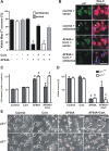
Functional inhibition of p21Waf1/Cip1 reverses the neuroprotective effect of corticosterone. A, Cortical neurons were transiently transfected with rat p21 antisense and sense oligonucleotides 24 h before pretreatment with 50 μm corticosterone (Cort.). Immunocytochemistry was performed with antibodies against p21 (green) or Map-2 (red). Transfection of either antisense or sense oligodeoxynucleotide did not influence the viability of control cultures. Nuclear morphology was assessed by Hoechst staining, and cell counts were performed as described in Materials and Methods. B, Immunocytochemical analysis of p21Waf1/Cip1 expression shows a marked loss of p21Waf1/Cip1 in AF64A- and Cort. (50 μm)-treated neurons after transfection with p21 antisense oligonucleotide with subsequent failure of the neuroprotective effect of corticosterone. Transfection with antisense oligodeoxynucleotide revealed neurons with pronounced (filled arrow), moderate (arrowhead), or weak (open arrow) effects on endogenous p21 levels after antisense transfection. Scale bar, 30 μm. C, Primary cortical neurons derived from p21Waf1/Cip1+/+ or p21Waf1/Cip1−/− mice were treated with 10 μm Cort. and 20 h later with AF64A. LDH release into the supernatant was measured after 72 h. #p < 0.001 versus corresponding control; *p < 0.001 versus AF64A plus Cort. in p21Waf1/Cip1−/− cultures; +p < 0.001 versus AF64A in p21Waf1/Cip1+/+ cultures. D, Naive cells were incubated with propidium iodide (PI; 48 h after AF64A), and digital images were taken with inverse fluorescence or phase contrast. Viable neurons were counted and shown as a percentage of all neurons as described in Materials and Methods. #p < 0.001 versus corresponding control; *p < 0.001 versus AF64A plus Cort. in p21Waf1/Cip1−/− cultures; +p < 0.001 versus AF64A in p21Waf1/Cip1+/+ cultures. E, Representative pictures in phase contrast and red fluorescent PI were merged and shown from cortical neurons derived from p21Waf1/Cip1+/+ and p21Waf1/Cip1−/− mice. Neurons were treated with Cort. (10 μm) and 20 h later with AF64A. PI staining was performed 48 h later. Scale bar, 30 μm.
Figure 6.
Figure 6.
Ectopic expression of p21Waf1/Cip1 is neuroprotective and enhances the positive effects of corticosterone. A, Rat cortical neurons were adenovirally transduced with p21Waf1/Cip1 or β-Gal as a control and/or 50 μm corticosterone (Cort.). At 20 h after transfection, AF64A was added. Cells were fixed 48 h after addition of AF64A. For immunostaining, cells were stained with the neuronal marker Map-2 (red) and the nuclear counterstain Hoechst (blue). Scale bar, 30 μm. B, Neurons were stained with antibodies raised against the neuronal marker Map-2 (red). Nuclear morphology was revealed by Hoechst staining. Cell counts of viable neurons were performed as described in Materials and Methods. *p < 0.001 versus AF64A plus Cort. plus β-Gal-treated cultures; +p < 0.05 versus AF64A plus p21Waf1/Cip1; one-way ANOVA followed by Tukey's post hoc test. C, Loss of dendritic network was prevented by additive treatment with ectopic p21Waf1/Cip1 and 50 μm Cort. Higher magnification of A revealed differences in the intensity of Map-2 staining and the dendritic network after treatment with either ectopic p21Waf1/Cip1 or p21Waf1/Cip1 plus Cort. Scale bar, 30 μm.

Similar articles

Cited by

References

    1. Ábrahám IM, Harkany T, Horvath KM, Luiten PGM. Action of glucocorticoids on survival of nerve cells: promoting neurodegeneration or neuroprotection? J Neuroendocrinol. 2001;13:749–760. - PubMed
    1. Amsterdam A, Tajima K, Sasson R. Cell-specific regulation of apoptosis by glucocorticoids. Biochem Pharmacol. 2002;64:843–850. - PubMed
    1. Asada M, Yamada T, Ichijo H, Delia D, Miyazono K, Fukumuro K, Mizutani S. Apoptosis inhibitory activity of cytoplasmic p21(Cip1/WAF1) in monocytic differentiation. EMBO J. 1999;18:1223–1234. - PMC - PubMed
    1. Bailly-Maitre B, de Sousa G, Boulukos K, Gugenheim J, Rahmani R. Dexamethasone inhibits spontaneous apoptosis in primary cultures of human and rat hepatocytes via Bcl-2 and Bcl-xL induction. Cell Death Differ. 2001;8:279–288. - PubMed
    1. Becker EB, Bonni A. Cell cycle regulation of neuronal apoptosis in development and disease. Prog Neurobiol. 2004;72:1–25. - PubMed

Publication types

MeSH terms

Substances