. 2007 Jul;27(13):4917-30.
doi: 10.1128/MCB.01789-06. Epub 2007 Apr 23.

Induction of Mxi1-SR alpha by FOXO3a contributes to repression of Myc-dependent gene expression

Affiliations

Induction of Mxi1-SR alpha by FOXO3a contributes to repression of Myc-dependent gene expression

Oona Delpuech et al. Mol Cell Biol. 2007 Jul.

Abstract

Forkhead transcription factors of the O class (FOXOs) are important targets of the phosphatidylinositol 3-kinase (PI3-kinase)/Akt pathway. FOXOs have been implicated in the regulation of cell cycle progression, oxidative stress resistance, and apoptosis. Using DNA microarrays, we analyzed the transcriptional response to FOXO3a activation by gene expression analysis in DLD-1 colon cancer cells stably expressing a FOXO3a.A3-ER fusion protein. We found that activation of FOXO3a resulted in repression of a number of previously identified Myc target genes. Furthermore, FOXO3a activation induced expression of several members of the Mad/Mxd family of transcriptional repressors, most notably Mxi1. The induction of Mxi1 by FOXO3a was specific to the Mxi1-SR alpha isoform and was mediated by three highly conserved FOXO binding sites within the first intron of the gene. Activation of FOXO3a in response to inhibition of Akt also resulted in activation of Mxi1-SR alpha expression. Silencing of Mxi1 by small interfering RNA (siRNA) reduced FOXO3a-mediated repression of a number of Myc target genes. We also observed that FOXO3a activation induced a switch in promoter occupancy from Myc to Mxi1 on the E-box containing promoter regions of two Myc target genes, APEX and FOXM1. siRNA-mediated transient silencing of Mxi1 or all Mad/Mxd proteins reduced exit from S phase in response to FOXO3a activation, and stable silencing of Mxi1 or Mad1 reduced the growth inhibitory effect of FOXO3a. We conclude that induction of Mad/Mxd proteins contributes to the inhibition of proliferation in response to FOXO3a activation. Our results provide evidence of direct regulation of Mxi1 by FOXO3a and imply an additional mechanism through which the PI3-kinase/Akt/FOXO pathway can modulate Myc function.


Figures
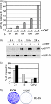
FIG. 1.
FIG. 1.
Activation of FOXO3a.A3-ER induces cell cycle arrest in DL23 cells. (A) Time course of induction of DBE-dependent transcription in response to FOXO3a activation. DL23 cells were transfected with 500 ng DBE-luc. At 24 h posttransfection, cells were treated with 100 nM 4-OHT or solvent (ethanol) for the indicated times. Values represent firefly luciferase activities relative to solvent-treated cells and are normalized to the activity of a cotransfected Renilla luciferase construct. (B) Time course of induction of cell cycle regulators. DL23 cells were treated with 100 nM 4-OHT or solvent (ethanol) for the indicated times, and total cell lysates were analyzed for the expression of p27KIP1 or cyclin A1 by immunoblotting. (C) DL23 cells were treated with solvent (light bars) or 100 nm 4-OHT (black bars) for 16 h prior to addition of 10 mM BrdU for 30 min. Proportions of BrdU-positive cells were determined by FACS analysis. (D) Colony formation assay. DL23 cells were seeded at clonal density and grown in the presence of 100 nM 4-OHT or solvent for 10 days. Cells were fixed and stained with crystal violet.
FIG. 2.
FIG. 2.
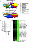
Activation of FOXO3a.A3-ER in DL23 cells results in inhibition of Myc-dependent transcription. (A) DL23 or DLD-1 cells were treated with 100 nM 4-OHT or solvent for 6 or 24 h. Relative mRNA abundance was measured by comparative hybridization to cDNA microarrays. Genes regulated in response to FOXO3a activation were grouped according to their GO annotations. The pie charts represent the number of genes in each class found to be induced or repressed in response to FOXO3a activation. (B) Repression of E-box-dependent transcription in response to FOXO3a activation. DL23 cells were transfected with 500 ng of a reporter construct containing a series of eight E-boxes (MycT-luc) or the corresponding control vector (CT-luc) prior to treatment with 4-OHT or solvent for 24 h. Values represent the relative activities of MycT-luc normalized to the activity of CT-luc and a cotransfected Renilla luciferase reporter. (C) Expression profile of Myc target genes in DL23 cells. The panel shows expression profiles of 18 cDNA clones which were found to be downregulated in response to FOXO3a.A3-ER activation and mapped to 10 Myc target genes. Green indicates downregulation relative to solvent-treated control cells. Results have been reproduced by qPCR.
FIG. 3.
FIG. 3.
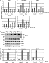
Changes in expression of members of the Myc/Max/Mxd network in response to FOXO3a activation. (A) DL23 and DLD-1 cells were treated with 100 nM 4-OHT or solvent for 6 or 24 h. Expression of Mad1, Mad2/Mxi1, Mad3, Mad4, and Max was determined by qPCR. It should be noted that CT values for Mad3 were very high, suggesting low abundance of the transcript. (B) DL23 cells were treated with 100 nM 4-OHT or solvent for 8, 10, 15, and 24 h, and total cell lysates were used to detect expression of Mxi1, Mad1, Max, and Myc by immunoblotting. Lysates from cells transfected with expression vectors for Mxi1-SRα, -SRβ, and -WR were loaded as size controls, and actin is shown as a loading control. (C) Mxi1 but not Mad1 or Mad4 is a direct target of FOXO3a. DL23 cells were stimulated with 100 nM 4-OHT in the presence of 2 μg/ml cycloheximide or dimethyl sulfoxide (DMSO) for 24 h. Expression of Mad1, Mad2/Mxi1, and Mad4 was determined by qPCR.
FIG. 4.
FIG. 4.
FOXO3a activation induces expression of Mxi1-SRα. (A) Schematic overview of the human Mxi1 locus. The human Mxi1 locus contains three transcriptional start sites that give rise to three transcripts (Mxi1-SRα, Mxi1-SRβ, and Mxi1-WR) that differ in their first exon. The locations of highly conserved DBEs are indicated by black bars. Sequences with similarity to DBEs but poorer conservation are indicated by gray bars. Luciferase reporter constructs used in panel C are depicted. (B) cDNA from DL23 or DLD-1 cells treated with 4-OHT for 6 or 24 h was used to perform isoform-specific qPCR. Values represent expression levels relative to solvent-treated controls. (C) DL23 cells were transfected with the luciferase reporter constructs indicated in panel A and treated with 100 nM 4-OHT or solvent for 24 h. Values represent luciferase activities relative to solvent-treated control and are normalized to the activity of a cotransfected Renilla luciferase construct.
FIG. 5.
FIG. 5.
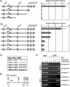
FOXO3a activates Mxi1-SRα through conserved DBEs located in the first intron. (A) Deletion analysis of the Mxi1-SRα promoter. Luciferase reporter constructs representing a series of deletions of the Mxi1-SRα downstream promoter were transfected into DL23 cells prior to induction with 4-OHT (dark bars) or solvent (light bars) for 24 h. Values represent luciferase activity relative to the activity of the full-length construct in solvent-treated cells and are normalized to the activity of a cotransfected Renilla luciferase reporter. (B) Mutation analysis of Mxi1-SRα downstream promoter. Luciferase reporter constructs in which the indicated DBEs were disrupted by mutation were transfected into DL23 cells prior to induction with 4-OHT for 24 h. Values represent luciferase activities relative to solvent-treated controls and are normalized to the activity of a cotransfected Renilla luciferase reporter. (C) Sequence of the DBEs located in the first intron of the Mxi1-SRα gene. Positions are given relative to the putative transcriptional start site. (D) Schematic representation of the regions amplified by primer pairs used in panel E. (E) FOXO binding to the DBE-containing region of the Mxi1 promoter. DL23 or MCF-10A cells expressing the FOXO3a.A3-ER fusion protein (M11) were treated with 100 nM 4-OHT for 2 or 4 h. Chromatin immunoprecipitations using a FOXO3a-specific antibody or unspecific serum (immunoglobulin G [IgG]) were analyzed using primer pairs depicted in panel D.
FIG. 6.
FIG. 6.
Chemical inhibition of Akt induces expression of Mxi1-SRα in DLD-1 cells. (A) DLD-1 cells were treated with 1 μg/ml triciribine or solvent (dimethyl sulfoxide [DMSO]) for 24 h and analyzed for expression of Mxi1-SRα, Akt, and FOXO3a by immunoblotting. Phosphorylation of Akt and FOXO3a was investigated using phospho-specific antibodies. (B) DL23 cells were transfected with a luciferase construct containing sequences of the Mxi1-SRα promoter, Mxi1-SRα(−441/+1292), and treated with 1 μg/ml triciribine or solvent (DMSO) for 24 h. Values were normalized to the activity of a cotransfected Renilla luciferase reporter.
FIG. 7.
FIG. 7.
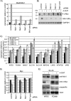
Induction of Mxi1 contributes to repression of Myc-dependent transcription in response to FOXO3a activation. (A) Silencing of Mxi1 expression using siRNA. DL23 cells were transfected with 100 nM of individual RNA oligonucleotides targeting Mxi1 (Mxi1-1, Mxi1-2, or Mxi1-3), a mixture of the three sequences (Mxi1-pool), or scrambled control prior to stimulation with 4-OHT for 24 h. Expression of Mxi1 after 24 h of 4-OHT (gray bars) or solvent (ethanol) treatment (black bars) was detected by qPCR. (B) Cells transfected with Mxi1 or scrambled control oligonucleotides were treated as for panel A. Expression levels of Mxi1-SRα and c-Myc were detected by immunoblotting. GAPDH is shown as a loading control. (C) Silencing of Mxi1 rescues repression of Myc target gene expression by FOXO3a. The same RNA samples shown in panel A were analyzed for expression of the same nine genes shown in Fig. 2C by qPCR. Values represent expression levels relative to solvent-treated control after transfection of scrambled control (back bars), a pool of three oligonucleotides targeting Mxi1 (dark gray bars), or individual oligonucleotides (light gray bars). (D) Expression of c-Myc mRNA in DL23 cells after silencing of Mxi1 by siRNA and subsequent activation of FOXO3a.A3-ER. Values represent relative mRNA abundance in 4-OHT-treated cells (gray bars) compared to solvent controls (black bars). (E) DL23 cells were treated with 100 nM 4-OHT for 24 h. Chromatin immunoprecipitations using antibodies specific for Mxi1 (top panel), c-Myc (second panel), or unspecific serum (immunoglobulin G [unspecific]) were analyzed by PCR using primers derived from the human APEX and FOXM1 promoters.
FIG. 8.
FIG. 8.
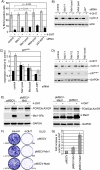
Induction of Mxi1-SRα and other Mad/Mxd proteins contributes to inhibition of proliferation by FOXO3a. (A) DL23 cells were transfected with siRNA oligonucleotides targeting Mxi1 (Mxi1-1 and Mxi1-pool), a mixture of oligonucleotides targeting all Mad/Mxd family members simultaneously (Mxd-pool), or p27KIP1. At 48 h posttransfection, cells were reseeded at lower density and treated with 100 nM 4-OHT (black bars) or solvent (gray bars) for 16 h. DNA synthesis was detected by incorporation of BrdU and FACS analysis. Values represent the means and standard deviations of three independent experiments. P values were determined by performing unpaired t tests with equal variance. (B) Cells were treated as for panel A and analyzed for expression of cyclin A by immunoblotting. Actin is shown as a loading control. (C) DL23 cells were transfected with the indicated siRNA oligonucleotides as for panel A and treated with 100 nM 4-OHT (gray bars) or solvent (black bars) for 24 h. Expression of p27KIP1 mRNA was quantified by qPCR. (D) DL23 cells were transfected with three different siRNA oligonucleotides targeting Mxi1 expression (Mxi1-1, Mxi1-2, or Mxi1-3) or a pool of all three sequences (Mxi1-pool). At 48 h posttransfection, cells were treated with 100 nM 4-OHT or solvent for 24 h. Expression of cyclin A and p27KIP1 was analyzed by immunoblotting. GAPDH is shown as a loading control. (E) DL23 cells stably expressing pMSCV-Mxi1 (left panel), pMSCV-Mad1 (right panel), or empty vector (pMSCV) were treated with 100 nM 4-OHT (+) or solvent (−) for 24 h. Expression of the FOXO3a.A3-ER fusion protein as well as Mxi1 or Mad was analyzed by immunoblotting. GAPDH is shown as a loading control. (F) DL23 cells stably expressing pMSCV-Mxi1, pMSCV-Mad1, or empty vector were seeded at clonal density and grown in the presence of 100 nM 4-OHT or solvent (ethanol) for 14 days. Cells were fixed and stained with crystal violet. Numbers of colonies are given in brackets. Note that colony number of the solvent-treated cultures was estimated by counting three segments of the plate. (G) Quantification of colony formation experiments, performed as for panel B. The values are normalized to the empty vector control and represent the results of two independent experiments.

Similar articles

Cited by

References

    1. Adhikary, S., and M. Eilers. 2005. Transcriptional regulation and transformation by Myc proteins. Nat. Rev. Mol. Cell Biol. 6:635-645. - PubMed
    1. Altomare, D. A., and J. R. Testa. 2005. Perturbations of the AKT signaling pathway in human cancer. Oncogene 24:7455-7464. - PubMed
    1. Aoki, M., H. Jiang, and P. K. Vogt. 2004. Proteasomal degradation of the FoxO1 transcriptional regulator in cells transformed by the P3k and Akt oncoproteins. Proc. Natl. Acad. Sci. USA 101:13613-13617. - PMC - PubMed
    1. Bakker, W. J., M. Blazquez-Domingo, A. Kolbus, J. Besooyen, P. Steinlein, H. Beug, P. J. Coffer, B. Lowenberg, M. von Lindern, and T. B. van Dijk. 2004. FoxO3a regulates erythroid differentiation and induces BTG1, an activator of protein arginine methyl transferase 1. J. Cell Biol. 164:175-184. - PMC - PubMed
    1. Barthel, A., D. Schmoll, K. D. Kruger, G. Bahrenberg, R. Walther, R. A. Roth, and H. G. Joost. 2001. Differential regulation of endogenous glucose-6-phosphatase and phosphoenolpyruvate carboxykinase gene expression by the forkhead transcription factor FKHR in H4IIE-hepatoma cells. Biochem. Biophys. Res. Commun. 285:897-902. - PubMed

Publication types

MeSH terms