. 2007 Jun 8;146(4):1504-12.
doi: 10.1016/j.neuroscience.2007.02.015. Epub 2007 Apr 19.

The critical role of voltage-dependent calcium channel in axonal repair following mechanical trauma

Affiliations

The critical role of voltage-dependent calcium channel in axonal repair following mechanical trauma

A Nehrt et al. Neuroscience. .

Abstract

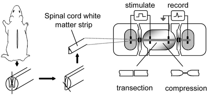
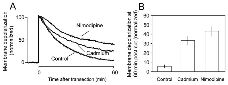
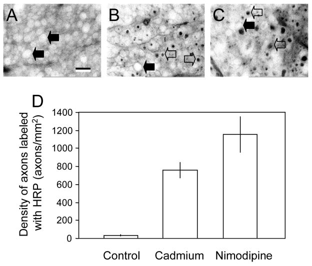
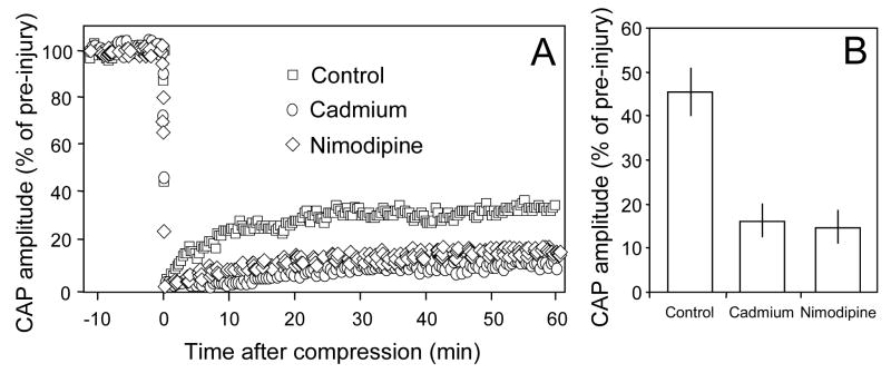
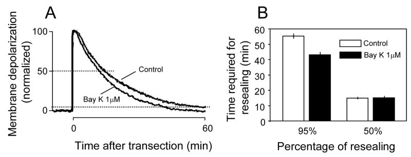
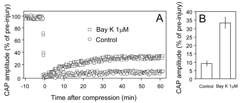
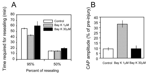
Membrane disruption following mechanical injury likely plays a critical role in the pathology of spinal cord trauma. It is known that intracellular calcium is a key factor that is essential to membrane resealing. However, the differential role of calcium influx through the injury site and through voltage dependent calcium channels (VDCC) has not been examined in detail. Using a well-established ex vivo guinea-pig spinal cord white matter preparation, we have found that axonal membrane resealing was significantly inhibited following transection or compression in the presence of cadmium, a non-specific calcium channel blocker, or nimodipine, a specific L-type calcium channel blocker. Membrane resealing was assessed by the changes of membrane potential and compound action potential (CAP), and exclusion of horseradish peroxidase 60 min following trauma. Furthermore, 1 microM BayK 8644, a VDCC agonist, significantly enhanced membrane resealing. Interestingly, this effect was completely abolished when the concentration of BayK 8644 was increased to 30 microM. These data suggest that VDCC play a critical role in membrane resealing. Further, there is likely an appropriate range of calcium influx through VDCC which ensures effective axonal membrane resealing. Since elevated intracellular calcium has also been linked to axonal deterioration, blockage of VDCC is proposed to be a clinical treatment for various injuries. The knowledge gained in this study will likely help us better understand the role of calcium in various CNS trauma, which is critical for designing new approaches or perhaps optimizing the effectiveness of existing methods in the treatment of CNS trauma.


Figures

Figure 1
Figure 1
Diagrams showing the method for guinea pig spinal cord white matter isolation (left) recording apparatus and injury technique (right). Once the spinal cord was removed from the vertebral column it was cut longitudinally down the center axis and then subdivided again to extract only ventral white matter strips. The isolated spinal cord tract is shown mounted in the middle of the center well, which is continuously perfused with oxygenated Krebs’ solution. The two end chambers containing the ends of the spinal cord segment contained isotonic KCl. These chambers were divided from the center chamber by two thinner gaps filled with flowing, isotonic sucrose solution. Stimuli were presented at the left with axonal activity recorded at the right. The two injury models used were a transection model in which the entire white matter strip was transected using micro scissors, or a crush model in which the white matter strips were injured using a pair of modified forceps.
Figure 2
Figure 2
Individual examples (A) and averaged membrane depolarization potential recordings (B) in response to transection under different calcium blocking conditions. A: There was a significant decrease in the rate of recovery of membrane repolarization when cadmium (100 μm) was added to the Krebs’ solution. Membrane repolarization was inhibited further when the specific L-type calcium channel blocker nimodipine (10 μm) was added to the Krebs’ solution. B: Average membrane depolarization at 60 minutes post injury. There was a significant increase in membrane depolarization when cadmium (33.13±5.04%, N=8) or nimodipine (43.13±4.8%, N=8) was added to the perfusion solution compared to Krebs’ alone (5.63±1.52%, n=8) (P < 0.001 when compared to control in both treatment groups, ANOVA). However, there is no significant difference between cadmium and nimodipine treated group (P>0.05, ANOVA).
Figure 3
Figure 3
Micrographs of vibratome sections showing HRP labeling (A, B, C) and the average density of labeled axons under different conditions (D). A: Micrographs of spinal cord strips stained for HRP at 60 minutes after transection in control conditions (2mM Ca2+ in Krebs’ solution, 37°C). B and C: Spinal cord strips stained with HRP at 60 minutes following transection and treated with either the general calcium channel blocker cadmium (B) or the L-type specific calcium channel blocker nimodipine (C). Notice the appearance of HRP-labeled axons in both conditions indicated by the open arrows while solid arrows indicate axons which have excluded HRP stain. D: Bar graph showing the quantification of HRP-labeled axons in different conditions. Note that there is a significant increase in the density of axons stained with HRP in axons treated with cadmium (758.4±83.92 axons/mm2, N=5) as well as nimodipine (1153±203.31 axons/mm2, n=5) compared to control conditions (34±8.32 axons/mm2, N=5). (P<0.01 when cadmium treated groups were compared to control, P<0.001 when nimodipine treated groups were compared to control, ANOVA). However, there is no statistical difference between cadmium and nimodipine treated group (P>0.05, ANOVA). Scale bar indicates a distance of 10 μm.
Figure 4
Figure 4
The response of the compound action potential amplitude to compression injury in 3 groups of spinal cord strips. A: Examples of CAP changes in response to compression injury in control conditions or in the presence of cadmium or nimodipine. The compound action potential was extinguished initially after the crush injury, but recovered and reached a new plateau level. Note CAP recovery was noticeably less when cadmium or nimodipine was present compared to control conditions. B: Average CAP recovery after injury in control and cadmium or nimodipine treated injury groups. The recovery plateau amplitude was approximately 2 ½ times greater in the control solution (45.61±5.18% of pre-injury, n=9) compared to the solution containing cadmium (16.87±3.69% of pre-injury, n=9) or nimodipine treated group (14.48±5.28%, n=4). The recovered CAP amplitude in both cadmium and nimodipine treated groups are significantly lowered that that in control (P<0.01, ANOVA). However, there is no significant difference between cadmium and nimodipine treated group (P>0.05, ANOVA).
Figure 5
Figure 5
Individual examples and averaged quantification of membrane potential recordings in response to transection injury in 2 different conditions. A: Individual examples of membrane potential recordings in response to transection injury. There was an obvious acceleration of membrane depolarization when BayK 8644 (1 μM) was added to the Krebs’ solution compared to the control condition. B: Examples of averaged quantification of membrane potential recordings in response to transection injury. The time it took for the membrane to reach 95% repolarization was significantly less in BayK 8644 (1 μM) treated groups compared to control rates (P<0.005, N=7 for both control and BayK8644 treated groups). Note that the time required for 50% of membrane repolarization was similar in both control and BayK 8644 (1 μM) treated (P>0.05).
Figure 6
Figure 6
The response of the compound action potential amplitude to compression injury in 2 groups of spinal cord strips. A: Individual examples of CAP changes in response to compression injury in control conditions or in the presence of BayK 8644 (1 μM). The compound action potential was almost extinguished after the crush injury, but recovered and reached a new plateau level. Note CAP recovery was noticeably higher when BayK 8644 (1 μM) was present compared to control conditions (2 mM Ca2+ and 37°C). B: Average CAP recovery after injury in control and BayK 8644 treated injury groups. The recovery plateau amplitude was approximately 3 times greater in the compression injury bathed in the Krebs’ solution containing BayK 8644 (1 μM) (9 ± 1.67% of pre, N=8) than in the control solution (33.25 ± 3.41% of pre, N=8). (P<0.001).
Figure 7
Figure 7
Averaged membrane potential recordings and average CAP recovery in response to transection (A) and crush injury (B) in control and in the presence of either 1 μm or 30 μm Bay K 8644. A: Data with Bay K 8644 1 μM was included here for ease of comparison. The time required for the membrane to repolarize to 50% was similar between the three groups. The time it took for the membrane to repolarize to 95% of pre injury levels was not significantly different between control conditions (55.4 ± 1.18min, N=9) and BayK 8644 (30 μM) treated groups (59.8 ± 8.2min, N=9). However, the repolarization rate was significantly faster when BayK 8644 1 μM was used compared to the other 2 groups. Note that the time required for 95% membrane resealing was similar to the time it took for control axons to reseal when BayK 8644 was increased from 1 μM to 30 μM. B: Average CAP recovery 60 minutes post crush injury was relatively equal when bathed in either the control Krebs’ solution (9 ± 1.67% of pre, N=5) or the BayK 8644 (30 μM) treated solution (9.53 ± 3.01% of pre, N=9, P>0.05). Note that the CAP recovery was reduced back to control levels when BayK 8644 was increased from 1 μM to 30 μM.

Similar articles

Cited by

References

    1. Agrawal SK, Nashmi R, Fehlings MG. Role of L- and N-type calcium channels in the pathophysiology of traumatic spinal cord white matter injury. Neuroscience. 2000;99:179–188. - PubMed
    1. Borgens RB, Shi R. Immediate recovery from spinal cord injury through molecular repair of nerve membranes with polyethylene glycol. FASEB J. 2000;14:27–35. - PubMed
    1. Borgens RB, Jaffe LF, Cohen MJ. Large and persistent electrical currents enter the transected lamprey spinal cord. Proc Natl Acad Sci USA. 1980;77:1209–1213. - PMC - PubMed
    1. Cano-Abad MF, Villarroya M, Garcia AG, Gabilan NH, Lopez MG. Calcium entry through L-type calcium channels causes mitochondrial disruption and chromaffin cell death. J Biol Chem. 2001;276:39695–39704. - PubMed
    1. Cocucci E, Racchetti G, Podini P, Rupnik M, Meldolesi J. Enlargeosome, an exocytic vesicle resistant to nonionic detergents, undergoes endocytosis via a nonacidic route. Mol Biol Cell. 2004;15:5356–5368. - PMC - PubMed

Publication types

MeSH terms

Substances