. 2007 Jun;27(12):4283-92.
doi: 10.1128/MCB.02196-06. Epub 2007 Apr 9.

DNA structure-induced recruitment and activation of the Fanconi anemia pathway protein FANCD2

Affiliations

DNA structure-induced recruitment and activation of the Fanconi anemia pathway protein FANCD2

A Sobeck et al. Mol Cell Biol. 2007 Jun.

Abstract

The Fanconi anemia (FA) pathway proteins are thought to be involved in the repair of irregular DNA structures including those encountered by the moving replication fork. However, the nature of the DNA structures that recruit and activate the FA proteins is not known. Because FA proteins function within an extended network of proteins, some of which are still unknown, we recently established cell-free assays in Xenopus laevis egg extracts to deconstruct the FA pathway in a fully replication-competent context. Here we show that the central FA pathway protein, xFANCD2, is monoubiquitinated (xFANCD2-L) rapidly in the presence of linear and branched double-stranded DNA (dsDNA) structures but not single-stranded or Y-shaped DNA. xFANCD2-L associates with dsDNA structures in an FA core complex-dependent manner but independently of xATRIP, the regulatory subunit of xATR. Formation of xFANCD2-L is also triggered in response to circular dsDNA, suggesting that dsDNA ends are not required to trigger monoubiquitination of FANCD2. The induction of xFANCD2-L in response to circular dsDNA is replication and checkpoint independent. Our results provide new evidence that the FA pathway discriminates among DNA structures and demonstrate that triggering the FA pathway can be uncoupled from DNA replication and ATRIP-dependent activation.


Figures

FIG. 1.
FIG. 1.
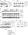
Monoubiquitination of xFANCD2 in response to dsDNA structures. (A) Presence of dsDNA fragments triggers formation of xFANCD2-L. dsDNA fragments were incubated in egg extracts for 30 min. One microliter of egg extract containing no DNA (circle with diagonal line) or 150 ng/μl dsDNA fragments was analyzed for xMre11, xFANCD2, and xFANCA by immunoblotting. Mobility shift of xMre11 was used as a positive control (6). Egg extracts contained the phosphatase inhibitor tautomycin to stabilize phosphorylated isoforms of xMre11. (B) xFANCD2-L induced by dsDNA fragments represents monoubiquitinated xFANCD2. DNA-free extracts (lanes 1, 3, 5, and 7) or extracts containing 160 ng/μl dsDNA fragments (lanes 2, 4, 6, and 8) were supplemented with either His-tagged human ubiquitin (His-Ub) (lanes 3, 4, 7, and 8) or an equal volume of H2O (lanes 1, 2, 5, and 6). Following incubation for 30 min, 1 μl of extract was directly analyzed for xFANCD2-L (lanes 1 to 4). His-tagged, ubiquitinated proteins were reisolated from DNA-containing or DNA-free extracts using Ni beads and analyzed for the presence of xFANCD2 by immunoblotting (lanes 7 and 8). Ni beads reisolated from DNA-free or DNA-containing extracts that did not contain His-Ub (lanes 5 and 6) were used as a negative control for direct binding of xFANCD2-S or -L to Ni beads. (C) xFANCD2-L is induced by linear and forked dsDNA structures. DNA structures ssDNA70 (lanes 2 and 3), dsDNA70 (lanes 4 and 5), Y-DNA70 (lanes 6 and 7), and forkDNA70 (lanes 8 and 9) were incubated in egg extracts for 30 min, and 1 μl of extract was analyzed for induction of xFANCD2-L or xMre11-PPP by immunoblotting. Symbols for each DNA substrate are explained in Table 1. For stabilization of xMre11-PPP, the phosphatase inhibitor tautomycin (T) was added to egg extracts where indicated (lanes 3, 5, 7, and 9).
FIG. 2.
FIG. 2.
xFANCD2-L associates with linear and branched dsDNA structures. (A) xFANCD2-L is recruited to DNA structures that trigger its activation. Bead-coupled ssDNA70 and forkDNA70 structures were incubated in egg extracts for 30 min, and 1 μl of extract was directly analyzed for induction of xFANCD2-L (lanes 1 to 3). Bead-DNA substrates were separated from the extract followed by analysis of bead-DNA (lanes 7 to 9) and extract supernatant (lanes 4 to 6) for xFANCD2 and xFANCA by immunoblotting. Empty beads were used as a negative control (lanes 1, 4, and 7). (B) xFANCD2-L associates with linear and branched dsDNA structures. Bead-coupled ssDNA70, dsDNA70, Y-DNA70, and forkDNA70 were incubated in egg extracts for 30 min, reisolated, and analyzed for bound xFANCD2 and xFANCA by immunoblotting. (C) xFANCD2-L associates with an HJ structure. Bead-coupled HJ-DNA68 or dsDNA68 was incubated in egg extract for 30 min, reisolated, and analyzed for xFANCA and xFANCD2 by immunoblotting. DNA substrate concentrations were as follows: HJ-DNA68 was incubated at 10 pmol/10 μl extract (lane 1), and dsDNA68 was incubated at 10 pmol/10 μl extract (lane 2), 20 pmol/10 μl extract (lane 3), or 30 pmol/10 μl extract (lane 4). The three different molar ratios between HJ-DNA68 and dsDNA68 are 1:1 (compare lanes 1 and 2), 1:2 (compare lanes 1 and 3), and 1:3 (compare lanes 1 and 4), respectively. One microliter of DNA-free extract was used as a size control for xFANCD2-S (lane 5).
FIG. 3.
FIG. 3.
DNA-induced formation and recruitment of xFANCD2-L are dependent on xFANCA but not on xMre11 or the xATR/xATRIP complex. (A) Formation of xFANCD2-L and xMre11-PPP in response to dsDNA fragments is xFANCA controlled. Egg extracts were depleted of xFANCA (lane 3), xFANCD2 (lane 4), or xMre11 (lane 1) or mock depleted (lane 2) and incubated with dsDNA fragments for 30 min. One microliter of extract was subsequently analyzed for xFANCA, xFANCD2, or xMre11 by immunoblotting. The inset (upper right corner) shows that xFANCD2 and xMre11 protein levels are not affected in an xFANCA-depleted extract (lane 2) compared to a mock-depleted extract (lane 1). (B) Recruitment of xFANCD2-L to forked DNA is dependent on xFANCA but not xATRIP. Egg extracts were depleted of xFANCA (upper panel, lanes 2 and 4) or xATRIP (lower panel, lanes 2 and 4). Mock-depleted extract was used as a control in all depletion experiments (lanes 1 and 3). Depleted extracts were incubated with bead-coupled ssDNA70 (lanes 1 and 2) or bead-coupled forkDNA70 (lanes 3 and 4) for 30 min and analyzed for the indicated proteins. One microliter of DNA-free egg extract was used as a control for protein size (lane 5).
FIG. 4.
FIG. 4.
dsDNA ends are not a major trigger for xFANC2-L formation. (A) Induction of xFANCD2-L occurs independently of dsDNA ends. Egg extracts were incubated for 30 min with 45 ng/μl of circular plasmid dsDNA (lane 1), fragmented plasmid dsDNA (lane 2), or linearized plasmid dsDNA (lane 3). DNA-free extract served as a negative control (lane 4). Following incubation, 1 μl of extract was analyzed for xFANCD2 and xMre11 by immunoblotting. (B) Linearized plasmid dsDNA induces xFANCD2-L earlier and at lower DNA concentrations than fragmented plasmid dsDNA. Egg extracts were incubated with three different concentrations of linearized or fragmented plasmid dsDNA (50 ng/μl, lanes 2, 5, and 8; 25 ng/μl, lanes 3, 6, and 9; 10 ng/μl, lanes 4 and 7). Aliquots were taken at 5 min, 15 min, and 25 min, and 1 μl of extract was analyzed for xFANCD2 and xMre11 by immunoblotting. One microliter of DNA-free extract was used as a negative control (lane 1). (C) High concentrations of circular plasmid dsDNA induce both xFANCD2-L and xMre11-PPP. Egg extracts were incubated for 30 min with increasing concentrations of circular plasmid dsDNA as indicated. Following incubation, 1 μl of extract was analyzed for xFANCD2 and xMre11 by immunoblotting. DNA-free extract incubated for 30 min served as a negative control (lane 5). (D) Low concentrations of nicked plasmid DNA induce xFANCD2-L and xMre11-PPP. Egg extracts were incubated for 30 min with 30 ng/μl (lanes 2 to 4) or 60 ng/μl (lanes 5 to 7) of linearized plasmid dsDNA (lanes 2 and 5), nicked plasmid dsDNA (lanes 3 and 6), or circular plasmid dsDNA (lanes 4 and 7). DNA-free extract served as a negative control (lane 1). Following incubation, 1 μl of extract was analyzed for xFANCD2 and xMre11 by immunoblotting. (Inset) For quality control of circular plasmid dsDNA (lanes 2 and 3), linear plasmid dsDNA (lanes 4 and 5), and nicked plasmid dsDNA (lanes 6 and 7), DNA samples (2.5 μg/lane) were analyzed by agarose gel electrophoresis. M, dsDNA size marker. (E) xFANCD2-L is induced by circular ssDNA. Egg extracts were incubated for 30 min with 160 ng/μl of circular plasmid dsDNA (lane 2) or circular plasmid ssDNA (lane 3). DNA-free extract served as a negative control (lane 1). Following incubation, 1 μl of extract was analyzed for xFANCD2 and xMre11 by immunoblotting.
FIG. 5.
FIG. 5.
Formation of xFANCD2-L in response to circular dsDNA is replication and checkpoint independent. (A and B) Circular dsDNA induces xFANCD2-L in a checkpoint-independent manner. (A) Egg extracts were incubated with 160 ng/μl circular plasmid dsDNA for 30 min in the absence (lanes 2 and 3) or presence (lanes 4 and 5) of caffeine. Where indicated, extracts were supplemented with the phosphatase inhibitor tautomycin to stabilize xMre11-PPP. Following incubation, 1 μl of extract was analyzed for xFANCD2 and xMre11 by immunoblotting. DNA-free extract was used as a negative control (lane 1). (B) Egg extracts depleted of xATRIP (lanes 2 and 4) or mock-depleted extracts (lanes 1 and 3) were incubated with 160 ng/μl circular plasmid dsDNA. Aliquots were taken at 20 min (lanes 1 and 2) and 60 min (lanes 3 and 4) and assayed for xFANCD2 by immunoblotting. DNA-free extract was used as a size control for xFANCD2 (lane 5). (C and D) Circular dsDNA induces xFANCD2-L in a replication-independent manner. (C) Egg extracts were incubated with 160 ng/μl of circular plasmid dsDNA for 30 min in the absence (lane 2) or presence (lane 3) of geminin and assayed for xFANCD2 and xMre11 by immunoblotting. DNA-free extract was used as a size control for xFANCD2 and xMre11 (lane 1). The efficiency of replication inhibition was measured in a parallel replication assay using an aliquot of the geminin-treated extract supplemented with Xenopus sperm chromatin (inset). (D) Nonactivated, M-phase egg extracts or CaCl2-activated, S-phase egg extracts were incubated with 160 ng/μl circular plasmid dsDNA. Extract aliquots were taken at the indicated time points and analyzed for xFANCD2 and xMre11.

Similar articles

Cited by

References

    1. Andreassen, P. R., A. D. D'Andrea, and T. Taniguchi. 2004. ATR couples FANCD2 monoubiquitination to the DNA-damage response. Genes Dev. 18:1958-1963. - PMC - PubMed
    1. Aquiles Sanchez, J., D. R. Wonsey, L. Harris, J. Morales, and L. J. Wangh. 1995. Efficient plasmid DNA replication in Xenopus egg extracts does not depend on prior chromatin assembly. J. Biol. Chem. 270:29676-29681. - PubMed
    1. Blow, J. J., and R. A. Laskey. 1986. Initiation of DNA replication in nuclei and purified DNA by a cell-free extract of Xenopus eggs. Cell 47:577-587. - PubMed
    1. Ciccia, A., C. Ling, R. Coulthard, Z. Yan, Y. Xue, A. R. Meetei, H. el Laghmani, H. Joenje, N. McDonald, J. P. de Winter, W. Wang, and S. C. West. 2007. Identification of FAAP24, a Fanconi anemia core complex protein that interacts with FANCM. Mol. Cell 25:331-343. - PubMed
    1. Conn, C. W., A. L. Lewellyn, and J. L. Maller. 2004. The DNA damage checkpoint in embryonic cell cycles is dependent on the DNA-to-cytoplasmic ratio. Dev. Cell 7:275-281. - PubMed

Publication types

MeSH terms