

A comparison of the influence of global functional loads vs. local contact anatomy on articular cartilage thickness at the knee
- PMID: 17418219
- PMCID: PMC2358971
- DOI: 10.1016/j.jbiomech.2007.02.005
A comparison of the influence of global functional loads vs. local contact anatomy on articular cartilage thickness at the knee
Abstract
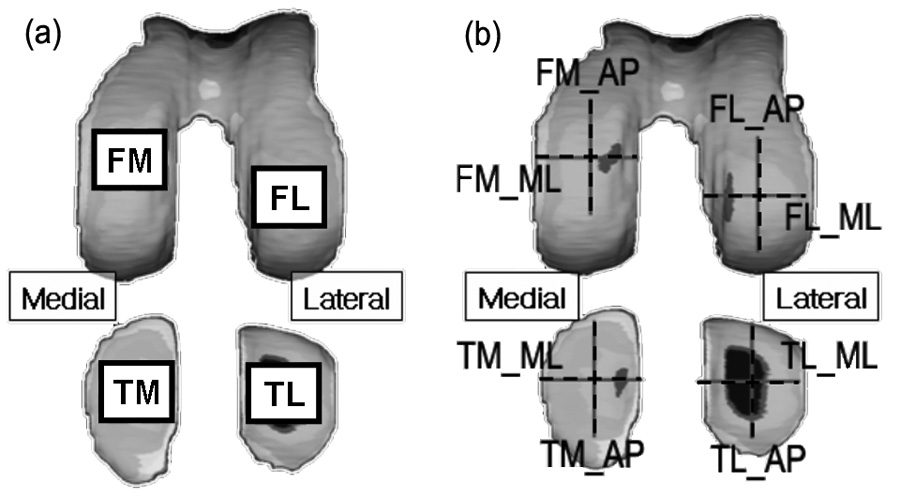
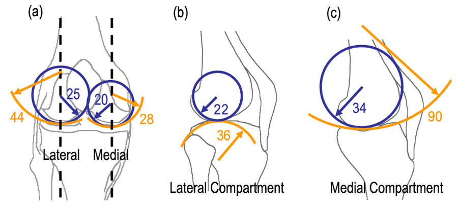
Cartilage contact geometry, along with joint loading, can play an important role in determining local articular cartilage tissue stress. Thus individual variations in cartilage thickness can be associated with both individual variations in joint loading associated with activities of daily living as well as individual differences in the anatomy of the contacting surfaces of the joint. The purpose of this study was to isolate the relationship between cartilage thickness predicted by individual variations in contact surface geometry based on the radii of the femur and tibia vs. cartilage thickness predicted by individual variations in joint loading. Knee magnetic resonance (MR) images and the peak knee adduction moments during walking were obtained from 11 young healthy male subjects (age 30.5+/-5.1 years). The cartilage thicknesses and surface radii of the femoral and tibial cartilage were measured in the weight-bearing regions of the medial and lateral compartments of three-dimensional models from the MR images. The ratio of contact pressure between the medial and lateral compartments was calculated from the radii of tibiofemoral contact surface geometries. The results showed that the medial to lateral pressure ratios were not correlated with the medial to lateral cartilage thickness ratios. However, in general, pressure was higher in the lateral than medial compartments and cartilage was thicker in the lateral than medial compartments. The peak knee adduction moment showed a significant positive linear correlation with medial to lateral thickness ratio in both femur (R(2)=0.43,P<0.01) and tibia (R(2)=0.32,P<0.01). The results of this study suggest that the dynamics of walking is an important factor to describe individual differences in cartilage thickness for normal subjects.
Figures


Similar articles
-
In-vivo time-dependent articular cartilage contact behavior of the tibiofemoral joint.Osteoarthritis Cartilage. 2010 Jul;18(7):909-16. doi: 10.1016/j.joca.2010.04.011. Epub 2010 Apr 29. Osteoarthritis Cartilage. 2010. PMID: 20434573 Free PMC article.
-
Knee adduction moment relates to medial femoral and tibial cartilage morphology in clinical knee osteoarthritis.J Biomech. 2015 Sep 18;48(12):3495-501. doi: 10.1016/j.jbiomech.2015.04.039. Epub 2015 May 6. J Biomech. 2015. PMID: 26141161
-
In vivo cartilage contact deformation in the healthy human tibiofemoral joint.Rheumatology (Oxford). 2008 Nov;47(11):1622-7. doi: 10.1093/rheumatology/ken345. Epub 2008 Sep 5. Rheumatology (Oxford). 2008. PMID: 18775967 Free PMC article. Clinical Trial.
-
In vivo morphometry and functional analysis of human articular cartilage with quantitative magnetic resonance imaging--from image to data, from data to theory.Anat Embryol (Berl). 2001 Mar;203(3):147-73. doi: 10.1007/s004290000154. Anat Embryol (Berl). 2001. PMID: 11303902 Review.
-
Articular Contact Mechanics from an Asymptotic Modeling Perspective: A Review.Front Bioeng Biotechnol. 2016 Nov 1;4:83. doi: 10.3389/fbioe.2016.00083. eCollection 2016. Front Bioeng Biotechnol. 2016. PMID: 27847803 Free PMC article. Review.
Cited by
-
The effects of defect size, orientation, and location on subchondral bone contact in oval-shaped experimental articular cartilage defects in a bovine knee model.Knee Surg Sports Traumatol Arthrosc. 2014 Jan;22(1):174-80. doi: 10.1007/s00167-012-2342-6. Epub 2012 Dec 19. Knee Surg Sports Traumatol Arthrosc. 2014. PMID: 23250200
-
Longitudinal Changes in the Total Knee Joint Moment After Anterior Cruciate Ligament Reconstruction Correlate With Cartilage Thickness Changes.J Orthop Res. 2019 Jul;37(7):1546-1554. doi: 10.1002/jor.24295. Epub 2019 Apr 25. J Orthop Res. 2019. PMID: 30977551 Free PMC article.
-
Knee Cartilage Thickness, T1ρ and T2 Relaxation Time Are Related to Articular Cartilage Loading in Healthy Adults.PLoS One. 2017 Jan 11;12(1):e0170002. doi: 10.1371/journal.pone.0170002. eCollection 2017. PLoS One. 2017. PMID: 28076431 Free PMC article.
-
Unload it: the key to the treatment of knee osteoarthritis.Knee Surg Sports Traumatol Arthrosc. 2011 Nov;19(11):1823-9. doi: 10.1007/s00167-011-1403-6. Epub 2011 Feb 5. Knee Surg Sports Traumatol Arthrosc. 2011. PMID: 21298256 Review.
-
Spatial analysis of magnetic resonance T1rho and T2 relaxation times improves classification between subjects with and without osteoarthritis.Med Phys. 2009 Sep;36(9):4059-67. doi: 10.1118/1.3187228. Med Phys. 2009. PMID: 19810478 Free PMC article.
References
-
- Andriacchi TP. Dynamics of knee malalignment. Orthopedic Clinics of North America. 1994;25:395–403. - PubMed
-
- Andriacchi TP, Alexander EJ, Toney MK, Dyrby C, Sum J. A point cluster method for in vivo motion analysis: applied to a study of knee kinematics. Journal of Biomechanical Engineering. 1998;120:743–749. - PubMed
-
- Andriacchi TP, Mündermann A, Smith RL, Alexander EJ, Dyrby CO, Koo S. A framework for the in vivo pathomechanics of osteoarthritis at the knee. Annals of Biomedical Engineering. 2004;32:447–457. - PubMed
-
- Carter DR, Orr TE, Fyhrie DP, Schurman DJ. Influences of mechanical stress on prenatal and postnatal skeletal development. Clinical Orthopaedics and Related Research. 1987;219:237–250. - PubMed
-
- Carter DR, Wong M. The role of mechanical loading histories in the development of diarthrodial joints. Journal of Orthopaedic Research. 1988;6:804–816. - PubMed
Publication types
MeSH terms
Grants and funding
LinkOut - more resources
Full Text Sources
