. 2007 Apr 15;120(Pt 8):1457-68.
doi: 10.1242/jcs.03436. Epub 2007 Mar 27.

Syntaxin 16 and syntaxin 5 are required for efficient retrograde transport of several exogenous and endogenous cargo proteins

Affiliations

Syntaxin 16 and syntaxin 5 are required for efficient retrograde transport of several exogenous and endogenous cargo proteins

Mohamed Amessou et al. J Cell Sci. .

Abstract

Retrograde transport allows proteins and lipids to leave the endocytic pathway to reach other intracellular compartments, such as trans-Golgi network (TGN)/Golgi membranes, the endoplasmic reticulum and, in some instances, the cytosol. Here, we have used RNA interference against the SNARE proteins syntaxin 5 and syntaxin 16, combined with recently developed quantitative trafficking assays, morphological approaches and cell intoxication analysis to show that these SNARE proteins are not only required for efficient retrograde transport of Shiga toxin, but also for that of an endogenous cargo protein - the mannose 6-phosphate receptor - and for the productive trafficking into cells of cholera toxin and ricin. We have found that the function of syntaxin 16 was specifically required for, and restricted to, the retrograde pathway. Strikingly, syntaxin 5 RNA interference protected cells particularly strongly against Shiga toxin. Since our trafficking analysis showed that apart from inhibiting retrograde endosome-to-TGN transport, the silencing of syntaxin 5 had no additional effect on Shiga toxin endocytosis or trafficking from TGN/Golgi membranes to the endoplasmic reticulum, we hypothesize that syntaxin 5 also has trafficking-independent functions. In summary, our data demonstrate that several cellular and exogenous cargo proteins use elements of the same SNARE machinery for efficient retrograde transport between early/recycling endosomes and TGN/Golgi membranes.


Figures
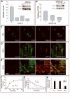
Figure 1
Figure 1
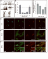
Specific inhibition of retrograde transport of STxB in Syn16 RNAi conditions. (A) Metabolic sulfation of internalized STxB-Sulf2 on HeLa cells over a 20 min period was visualized by immunoprecipitation from cells that had been transfected for 72 hours with three different siRNAs against Syn16 or with scrambled (Sc) siRNA (200 nM each). Data are expressed as percentages of STxB-Sulf2 sulfation for each experimental condition, using sulfation of STxB-Sulf2 on mock-transfected cells as the reference (set to 100%), and normalized for total sulfation counts (see Materials and Methods and Figures 2C). Inset: Western blotting confirmed the down-modulation of Syn16 in these conditions. Tubulin was used as a loading control. (B) Sulfation analysis as in (A) of STxB-Sulf2 applied to HeLa cells that had been transfected for 72 hours with increasing doses of anti-Syn16 siRNA #3. Inset: Western blot of Syn16 in the indicated conditions. (C-E) Immunofluorescence analysis of retrograde STxB transport in (C) mock-transfected control (CTL) and (D-E) Syn16 RNAi cells (siRNA sequence #3 at 200 nM). In all conditions fluorophore-labeled STxB (green) was internalized for 45 min at 37°C. Cells were fixed and labeled for Syn16 (blue), the Golgi marker CTR433 (red, D), or TfR (red, E). Under Syn16 RNAi conditions, STxB accumulation in the Golgi was reduced, and the protein appeared in peripheral structures where it co-distributed with the TfR. Space bar = 10 μm. (F-H) Specificity controls for the Syn16 RNAi. (F) EGF degradation. 2.5 ng/ml of iodine-labeled EGF was incubated for the indicated times with HeLa cells in the indicated conditions. Lysosomal degradation was measured by appearance of TCA-soluble counts in the culture medium (G) Tf recycling. Biotin-tagged Tf was internalized at 37°C for 40 min into HeLa cells in the indicated conditions. Cells were washed, and further incubated as shown at 37°C. Remaining cell associated Tf was determined by ELISA. In F-G, means of 3-4 independent experiments are shown. (H) VSVG transport in the biosynthetic and/or secretory pathway. The trafficking of newly synthesized VSVG-ts045 to the plasma membrane was analyzed by cell surface biotinylation after a temperature shift from 37°C to 32°C. BFA was used as a specificity control.
Figure 2
Figure 2
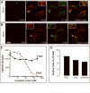
Syn5 RNAi effect on cell entry by STxB. (A) Western blot analysis of target protein expression under variable RNAi conditions. Note that for Syn5, two different isoforms of the protein were detected. Sc = scrambled. (B) Sulfation was monitored over 20 min following addition of STxB-Sulf2 to HeLa cells that had been transfected for 72 hours with 200 nM scrambled siRNA or siRNAs targeting the indicated proteins. For Syn5, two independent siRNA sequences were used that gave similar results. For Syn16, sequence #3 was used. Data are expressed as percentages of STxB-Sulf2 sulfation for each experimental condition, using sulfation of STxB-Sulf2 on mock-transfected cells as the reference (set to 100%), and normalized for total sulfation counts (see (C)). RNAi against the late endosomal Syn7 was taken as a specificity control. Means ± s.e.m. of 3-5 determinations are shown. (C) Sulfation levels on endogenous proteins under the indicated experimental conditions. (D-F) Immunofluorescence analysis of retrograde STxB transport in mock-transfected control (CTL, (D)) or Syn5 RNAi cells (E-F). In all conditions, fluorophore-labeled STxB (green) was internalized for 45 min at 37°C. Cells were fixed and labeled for Syn5 (blue), the Golgi marker CTR433 (red), or TfR (red). Under Syn5 RNAi conditions, STxB accumulation in the Golgi was reduced, and the protein appeared in peripheral structures where it codistributed with the TfR. Note that Golgi membranes were partly dispersed in Syn5 RNAi cells (CTR433). Space bar = 10 μm.
Figure 3
Figure 3
Analysis of endocytosis and TGN/Golgi-to-ER transport in Syn5 RNAi cells. (A) Glycosylation analysis. Iodinated STxB-Glyc-KDEL was incubated with mock-transfected or RNAi-transfected HeLa cells for 4 hours. The percentage of glycosylated STxB-Glyc-KDEL was determined from gels, as described (Johannes et al., 1997; Mallard and Johannes, 2003). Glycosylation of STxB-Glyc-KDEL in mock-transfected cells was taken as the maximal possible (100%) value. Means ± s.e.m. of 2-3 experiments. (B-C) The endocytosis of STxB (B) and Tf (C) was determined on the same cells. The data represent the percentage of cell-surface inaccessible biotin-tagged STxB or Tf at each time point. Means ± s.e.m. of 4 experiments. (D-E) Quantitative analysis of TGN/Golgi-to-ER transport. (D) STxB-Sulf-Glyc-KDEL was internalized into mock-transfected (± tunicamycin) or siRNA-transfected HeLa cells for 2 hours in the presence of radioactive sulfate and then chased for 4 hours at 37°C. Cells were lysed, STxB-Sulf-Glyc-KDEL immunoprecipitated, and analyzed by SDS-PAGE. The uppermost band (n°1) corresponds to the glycosylation product which disappears upon tunicamycin treatment. See text for details. (E) Quantitative analysis by Phosphoimager of the mean of 2-3 experiments ± s.e.m., as shown in (D). The data express the percentages of sulfated STxB-Sulf-Glyc-KDEL that also become glycosylated in each experimental condition.
Figure 4
Figure 4
Retrograde transport of different cargo proteins in Syn5 and Syn16 RNAi cells. (A) Constructs used for sulfation analysis. A genetic fusion of STxB with tandem sulfation sites was made as described (Mallard et al., 1998). For CTxB, a tandem sulfation site peptide was chemically coupled to primary amines. In the case of MPR, the tandem sulfation site peptide was chemically coupled to an anti-GFP antibody recognizing MPR-GFP protein in stably transfected HeLa cells (Waguri et al., 2003). (B) Sulfation analysis of sulfation site peptide-coupled CTxB and MPR. HeLa cells were incubated for 40 min with the indicated proteins in the presence of radioactive sulfate with or without BFA, followed by immunoprecipitation and autoradiography. For CTxB, the arrow indicates sulfated toxin B-subunit, while the upper bands that are visible with or without BFA, originate from contaminating endogenous sulfoproteins at the level of the load. (C-D) STxB-Sulf2, CTxB-Sulf2, and sulfation-site coupled anti-GFP-MPR antibody were incubated for 40 min in the presence of radioactive sulfate with HeLa cells that had been transfected with (C) Syn16 siRNA #3 or (D) Syn5 siRNA #1. Sulfated proteins were immunoprecipitated and analyzed by SDS-PAGE and autoradiography. Sulfation signals are expressed as normalized percentages of sulfation observed in mock-transfected control cells.
Figure 5
Figure 5
Co-distribution analysis of STxB and CTxB. (A-B) Immunofluorescence analysis of STxB (red) and CTxB (green) distribution in (A) Syn16 or (B) Syn5 RNAi cells after internalization for 45 min at 37°C. (C) Competition analysis. Iodinated STxB was incubated with HeLa cells in the presence of increasing concentrations of non-labeled CTxB or STxB. The amount of cell-associated radioactivity was determined in each condition. Note that CTxB did not displace the iodinated STxB from its receptor. (D) Binding analysis. HeLa cells were incubated on ice with 1 μM Alexa Fluor-488-labeled CTxB and 1 μM Cy3-labeled STxB. The cells were washed, fixed, and numbers of cells positive for one or the other marker were counted. Double-positive cells are roughly present at numbers as expected if GM1 and Gb3 expression are independent events.
Figure 6
Figure 6
Syn5 and Syn16 RNAi effects on cell intoxication by STx, CTx, and ricin. (A) Mock-transfected control cells or HeLa cells transfected with the indicated siRNAs were treated for 1 hour at 37°C with the indicated doses of STx. Protein biosynthesis was then determined by measuring the incorporation of radiolabeled methionine into acid-precipitable material. Data are presented as percentages of proteins biosynthesis in the indicated conditions, when compared to non-toxin treated cells. (B) Cells were treated for 1 hour with the indicated doses of STx and analyzed as described in (A). (C) Mock-transfected or RNAi-transfetced cells were treated with increasing doses of CTx, and cAMP production was measured to detect CTx arrival in the cytosol. (D) Mock-transfected or RNAi-transfetced cells were treated for 1 hour with the indicated doses of ricin, and analyzed as described in (A). (E) Intoxication of HeLa cells by unnicked (STx) or pre-nicked STx (Nicked) in control mock-transfected control cells or Syn5 RNAi cells (siSyn5). Pre-nicking did not alter the Syn5 RNAi-mediated protection of cells against the toxin. (A-E) Means of 2-4 experiments.

Similar articles

Cited by

References

    1. Amessou M, Popoff V, Yelamos B, Saint-Pol A, Johannes L. Recent methods for studying retrograde transport. Curr. Protocols Cell. Biol. in press. - PubMed
    1. Bennett MK, Garcia-Arraras JE, Elferink LA, Peterson K, Fleming AM, Hazuka CD, Scheller RH. The syntaxin family of vesicular transport receptors. Cell. 1993;74:863–873. - PubMed
    1. Bock JB, Lin RC, Scheller RH. A new syntaxin family member implicated in targeting of intracellular transport vesicles. J. Biol. Chem. 1996;271:17961–17965. - PubMed
    1. Chen YA, Scheller RH. SNARE-mediated membrane fusion. Nat. Rev. Mol. Cell Biol. 2001;2:98–106. - PubMed
    1. Dascher C, Matteson J, Balch WE. Syntaxin 5 regulates endoplasmic reticulum to Golgi transport. J. Biol. Chem. 1994;269:29363–29366. - PubMed

Publication types