Prostate cancer specific integrin alphavbeta3 modulates bone metastatic growth and tissue remodeling
- PMID: 17369840
- PMCID: PMC2753215
- DOI: 10.1038/sj.onc.1210429
Prostate cancer specific integrin alphavbeta3 modulates bone metastatic growth and tissue remodeling
Abstract
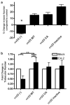
The management of pain and morbidity due to the spreading and growth of cancer within bone remains to be a paramount problem in clinical care. Cancer cells actively transform bone, however, the molecular requirements and mechanisms of this process remain unclear. This study shows that functional modulation of the alphavbeta3 integrin receptor in prostate cancer cells is required for progression within bone and determines tumor-induced bone tissue transformation. Using histology and quantitative microCT analysis, we show that alphavbeta3 integrin is required not only for tumor growth within the bone but for tumor-induced bone gain, a response resembling bone lesions in prostate cancer patients. Expression of normal, fully functional alphavbeta3 enabled tumor growth in bone (incidence: 4/4), whereas alphavbeta3 (-), inactive or constitutively active mutants of alphavbeta3 did not (incidence: 0/4, 0/6 and 1/7, respectively) within a 35-day-period. This response appeared to be bone-specific in comparison to the subcutis where tumor incidence was greater than 60% for all groups. Interestingly, bone residing prostate cancer cells expressing normal or dis-regulated alphavbeta3 (either inactive of constitutively active), but not those lacking beta3 promoted bone gain or afforded protection from bone loss in the presence or absence of histologically detectable tumor 35 days following implantation. As bone is replete with ligands for beta3 integrin, we next demonstrated that alphavbeta3 integrin activation on tumor cells is essential for the recognition of key bone-specific matrix proteins. As a result, prostate cancer cells expressing fully functional but not dis-regulated alphavbeta3 integrin are able to control their own adherence and migration to bone matrix, functions that facilitate tumor growth and control bone lesion development.
Figures


Similar articles
-
Tumor-specific expression of alphavbeta3 integrin promotes spontaneous metastasis of breast cancer to bone.Breast Cancer Res. 2006;8(2):R20. doi: 10.1186/bcr1398. Epub 2006 Apr 11. Breast Cancer Res. 2006. PMID: 16608535 Free PMC article.
-
Inhibition of alpha(v)beta3 integrin reduces angiogenesis, bone turnover, and tumor cell proliferation in experimental prostate cancer bone metastases.Clin Exp Metastasis. 2003;20(5):413-20. doi: 10.1023/a:1025461507027. Clin Exp Metastasis. 2003. PMID: 14524530
-
Prostate cancer sheds the αvβ3 integrin in vivo through exosomes.Matrix Biol. 2019 Apr;77:41-57. doi: 10.1016/j.matbio.2018.08.004. Epub 2018 Aug 8. Matrix Biol. 2019. PMID: 30098419 Free PMC article.
-
Avβ3 integrin: Pathogenetic role in osteotropic tumors.Crit Rev Oncol Hematol. 2015 Oct;96(1):183-93. doi: 10.1016/j.critrevonc.2015.05.018. Epub 2015 Jun 23. Crit Rev Oncol Hematol. 2015. PMID: 26126493 Review.
-
Dynamic process of prostate cancer metastasis to bone.J Cell Biochem. 2004 Mar 1;91(4):706-17. doi: 10.1002/jcb.10664. J Cell Biochem. 2004. PMID: 14991762 Review.
Cited by
-
Targeting bone physiology for the treatment of metastatic prostate cancer.Clin Adv Hematol Oncol. 2013 Mar;11(3):134-43. Clin Adv Hematol Oncol. 2013. PMID: 23598981 Free PMC article. Review.
-
Tumor Dormancy: Biologic and Therapeutic Implications.World J Oncol. 2022 Feb;13(1):8-19. doi: 10.14740/wjon1419. Epub 2022 Feb 8. World J Oncol. 2022. PMID: 35317328 Free PMC article. Review.
-
Thy-1 (CD90)-Induced Metastatic Cancer Cell Migration and Invasion Are β3 Integrin-Dependent and Involve a Ca2+/P2X7 Receptor Signaling Axis.Front Cell Dev Biol. 2021 Jan 12;8:592442. doi: 10.3389/fcell.2020.592442. eCollection 2020. Front Cell Dev Biol. 2021. PMID: 33511115 Free PMC article.
-
Platelets govern pre-metastatic tumor communication to bone.Oncogene. 2013 Sep 5;32(36):4319-24. doi: 10.1038/onc.2012.447. Epub 2013 Sep 15. Oncogene. 2013. PMID: 23069656 Free PMC article.
-
Integrins and metastasis.Cell Adh Migr. 2013 May-Jun;7(3):251-61. doi: 10.4161/cam.23840. Epub 2013 Apr 5. Cell Adh Migr. 2013. PMID: 23563505 Free PMC article. Review.
References
-
- Byzova TV, Kim W, Midura RJ, Plow EF. Activation of integrin alpha(V)beta(3) regulates cell adhesion and migration to bone sialoprotein. Exp Cell Res. 2000;254:299–308. - PubMed
-
- Chen YP, Djaffar I, Pidard D, Steiner B, Cieutat AM, Caen JP, et al. Ser-752—>Pro mutation in the cytoplasmic domain of integrin beta 3 subunit and defective activation of platelet integrin alpha IIb beta 3 (glycoprotein IIb-IIIa) in a variant of Glanzmann thrombasthenia. Proc Natl Acad Sci USA. 1992;89:10169–10173. - PMC - PubMed
Publication types
MeSH terms
Substances
Grants and funding
LinkOut - more resources
Full Text Sources
Other Literature Sources
Medical
Research Materials

