

Monosynaptic restriction of transsynaptic tracing from single, genetically targeted neurons
- PMID: 17329205
- PMCID: PMC2629495
- DOI: 10.1016/j.neuron.2007.01.033
Monosynaptic restriction of transsynaptic tracing from single, genetically targeted neurons
Abstract
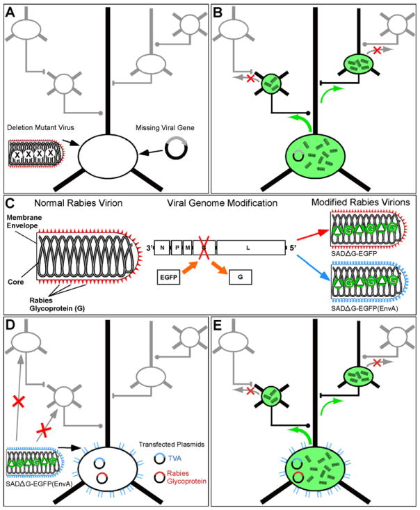
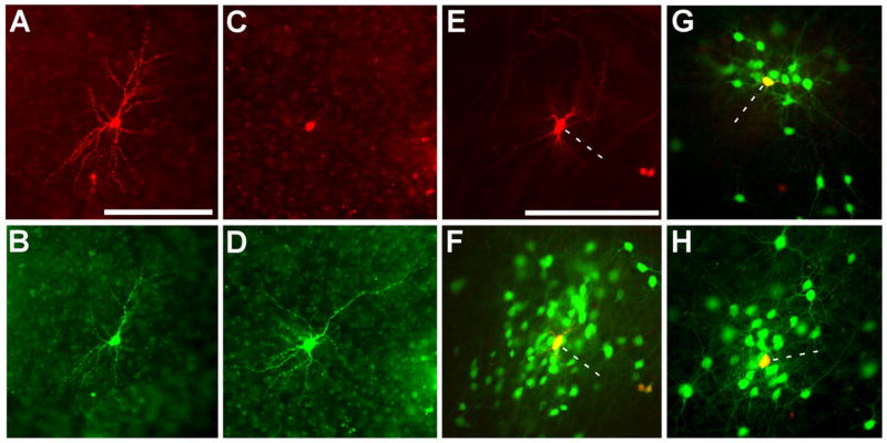
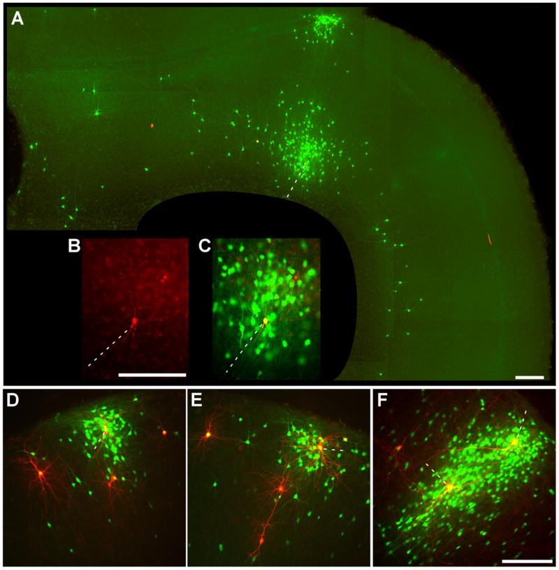
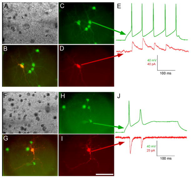
There has never been a wholesale way of identifying neurons that are monosynaptically connected either to some other cell group or, especially, to a single cell. The best available tools, transsynaptic tracers, are unable to distinguish weak direct connections from strong indirect ones. Furthermore, no tracer has proven potent enough to label any connected neurons whatsoever when starting from a single cell. Here we present a transsynaptic tracer that crosses only one synaptic step, unambiguously identifying cells directly presynaptic to the starting population. Based on rabies virus, it is genetically targetable, allows high-level expression of any gene of interest in the synaptically coupled neurons, and robustly labels connections made to single cells. This technology should enable a far more detailed understanding of neural connectivity than has previously been possible.
Figures
Similar articles
-
Rabies virus-based barcoded neuroanatomy resolved by single-cell RNA and in situ sequencing.Elife. 2024 Feb 6;12:RP87866. doi: 10.7554/eLife.87866. Elife. 2024. PMID: 38319699 Free PMC article.
-
Tracing synaptic connectivity onto embryonic stem cell-derived neurons.Stem Cells. 2012 Oct;30(10):2140-51. doi: 10.1002/stem.1185. Stem Cells. 2012. PMID: 22996827 Free PMC article.
-
Transgenic targeting of recombinant rabies virus reveals monosynaptic connectivity of specific neurons.J Neurosci. 2010 Dec 8;30(49):16509-13. doi: 10.1523/JNEUROSCI.2442-10.2010. J Neurosci. 2010. PMID: 21147990 Free PMC article.
-
G gene-deficient single-round rabies viruses for neuronal circuit analysis.Virus Res. 2016 May 2;216:41-54. doi: 10.1016/j.virusres.2015.05.023. Epub 2015 Jun 8. Virus Res. 2016. PMID: 26065596 Review.
-
The role of neural activity in cortical axon branching.Neuroscientist. 2006 Apr;12(2):102-6. doi: 10.1177/1073858405281673. Neuroscientist. 2006. PMID: 16514007 Review.
Cited by
-
From functional architecture to functional connectomics.Neuron. 2012 Jul 26;75(2):209-17. doi: 10.1016/j.neuron.2012.06.031. Neuron. 2012. PMID: 22841307 Free PMC article. Review.
-
Analyzing the structure and function of neuronal circuits in zebrafish.Front Neural Circuits. 2013 Apr 23;7:71. doi: 10.3389/fncir.2013.00071. eCollection 2013. Front Neural Circuits. 2013. PMID: 23630467 Free PMC article. Review.
-
The Insula Cortex Contacts Distinct Output Streams of the Central Amygdala.J Neurosci. 2020 Nov 11;40(46):8870-8882. doi: 10.1523/JNEUROSCI.0567-20.2020. Epub 2020 Oct 13. J Neurosci. 2020. PMID: 33051345 Free PMC article.
-
AAV9-Retro mediates efficient transduction with axon terminal absorption and blood-brain barrier transportation.Mol Brain. 2020 Oct 14;13(1):138. doi: 10.1186/s13041-020-00679-1. Mol Brain. 2020. PMID: 33054827 Free PMC article.
-
Towards Cell and Subtype Resolved Functional Organization: Mouse as a Model for the Cortical Control of Movement.Neuroscience. 2020 Dec 1;450:151-160. doi: 10.1016/j.neuroscience.2020.07.054. Epub 2020 Aug 6. Neuroscience. 2020. PMID: 32771500 Free PMC article. Review.
References
-
- Barnard RJ, Elleder D, Young JA. Avian sarcoma and leukosis virus-receptor interactions: from classical genetics to novel insights into virus-cell membrane fusion. Virology. 2006;344:25–29. - PubMed
-
- Bates P, Young JA, Varmus HE. A receptor for subgroup A Rous sarcoma virus is related to the low density lipoprotein receptor. Cell. 1993;74:1043–1051. - PubMed
-
- Borrell V, Yoshimura Y, Callaway EM. Targeted gene delivery to telencephalic inhibitory neurons by directional in utero electroporation. J Neurosci Methods. 2005;143:151–158. - PubMed
-
- Boyden ES, Zhang F, Bamberg E, Nagel G, Deisseroth K. Millisecond-timescale, genetically targeted optical control of neural activity. Nat Neurosci. 2005;8:1263–1268. - PubMed
Publication types
MeSH terms
Substances
Grants and funding
LinkOut - more resources
Full Text Sources
Other Literature Sources
Research Materials
