. 2007 Jun 20;363(1):91-103.
doi: 10.1016/j.virol.2007.01.016. Epub 2007 Feb 21.

Anti HIV-1 virucidal activity of polyamide nucleic acid-membrane transducing peptide conjugates targeted to primer binding site of HIV-1 genome

Affiliations

Anti HIV-1 virucidal activity of polyamide nucleic acid-membrane transducing peptide conjugates targeted to primer binding site of HIV-1 genome

Snehlata Tripathi et al. Virology. .

Abstract

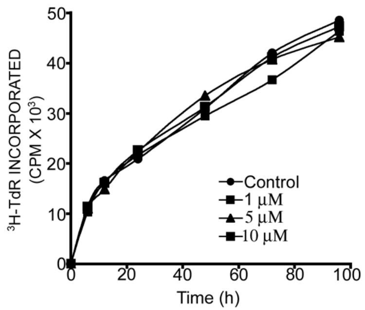
We have shown that polyamide nucleic acids (PNAs) targeted to the PBS (PNA(PBS)) and A-loop (PNA(A-loop)) sequences, when transfected into cells, inhibit HIV-1 replication by blocking the initiation of reverse transcription via destabilizing tRNA(3)(Lys) primer from the viral genome. Here we demonstrate that both PNA(PBS) and PNA(A-loop) conjugated with the membrane-transducing peptide (MTD) vectors penetratin and Tat are rapidly taken up by cells and inhibit HIV-1 replication. Moreover, MTD peptide conjugates of PNA(PBS) and PNA(A-loop) displayed potent virucidal activity against HIV-1. Brief exposure of HIV-1 virions to these conjugates rendered them noninfectious. The IC(50) values for virucidal activity were in the range of approximately 50 nM; IC(50) values for inhibition of HIV-1 replication/infection were 0.5 microM-0.7 microM. The virucidal property of these conjugates suggests that a cocktail of anti-HIV-1 PNA-MTD peptide conjugates targeting critical regions of the HIV-1 genome could serve as a prophylactic agent for inactivating HIV-1 virions after exposure to HIV-1.


Figures

Figure 1
Figure 1
The sequences of different PNA-peptide conjugates used.
Figure 2
Figure 2. Binding affinity of PNAPBS-MTD peptide conjugates to their target sequences
The gel mobility shift assay was performed to assess the binding affinity of the PNAPBS-MTD peptide conjugates in comparison to that of unconjugated naked PNAPBS control. PNAPBS-Penetratin, PNAPBS-Tat, and unconjugated naked PNAPBS were incubated at varying concentrations with 10 nM of internally 32P-labeled U5-PBS RNA transcript in the binding buffer for 30 min at room temperature. The incubated samples were then loaded on a pre-run 8% native polyacrylamide gel. The RNA: PNA complex was resolved and separated from free RNA by running at 150 V for 3 h. Lane 1 represents U5-PBS RNA alone; lanes 2 to 7 represent increasing amounts of PNAPBS or its MTD conjugates, ranging through 2.5, 5.0, 7.5, 10, 15, and 20 nM. (A) PNAPBS alone; (B) PNAPBS-Penetratin; (C) PNAPBS-Tat.
Figure 3
Figure 3. Flow cytometry analysis of uptake of anti-HIV-1 PNAPBS-MTD peptide conjugates
CEM cells (0.5 × 10⁶) were incubated with increasing concentrations of PNAPBS-Tat and PNAPBS-Penetratin for 1 min at 4°C and 37°C. After washing thoroughly with phosphate buffered saline solution, the cells were resuspended in RPMI media with 2% FCS. (A) Flow cytometry data at 37°C in the presence of propidium iodide (B) The percent of FITC-uptake per 104 cells as a function of the concentration of PNAPBS-MTD peptide conjugates; (C) The flow cytometry data after trypsin treatment for 10 min; (D) The percent uptake per 104 cells after trypsin treatment.
Figure 4
Figure 4
Fluorescence microscopy of the uptake of fluorescein-labeled PNAPBS-penetratin in CEM cells. The cells were incubated with fluorescein-labeled PNAPBS-penetratin conjugate for 30 min and then fixed and stained as described in the Materials and Methods. An aliquot of the cells was examined using the ZEISS Axiovert 200M microscope. Panel (a)-nucleus stained with DAPI; Panel (b)- cell cytoplasm showing bright green fluorescence of the fluorescent PNA; Panel (c)-nuclei merged with cell cytoplasm.
Figure 5
Figure 5. Uptake kinetics of PNAPBS-MTD peptide conjugates
CEM cells (0.5 ×10⁶) were incubated with either I125-labeled PNAPBS-Penetratin or PNAPBS-Tat conjugate for 30 sec at room temperature. The cells were filtered on glass fiber filters (GF-B) and washed with phosphate buffered saline. The incorporated radioactivity was counted in a gamma counter. The cooperative index ([S]0.9/[S]0.1) was determined by plotting the uptake of conjugate against the log of conjugate concentration as shown in the left panel. In the right panel, the log of the conjugate concentration was plotted against log V/Vmax-V to obtain a Hill plot. The value for Hill coefficient (nH), as well as [S] 0.5, denoting the concentration of conjugate at which uptake velocity is 0.5 Vmax, was determined from the slope of the Hill plot.
Figure 6
Figure 6. The antiviral efficacy of PNAPBS- and PNAA-loop-MTD peptide conjugates
T lymphocyte cells were first infected with pseudo-HIV-1 virions for 2 h in the presence of varying concentrations of either PNAPBS-MTD peptide conjugates (a) or PNAA-loop-MTD conjugates (b). Unconjugated PNA and peptides alone were included as control. The infected cells were further grown in the presence of increasing amounts of respective PNA-MTD conjugates for 48 h. The cells were harvested, washed, and lysed. An aliquot of the cell lysate was examined for luciferase activity and the percentage of inhibition of luciferase expression in treated cells was determined with respect to that in untreated control. Median-effect plots and dose-effect curves were calculated. The dose median values (IC50) for individual PNAPBS- and PNAA-Loop-MTD peptide conjugates are indicated. Antiviral efficacy was also examined in PHA stimulated peripheral blood mononuclear lymphocyte (PBMC) cells using PNAPBS-peptides (c) and the infected cells were grown for seven days before harvesting. The cell lysate was examined for luciferase activity as described above. Naked PNA and peptide alone had no effect on antiviral efficacy as naked PNA are found to be incompetent of cellular uptake and presence of peptide had no effect on viral infectivity. In both these cases the levels of luciferase expression was found to be similar to the untreated virions, therefore no values can be generated to put in the CalcuSyn software.
Figure 7
Figure 7. Dose-effect curve (IC50) of virucidal activity of individual PNAPBS- and PNAA-loop-MTD peptide conjugates
The HIV-1 virions (equivalent to 100 ng of p24) were first incubated with increasing concentrations of individual PNA-MTD peptide conjugates for 2 h in complete RPMI medium containing 10% FCS. Naked PNA and peptides alone were included as control. The pretreated virions were then used to infect the CEM cells. The infected cells were grown for 48 h and the extent of infection was determined by measuring the levels of luciferase reporter enzyme in cell lysate. Dose-effect curves and median-effect plots (IC50) were calculated. Dose-effect curves for virucidal activity of individual PNAA-Loop-MTD peptide conjugate (A), and individual PNAPBS-MTD peptide conjugates in the presence of 10% FCS (B), 60% FCS (C) and 100% FCS (D). No values could be generated for naked PNA and peptide alone.
Figure 8
Figure 8. Endogenous RT activity of HIV-1 virions after brief exposure to individual PNAPBS- and PNAA-loop-MTD peptide conjugates
The virion particles pretreated with individual PNA-MTD peptide conjugates at 250 nM (lane 1) and 500 nM (lane 2) concentrations were centrifuged at 100,000 g through a 20% sucrose cushion to separate them from free PNA-MTD peptide conjugates. The scrambled PNA-MTD-peptide conjugate was also used separately as the negative control. The virion pellets were disrupted in disruption buffer. Aliquots of the disrupted virions were examined for endogenous RT activity. The reaction products, after phenol-chloroform extraction and ethanol precipitation, were resolved on 6% denaturing polyacrylamide gel. The control (C) represents the endogenous RT activity in untreated disrupted HIV-1 virions showing products resulting from the strand transfer step. The lane marked M represents the DNA markers corresponding to 90, 250, and 490 bases.
Figure 9
Figure 9
Effect of PNA-peptide conjugate on [3H] thymidine incorporation into cellular DNA in CEM cells: CEM cells were grown in the absence and presence of PNAPBS-penetratin (1 μM, 5 μM and 10 μM). The culture media was supplemented with 10 μCi of [methyl-3H] Thymidine/mL. The cells were harvested at 6, 12, 18, 24, 48, 72 and 96 h. The cells were assayed for the amount of [3H]-Thymidine (TdR) incorporated into DNA by using TCA precipitation assay.

Similar articles

Cited by

References

    1. Argyris EG, Dornadula G, Nunnari G, Acheampong E, Zhang C, Mehlman K, Pomerantz RJ, Zhang H. Inhibition of endogenous reverse transcription of human and nonhuman primate lentiviruses: Potential for development of lentivirucides. Virology. 2006 doi: 10.1016/j.viro.2006.06.014. - DOI - PMC - PubMed
    1. Bestman-Smith J, Piret J, Desormeaux A, Tremblay MJ, Omar RF, Bergeron MG. Sodium lauryl sulfate abrogates human immunodeficiency virus infectivity by affecting viral attachment. Antimicrob Agents Chemother. 2001;45:2229–2237. - PMC - PubMed
    1. Bourinbaiar AS, Lee-Huang S. Comparative in vitro study of contraceptive agents with anti-HIV activity: gramicidin, nonoxynol-9, and gossypol. Contraception. 1994;49:131–137. - PubMed
    1. Brooks H, Lebleu B, Vives E. Tat peptide-mediated cellular delivery: back to basics. Adv Drug Deliv Rev. 2005;57:559–577. - PubMed
    1. Chaubey B, Tripathi S, Ganguly S, Harris D, Casale RA, Pandey VN. A PNA-transportan conjugate targeted to the TAR region of the HIV-1 genome exhibits both antiviral and virucidal properties. Virology. 2005;331:418–428. - PubMed

Publication types

MeSH terms