. 2007 Mar 7;26(5):1279-91.
doi: 10.1038/sj.emboj.7601584. Epub 2007 Feb 22.

A minimal CENP-A core is required for nucleation and maintenance of a functional human centromere

Affiliations

A minimal CENP-A core is required for nucleation and maintenance of a functional human centromere

Yasuhide Okamoto et al. EMBO J. .

Abstract

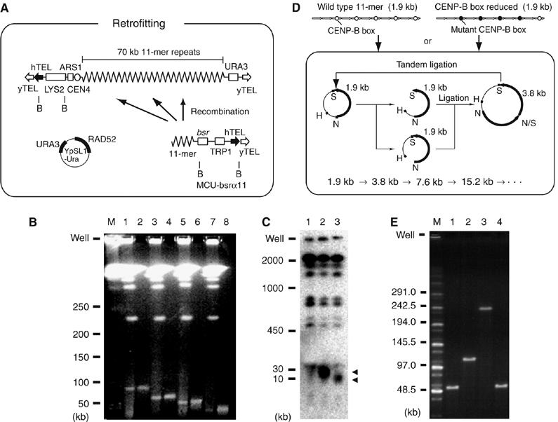
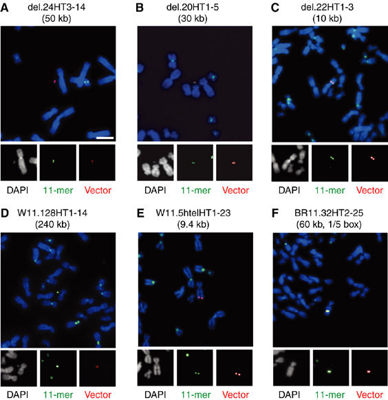
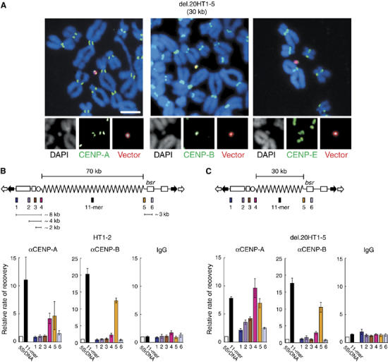
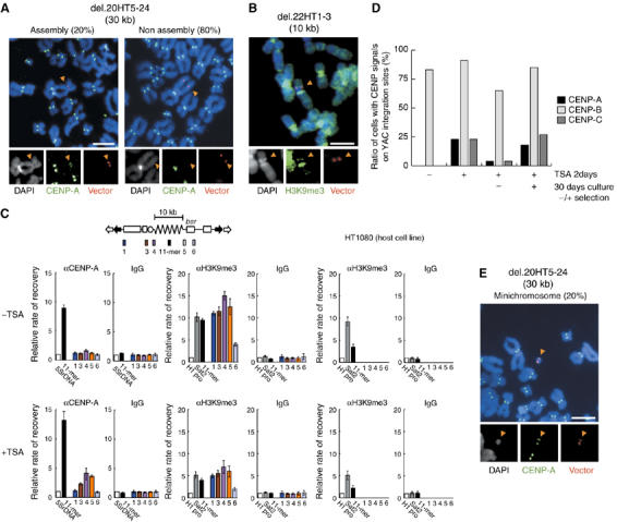
Chromatin clusters containing CENP-A, a histone H3 variant, are found in centromeres of multicellular eukaryotes. This study examines the ability of alpha-satellite (alphoid) DNA arrays in different lengths to nucleate CENP-A chromatin and form functional kinetochores de novo. Kinetochore assembly was followed by measuring human artificial chromosome formation in cultured human cells and by chromatin immunoprecipitation analysis. The results showed that both the length of alphoid DNA arrays and the density of CENP-B boxes had a strong impact on nucleation, spreading and/or maintenance of CENP-A chromatin, and formation of functional kinetochores. These effects are attributed to a change in the dynamic balance between assembly of chromatin containing trimethyl histone H3-K9 and chromatin containing CENP-A/C. The data presented here suggest that a functional minimum core stably maintained on 30-70 kb alphoid DNA arrays represents an epigenetic memory of centromeric chromatin.


Figures

Figure 1
Figure 1
Construction of YACs and BACs with shorter or longer alphoid DNA inserts. (A) The method for construction of deleted YACs using homologous recombination in yeast. B indicates the BamHI site. (B) Constructs were analyzed by PFGE, followed by ethidium bromide staining. Gel-purified YAC DNA was analyzed in lanes 2, 4, 6 and 8. Lanes 1 and 2, α7C5htel; lanes 3 and 4, del.24; lanes 5 and 6, del.20; lanes 7 and 8, del.22; M indicates DNA size marker. (C) HT1080 genomic DNA transformed with the YAC constructs were digested with BamHI and analyzed by PFGE, followed by Southern blot using a 32P-labeled 11-mer probe. The arrowheads indicate input alphoid DNAs. Lane 1, HT1080; lane 2, del.20HT1–5; lane 3, del.22HT1–3. (D) BACs with alphoid DNA with wild-type or mutant CENP-B boxes were constructed as shown. H, N and S indicate HindIII, NheI and SpeI sites, respectively. (E) BACs were digested with NheI and SpeI and analyzed by PFGE, followed by ethidium bromide staining. Lane 1, pWTR11.32; lane 2, pWTR11.64; lane 3, pWTR11.128; lane 4, pCBB11.1.32. M indicates DNA size marker.
Figure 2
Figure 2
FISH analysis of HAC formation and integration. Metaphase cells were prepared from cell lines with non-integrated (A: del.24HT3–14, B: del.20HT1–5 and D: W11.128HT1–14) and integrated YACs/BACs (C: del.22HT1–3, E: W11.5htelHT1–23 and F: BR11.1.32HT2–25). FISH analysis was carried out using probes for the α21-I alphoid 11-mer (green) and YAC vector sequences (red). Chromosomes were counterstained with DAPI (white and blue in merged panels). Scale bar, 10 μm.
Figure 3
Figure 3
Analysis of CENP-A chromatin in HACs. (A) Indirect immunostaining and FISH analysis of del.20HT1–5. Green fluorescent secondary antibodies were used to detect CENP-A, -B or -E. Red fluorescent signal indicates hybridization to YAC vector probes. Scale bar, 10 μm. (B, C) ChIP and real-time PCR analysis of HAC cell lines 7C5HT1–2 and del.20HT1–5. The diagrams of YAC structures indicate one unit of the concatenated multimers found in HACs. Relative enrichment of YAC regions 1–6 was quantified as described. Results were normalized to 5S ribosomal DNA and are presented as a bar graph. Normal IgG was used as a negative control. Error bars indicate s.e.m. (n=3). Enrichment of CENP-A in regions 4 and 5 in (B) and in regions 2–5 in (C) was statistically significant (P<0.01) by χ2 test, relative to values with normal IgG; values for regions 1, 2, 3 and 6 in (B), and regions 1 and 6 in (C) were not statistically significant (P>0.10) by χ2 test.
Figure 4
Figure 4
Analysis of CENP-A and H3K9me3 chromatin in alphoid DNA arrays. (A) Detection of CENP-A in 30 kb alphoid DNA arrays in del.20HT5–24 cell line. (B) Detection of H3K9me3 in 10 kb alphoid DNA array in del.22HT1–3 cell line. (A, B) Indirect immunostaining (green) and FISH (red) were carried out. Arrowheads indicate YAC integration site. Scale bar, 10 μm. (C) ChIP and real-time PCR analysis of CENP-A and H3K9me3 in 10 kb alphoid DNA array in del.22HT1–3 cells and parental HT1080 cells before or after treatment with TSA. The diagram of the YAC structure indicates one unit of the concatenated multimers found at ectopic chromosomal insertion sites. Enrichment of CENP-A or H3K9me3 was calculated as described. Results were normalized to 5S ribosomal DNA in CENP-A panels and H1 promoter in H3K9me3 panels, and are presented as a bar graph. Normal IgG was used as a negative control. DNA probes were for 11-mer, Sat2, regions 1–6 on the YAC and the H1 promoter. Error bars indicate s.e.m (n=3). Enrichment of CENP-A in regions 3, 4 and 5 was statistically significant in TSA treated cells (P<0.01) by χ2 test, relative to values with normal IgG; values in regions 1 and 6 were not statistically significant (P>0.10) by χ2 test. (D) Cells were treated with TSA for 2 days, incubated for 30 days in the presence or absence of blastocidin S and analyzed for CENPs assembly by immunofluorescence and FISH. The fraction of del.22HT1–3 cells positive for CENPs assembly are indicated. (E) Stable reformed minichromosomes (20% of cells) were detected by activated centromere signal (CENP-A, green) in the 30 kb alphoid DNA array in TSA-treated del.20HT5–24 cells. YAC probe is shown in red. Scale bars, 10 μm.
Figure 5
Figure 5
ChIP analysis of CENP-A, CENP-B and H3K9me3 in alphoid DNA arrays with wild-type or mutant CENP-B boxes. (A) Schematic diagram of synthetic alphoid DNA 11-mers. (a) Synthetic wild-type 11-mer constructs with different lengths. (b) Synthetic 11-mer with mutant CENP-B boxes. (c) Synthetic 11-mer with four mutant CENP-B boxes. The primer sites (arrow heads) used for the competitive PCR are shown. N/S indicates cohesive end of the ligated NheI and SpeI sites. The 17-bp motif of CENP-B box is shown as wild-type or mutant. The EcoRV site exists only on the mutant CENP-B box. (B) Control for competitive PCR. W0210R-1 cells carrying wild-type CENP-B boxes and M1319 cells carrying mutant CENP-B boxes were mixed in the indicated ratios (16:1–1:16). Competitive PCR was carried out with genomic DNA, and EcoRV-digested PCR products were analyzed by electrophoresis. (C) ChIP and competitive PCR analyses assays were carried out as described. I indicates input DNA. Antibodies to CENP-A, CENP-B or normal mouse IgG were used for immunoprecipitation. Fold enrichment was calculated by the ratio of the upper and lower bands and normalized to input DNA. (a), (b) and (c) are the DNA fragments corresponding to the alphoid constructs (a), (b) and (c), respectively, in panel A. (D) ChIP and real-time PCR analysis of H3K9me3 assembly on the synthetic alphoid DNA array in W11.128HT4–3, W11.128HT1–14 and W0210R-8 cells. Enrichment of H3K9me3 was calculated as described. Results were normalized to the H1 promoter DNA and are presented as a bar graph. Normal IgG was used as a negative control. DNA probes were for Sat2, endogenous 11-mer, synthetic 11-mer and the H1 promoter. Error bars indicate s.e.m. (n=3).
Figure 6
Figure 6
Hypothetical models for assembly and spreading of CENP-A chromatin. (A) Chromatin assembly on the 30 kb alphoid HAC. The CENP-A chromatin (red line) spread into the YAC vector arm regions (red arrow) following nucleation of the assembly on the insert 30 kb alphoid DNA array, and was maintained. Therefore, the functional centromere core of CENP-A chromatin (at least 30 kb plus spreading) may exist as an epigenetic memory. (B) Chromatin assembly on the short 10 kb alphoid integration site. The multimer of the 10 kb alphoid YACs acquired strong heterochromatic H3K9me3 state (blue line) at its integration site. CENP-A chromatin could nucleate the assembly if the heterochromatin structure was compulsorily broken by TSA treatment. However, this kind of CENP-A assembly without fulfilling the functional length was unstable, and thus, dynamically encroached by the spreading of H3K9me3 (blue arrow) under the non-selective culture. (C) Chromatin assembly on the CENP-B box-reduced alphoid BAC at the integration site. CENP-A nucleosomes can assemble on an alphoid unit containing only one CENP-B box in the 11-mer unit. However, such a CENP-A nucleosome cannot spread and generate a functional centromere core unit on the 60 kb CENP-B box-reduced alphoid DNA. (D) Chromatin assembly on the long 240 kb alphoid HAC or the integration site. The 240 kb alphoid BAC also has an ability to form a HAC, but a high level of H3K9me3 also covered the integration site of the 240 kb alphoid BAC.

Similar articles

Cited by

References

    1. Amor DJ, Choo KH (2002) Neocentromeres: role in human disease, evolution, and centromere study. Am J Hum Genet 71: 695–714 - PMC - PubMed
    1. Amor DJ, Kalitsis P, Sumer H, Choo KH (2004) Building the centromere: from foundation proteins to 3D organization. Trends Cell Biol 14: 359–368 - PubMed
    1. Ando S, Yang H, Nozaki N, Okazaki T, Yoda K (2002) CENP-A, -B, and -C chromatin complex that contains the I-type alpha-satellite array constitutes the prekinetochore in HeLa cells. Mol Cell Biol 22: 2229–2241 - PMC - PubMed
    1. Baer M, Nilsen TW, Costigan C, Altman S (1990) Structure and transcription of a human gene for H1 RNA, the RNA component of human RNase P. Nucleic Acids Res 18: 97–103 - PMC - PubMed
    1. Basu J, Stromberg G, Compitello G, Willard HF, Van Bokkelen G (2005) Rapid creation of BAC-based human artificial chromosome vectors by transposition with synthetic alpha-satellite arrays. Nucleic Acids Res 33: 587–596 - PMC - PubMed

Publication types

MeSH terms