. 2007 Apr;18(4):1437-46.
doi: 10.1091/mbc.e06-07-0593. Epub 2007 Feb 14.

Cytokine stimulation promotes glucose uptake via phosphatidylinositol-3 kinase/Akt regulation of Glut1 activity and trafficking

Affiliations

Cytokine stimulation promotes glucose uptake via phosphatidylinositol-3 kinase/Akt regulation of Glut1 activity and trafficking

Heather L Wieman et al. Mol Biol Cell. 2007 Apr.

Abstract

Cells require growth factors to support glucose metabolism for survival and growth. It is unclear, however, how noninsulin growth factors may regulate glucose uptake and glucose transporters. We show that the hematopoietic growth factor interleukin (IL)3, maintained the glucose transporter Glut1 on the cell surface and promoted Rab11a-dependent recycling of intracellular Glut1. IL3 required phosphatidylinositol-3 kinase activity to regulate Glut1 trafficking, and activated Akt was sufficient to maintain glucose uptake and surface Glut1 in the absence of IL3. To determine how Akt may regulate Glut1, we analyzed the role of Akt activation of mammalian target of rapamycin (mTOR)/regulatory associated protein of mTOR (RAPTOR) and inhibition of glycogen synthase kinase (GSK)3. Although Akt did not require mTOR/RAPTOR to maintain surface Glut1 levels, inhibition of mTOR/RAPTOR by rapamycin greatly diminished glucose uptake, suggesting Akt-stimulated mTOR/RAPTOR may promote Glut1 transporter activity. In contrast, inhibition of GSK3 did not affect Glut1 internalization but nevertheless maintained surface Glut1 levels in IL3-deprived cells, possibly via enhanced recycling of internalized Glut1. In addition, Akt attenuated Glut1 internalization through a GSK3-independent mechanism. These data demonstrate that intracellular trafficking of Glut1 is a regulated component of growth factor-stimulated glucose uptake and that Akt can promote Glut1 activity and recycling as well as prevent Glut1 internalization.


Figures
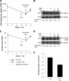
Figure 1.
Figure 1.
IL3 regulates intracellular trafficking of Glut1 to the cell surface independent of new protein synthesis. (A) Glucose uptake was measured in FL5.12 cells in IL3 or cultured in IL3-free media for 4 h followed by 1 h with IL3 restored. CHX at 10 μg/ml was added to some samples at 3 h. (B) Total endogenous Glut1 expression was measured by immunoblot with actin as a loading control (R, IL3 restored). (C) FL5.12 cells expressing exofacially FLAG-tagged Glut1 were cultured as described in A. Mean cell surface fluorescence for FLAG-Glut1 was determined at indicated times. (D) Total FLAG-Glut1 levels were analyzed by immunoblot with actin as a loading control (R, IL3 restored). (E) Cell surface FLAG-Glut1 was normalized to total FLAG expression. Standard deviations of triplicate samples are shown. Total levels of Glut1 and FLAG were normalized to actin and are shown below the respective immunoblots. Representative results are shown for three or more repeated experiments.
Figure 2.
Figure 2.
Growth factor attenuates Glut1 internalization and promotes Rab11a-dependent recycling of intracellular Glut1. (A) FLAG-Glut1 cells were cultured in IL3 or withdrawn from IL3 for 6 h. Cells were stained with anti-FLAG antibody, washed, and cultured at 37°C for indicated times before staining with fluorescent secondary antibody. (B and C) FLAG-Glut1 cells were transiently transfected with control or dominant-negative Rab11a (Rab11aS25N/dnRab11a) vector and cultured in IL3 or in the absence of IL3 for 6 h. (B) Total FLAG-Glut1 expression levels were measured by immunoblot with actin as a loading control. Total levels of FLAG were normalized to actin and are shown below the immunoblot. (C) Surface FLAG-Glut1 levels were measured by flow cytometry and normalized to total FLAG expression. Mean and SD of triplicate samples are shown. Asterisk (*) indicates p < 0.005 within the experiment.
Figure 3.
Figure 3.
PI3K activity is required to promote Glut1 cell surface localization. FLAG-Glut1–expressing cells were cultured in the presence or absence of IL3 for 8 h with a vehicle control or 10 μM LY294002. (A) Cells were stained with a FLAG antibody, and mean surface levels were analyzed with flow cytometry. (B) Total FLAG expression was measured by immunoblot and Bcl-2 expression was measured for loading control. (C) FLAG-Glut1 cells were cultured in the absence of IL3 for 3 h. Vehicle control, 10 μM LY294002, and 10 μg/ml CHX were added 1 h before readdition of IL3. After an additional hour, cells were stained with FLAG antibody, and surface levels of FLAG-Glut1 were compared before and after IL3 add-back by flow cytometry. The differences in mean fluorescence of surface FLAG-Glut1 before and after IL3 readdition are shown. Mean fluorescence and SD of triplicate samples are shown. Asterisk (*) indicates p value <0.05 within the experiment. Representative results are shown for three or more repeated experiments.
Figure 4.
Figure 4.
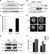
Activated Akt regulates Glut1 trafficking to maintain surface Glut1 levels after IL3 withdrawal. (A) FL5.12 cells were withdrawn from IL3 for 6 h, restimulated with IL3 for various times, and immunoblotted for phospho-Akt, total Akt1 and actin. (B) Cells were transiently transfected with control or myrAkt vectors and cultured in the presence or absence of IL3 for 6 h. Phosphorylation levels of both mTOR and Akt were analyzed by immunoblot with actin as a loading control. (C) FL5.12 cells were transfected with either Bcl-xL or myrAkt expression vectors and cultured in the presence of IL3 for 18 h followed by a growth factor-withdrawal for 24 h and glucose uptake was measured. (D) Control or myrAkt-expressing cells were transfected with GFP-Glut1, and then they were cultured in the presence or absence of IL3 for 8 h, and GFP was visualized by fluorescence microscopy. Bar, 10 μm. (E and F) FLAG-Glut1 cells were transiently transfected with control vector (C), myrAkt (A), or a phospho-mimetic aspartate mutant of Akt (DD). Cells were cultured in the presence and absence of IL3 for 6 h. (E) Total FLAG-Glut1 and Akt1 levels were analyzed by immunoblot with actin as a loading control. (F) Surface levels of FLAG-Glut1 were measured by flow cytometry. Standard deviations of triplicate samples are shown. Representative results are shown for three or more repeated experiments.
Figure 5.
Figure 5.
mTOR/RAPTOR does not regulate Glut1 cell surface localization but promotes Glut1 activity. (A and B) FL5.12 cells stably expressing FLAG-Glut1 were transfected with either Bcl-xL or myrAkt expression vectors and cultured in the presence of IL3 for 18 h. A vehicle control or 25 nM rapamycin was added at the time of growth factor withdrawal, and cells were cultured in the absence of IL3 for an additional 24 h. Mean surface levels of TfR (A) and FLAG-Glut1 (B) levels were analyzed with flow cytometry. (C and D) FL5.12 cells were transfected and cultured as described in A. Total endogenous Glut1 was measured via immunoblot with actin as a loading control (C), and glucose uptake was measured (D). Mean and SD of triplicate samples are shown. Asterisk (*) indicates p < 0.05 within the experiment. Representative results are shown for three or more repeated experiments.
Figure 6.
Figure 6.
GSK3 activity promotes Glut1 surface localization, but it does not affect internalization. (A and B) FLAG-Glut1–expressing cells were transfected with control or myrAkt vectors. Cells were treated with a vehicle control or 10 μM SB216763 for 12 h before they were cultured in the presence or absence of IL3 for an additional 6 h. (A) Total levels of FLAG-Glut1 and phospho-GSK3 α/β were measured by immunoblot with actin as a loading control. Lanes are shown from the same blot and were uniformly contrasted and digitally rearranged (marked by black line) for ease of viewing. (B) Mean surface levels of FLAG-Glut1 were analyzed by flow cytometry. (C) Cells stably expressing FLAG-Glut1 were treated and cultured as described in A and B. Cells were stained with anti-FLAG antibody, washed, and cultured at 37°C for indicated times before staining with fluorescent secondary antibody. Mean and SD for triplicate samples are shown. Asterisk (*) indicates p < 0.005 within the experiment. Representative results are shown for three or more repeated experiments.
Figure 7.
Figure 7.
Akt prevents internalization of Glut1 upon growth factor withdrawal independently of recycling. (A) FLAG-Glut1 cells were transiently transfected with control, dominant-negative Rab11a (Rab11aS25N, dnRab11a), and/or myrAkt vectors. Cells were cultured in IL3 or were withdrawn from IL3 for 6 h, and mean surface FLAG-Glut1 fluorescence levels were measured. (B) FLAG-Glut1 cells were transfected with control, constitutively active GSK3 (GSK3 S9A) and/or myrAkt constructs for 18 h and then cultured in the presence or absence of IL3 for an additional 6 h. Surface FLAG-Glut1 was measured by flow cytometry. (C) Cells stably expressing FLAG-Glut1 were transfected with control or myrAkt vectors and cultured in the presence of absence of IL3 for 6 h. Cells were stained with anti-FLAG antibody, washed, and cultured at 37°C for indicated times before staining with fluorescent secondary antibody. Mean and SD for triplicate samples are shown. Asterisk (*) indicates p < 0.05 within the experiment. Representative results are shown of three or more repeated experiments.

Similar articles

Cited by

  • Genetic heterogeneity of diffuse large B-cell lymphoma.
    Zhang J, Grubor V, Love CL, Banerjee A, Richards KL, Mieczkowski PA, Dunphy C, Choi W, Au WY, Srivastava G, Lugar PL, Rizzieri DA, Lagoo AS, Bernal-Mizrachi L, Mann KP, Flowers C, Naresh K, Evens A, Gordon LI, Czader M, Gill JI, Hsi ED, Liu Q, Fan A, Walsh K, Jima D, Smith LL, Johnson AJ, Byrd JC, Luftig MA, Ni T, Zhu J, Chadburn A, Levy S, Dunson D, Dave SS. Zhang J, et al. Proc Natl Acad Sci U S A. 2013 Jan 22;110(4):1398-403. doi: 10.1073/pnas.1205299110. Epub 2013 Jan 4. Proc Natl Acad Sci U S A. 2013. PMID: 23292937 Free PMC article.
  • Metabolic regulation of T lymphocytes.
    MacIver NJ, Michalek RD, Rathmell JC. MacIver NJ, et al. Annu Rev Immunol. 2013;31:259-83. doi: 10.1146/annurev-immunol-032712-095956. Epub 2013 Jan 3. Annu Rev Immunol. 2013. PMID: 23298210 Free PMC article. Review.
  • Signal transduction in cancer.
    Sever R, Brugge JS. Sever R, et al. Cold Spring Harb Perspect Med. 2015 Apr 1;5(4):a006098. doi: 10.1101/cshperspect.a006098. Cold Spring Harb Perspect Med. 2015. PMID: 25833940 Free PMC article. Review.
  • Changing the energy of an immune response.
    Delmastro-Greenwood MM, Piganelli JD. Delmastro-Greenwood MM, et al. Am J Clin Exp Immunol. 2013 Feb 27;2(1):30-54. Print 2013. Am J Clin Exp Immunol. 2013. PMID: 23885324 Free PMC article.
  • Membrane transporters in cell physiology, cancer metabolism and drug response.
    Alam S, Doherty E, Ortega-Prieto P, Arizanova J, Fets L. Alam S, et al. Dis Model Mech. 2023 Nov 1;16(11):dmm050404. doi: 10.1242/dmm.050404. Epub 2023 Dec 1. Dis Model Mech. 2023. PMID: 38037877 Free PMC article. Review.

References

    1. Asano T., et al. The role of N-glycosylation of GLUT1 for glucose transport activity. J. Biol. Chem. 1991;266:24632–24636. - PubMed
    1. Barnes K., McIntosh E., Whetton A. D., Daley G. Q., Bentley J., Baldwin S. A. Chronic myeloid leukaemia: an investigation into the role of Bcr-Abl-induced abnormalities in glucose transport regulation. Oncogene. 2005;24:3257–3267. - PubMed
    1. Barthel A., Okino S. T., Liao J., Nakatani K., Li J., Whitlock J. P., Jr, Roth R. A. Regulation of GLUT1 gene transcription by the serine/threonine kinase Akt1. J. Biol. Chem. 1999;274:20281–20286. - PubMed
    1. Bentley J., Itchayanan D., Barnes K., McIntosh E., Tang X., Downes C. P., Holman G. D., Whetton A. D., Owen-Lynch P. J., Baldwin S. A. Interleukin-3-mediated cell survival signals include phosphatidylinositol 3-kinase-dependent translocation of the glucose transporter GLUT1 to the cell surface. J. Biol. Chem. 2003;278:39337–39348. - PubMed
    1. Cham C. M., Gajewski T. F. Glucose availability regulates IFN-gamma production and p70S6 kinase activation in CD8+ effector T cells. J. Immunol. 2005;174:4670–4677. - PubMed

Publication types

MeSH terms

LinkOut - more resources