. 2007 Feb 20;104(8):2715-20.
doi: 10.1073/pnas.0610296104. Epub 2007 Feb 13.

The EGF repeat and discoidin domain protein, SED1/MFG-E8, is required for mammary gland branching morphogenesis

Affiliations

The EGF repeat and discoidin domain protein, SED1/MFG-E8, is required for mammary gland branching morphogenesis

Michael A Ensslin et al. Proc Natl Acad Sci U S A. .

Abstract

SED1, also known as MFG-E8, is a secreted protein composed of two EGF repeats (the second of which contains an RGD motif) and two discoidin/Factor V/VIII C domains. SED1 is expressed by a wide range of cell types, where it participates in diverse cellular interactions, such as sperm binding to the egg coat and macrophage recognition of apoptotic lymphocytes. Although SED1 was originally identified as a milk protein, its function in the mammary gland remains unclear; suggested functions include inhibition of viral infection and clearance of apoptotic cells during mammary gland involution. We report here that SED1 has an unexpected obligatory role during mammary gland development. Unlike that seen in WT glands, SED1-null glands show severely reduced branching from epithelial ducts and from terminal end buds, which are thin and poorly developed. SED1 is expressed by both luminal and myoepithelial cells in the developing epithelial duct, and binds to alpha(v) integrin receptors on myoepithelial cells leading to MAPK activation and cell proliferation. The absence of SED1 leads to greatly reduced levels of activated MAPK and a concomitant reduction in cell proliferation and branching throughout the epithelial tree. These results suggest that SED1 contributes, at least partly, to the intercellular signaling between luminal and myoepithelial cells that is required for branching morphogenesis.


Conflict of interest statement

The authors declare no conflict of interest.

Figures

Fig. 1.
Fig. 1.
Mammary glands from SED1-null mice fail to undergo normal branching morphogenesis. (A) Photomicrographs of delipidated whole-mount mammary glands stained with Carmine to visualize the ductal tree. Bulbous TEBs occur at the growing ends of WT ducts (3 weeks, arrowheads) but are not evident in SED1-null glands. WT TEBs have multiple cell layers that undergo continued proliferation and bifurcation, unlike TEBs in SED1-null glands that are reduced to a few cell layers (compare 5 weeks, brackets). Paired SED1+/+ and SED1−/− images are derived from littermate females and are shown at identical magnification. Lower-magnification images (5 weeks, 10 weeks) illustrate the overall branching phenotype not evident at higher magnifications. (B) Mammary duct branching progresses throughout development of WT glands, whereas branching remains dramatically reduced in SED1-null females (+/+ vs. −/−, P < 0.004 for all ages). (C) During pregnancy (5 days postcoitum), WT glands undergo another round of side branching not seen in SED1-null glands. The residual alveolar tissue that is present in SED1-null glands is responsive to lactogenic hormones, although the acini are sparse (1 dpp, whole mount). WT acini are engorged with milk secretions, whereas SED1-null acini are distended with a thin-walled epithelium (1 dpp, section). [Scale bars, 1.0 mm in all whole-mount images and 0.1 mm for histological sections (5 weeks, 1 dpp)].
Fig. 2.
Fig. 2.
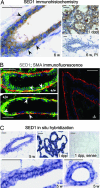
SED1 is expressed in the mammary epithelium during development. (A) SED1 immunoreactivity is seen at the earliest stages of ductal development associated with both luminal and myoepithelial cells. Immunoreactivity increases during development and reaches peak levels at ≈5–6 weeks, when both luminal and myoepithelial cells are embedded in SED1 immunoreactivity (arrowheads denote myoepithelial cells based on nuclear architecture, apposition to the basal lamina, and SMA reactivity). SED1 expression remains stable or decreases slightly until the onset of lactation, when it is abundantly expressed in the secretory acini (1 dpp). No immunoreactivity is seen with preimmune sera (PI). (B) Double-label immunofluorescence in WT glands (+/+) illustrates SED1 immunoreactivity (Alexa Fluor 488, green) associated with both luminal and myoepithelial cells, as seen by colocalization with SMA (Alexa Fluor 594, red) (arrowheads). (C) In situ hybridization confirms SED1 transcription during gland development (5 weeks) and is up-regulated during lactation (1 dpp). Sense probes produced no hybridization. The double layer of hybridization is consistent with SED1 being synthesized by both epithelial cell types. (Scale bars, 0.05 mm in all images.)
Fig. 3.
Fig. 3.
SED1-null mammary glands show abnormal cellular architecture and greatly reduced cell proliferation. (A) Myoepithelial cells in WT glands have a characteristic squamous morphology as they surround the ductal epithelium and acini (1 dpp, 3 days postweaning). However, myoepithelial cells in SED1-null glands are bulbous and discontinuous around the ductal epithelium (arrowheads). Myoepithelial cells are identified by SMA immunocytochemistry; nuclei are visualized with DAPI. (Scale bars, 0.05 mm in all images.) (B) Cell proliferation, as judged by BrdU incorporation (brown nuclei, arrowheads), is abundant in the epithelial and mesenchymal compartments of WT ducts (5 weeks shown) but is greatly reduced in SED1-null glands (P = 0.003). (Scale bars, 0.1 mm in all images.) (C) The degree of apoptosis, as assessed by TUNEL staining, is low and similar during early development (5 weeks) of both gland genotypes (red nuclei, arrowhead). No TUNEL-positive nuclei are seen in control incubations. (Scale bars, 0.05 mm in all images.)
Fig. 4.
Fig. 4.
SED1 participates in RGD-dependent adhesion required for branching morphogenesis. (A) WT organoids cultured in 3D collagen gels produce highly branched ductal trees (arrowheads, Upper) and are characterized by a multilayered epithelia invading the collagenous matrix (Lower). In contrast, organoids from age-matched SED1-null females remain viable during the same culture period but fail to extend branches and possess thin-walled epithelia. SED1 immunoreactivity is detected within the lumen (red arrowhead) of WT organoids and in the adjacent extracellular matrix (black arrowheads); no SED1 immunoreactivity is detectable in SED1-null organoids. [Scale bars, 0.1 mm for images (Upper), 0.05 mm for Lower.] (B) WT organoids adhere and grow out on 2D Matrigel substrates, whereas SED1-null cells adhere but grow out poorly, consistent with SED1 contributing to a normal adhesive phenotype. Heat-inactivated anti-SED1 antiserum (1:100) prevents outgrowth and spreading of WT cells, thus recapitulating the SED1-null phenotype. The addition of preimmune (PI) reagents had no effect on epithelial cell outgrowth. Nuclei stained with Syto24 (green). (Scale bars, 0.2 mm for all images.) (C) Recombinant SED1 supports mammary epithelial cell adhesion and is inhibited by competitive RGD containing peptides (0.5 mM), whereas RAD peptides have no effect. (D) RGD-dependent inhibition of single cell adhesion to SED1 matrices is dose-dependent, whereas RAD peptides fail to inhibit adhesion (10-min adhesion assay shown). (Error bars, SD.) (E) The presence of the 120-kDa αv integrin subunit was confirmed by immunoblotting lysates of WT organoids; nonimmune (ni) serum produced no reactivity. The antiserum localizes αv to the myoepithelial cells (arrowheads) surrounding the developing luminal cells. (F) The inclusion of anti-αv antiserum produced a dose-dependent inhibition of single-cell adhesion to SED1 matrices (10- and 20-min assays shown); ni serum had no effect. (Error bars, SD.)
Fig. 5.
Fig. 5.
The loss of SED1 is associated with reduced MAPK activation in myoepithelial cells. (A) The level of total MAPK protein is similar between WT and SED1-null organoids as assessed by immunoblotting, whereas the activated form of MAPK is greatly diminished in SED1-null organoids. Immunoblotting α-tubulin served as a protein-loading control. Densitometry analysis indicates that activation of the 44-kDa MAPK is reduced by nearly 50%, whereas activation of the 42-kDa isoform is closer to normal levels. (B) Immunocytochemistry reveals total MAPK (brown stain) in both the luminal and myoepithelial compartments in both WT and SED1-null organoids, but activated MAPK (black arrowheads) is confined primarily to the myoepithelial compartment, consistent with the expression of the αv integrin on myoepithelial cells. In contrast, MAPK activation is greatly reduced in the epithelial compartment of SED1-null organoids (P < 0.001). (Scale bars, 0.05 mm in all images.) (C) The identity of cells showing activated MAPK was assessed by overlaying images of phospho-MAPK-positive cells (immunocytochemistry) and SMA (immunofluorescence). Most cells with activated MAPK colocalize with SMA, indicative of myoepithelial cells (white arrowheads), whereas others lie adjacent to or outside of the SMA reactivity (pink arrowheads), suggesting they may be nonepithelial cells, epithelial precursors, or myoepithelial cell fragments from adjacent sections. (Scale bars, 0.05 mm in all images.) (D) The addition of rSED1 to primary epithelial cell cultures leads to a transient activation of MAPK. It is unknown why SED1 leads to activation of both MAPK isoforms in vitro, whereas the 44-kDa isoform is more sensitive to SED1 in vivo (A). (E) Hypothetical model to account for SED1 function during mammary gland morphogenesis. SED1 is secreted by myoepithelial and/or basally from luminal epithelial cells. The RGD motif within the second EGF repeat anchors SED1 to myoepithelial cells by the αvβ5/3 integrin, whereas adhesion to luminal epithelial cells is thought to be mediated by intercalation of the discoidin/C domains into the phospholipid bilayers (5, 14). Ligation of αvβ5/3 integrin induces MAPK activation in myoepithelial cells, leading to proliferation of the epithelial compartment, duct elongation, and branching.

Similar articles

Cited by

References

    1. Bissell MJ, Rizki A, Mian IS. Curr Opin Cell Biol. 2003;15:753–762. - PMC - PubMed
    1. Stubbs JD, Lekutis C, Singer KL, Bui A, Yuzuki D, Srinivasan U, Parry G. Proc Natl Acad Sci USA. 1990;87:8417–8421. - PMC - PubMed
    1. Larocca D, Peterson JA, Urrea R, Kuniyoshi J, Bistrain AM, Ceriani RL. Cancer Res. 1991;51:4994–4998. - PubMed
    1. Couto JR, Taylor MR, Godwin SG, Ceriani RL, Peterson JA. DNA Cell Biol. 1996;15:281–286. - PubMed
    1. Andersen MH, Berglund L, Rasmussen JT, Petersen TE. Biochemistry. 1997;36:5441–5446. - PubMed

Publication types

MeSH terms

LinkOut - more resources