. 2007 Feb 12;176(4):459-71.
doi: 10.1083/jcb.200606077. Epub 2007 Feb 5.

Activation of endosomal dynein motors by stepwise assembly of Rab7-RILP-p150Glued, ORP1L, and the receptor betalll spectrin

Affiliations

Activation of endosomal dynein motors by stepwise assembly of Rab7-RILP-p150Glued, ORP1L, and the receptor betalll spectrin

Marie Johansson et al. J Cell Biol. .

Abstract

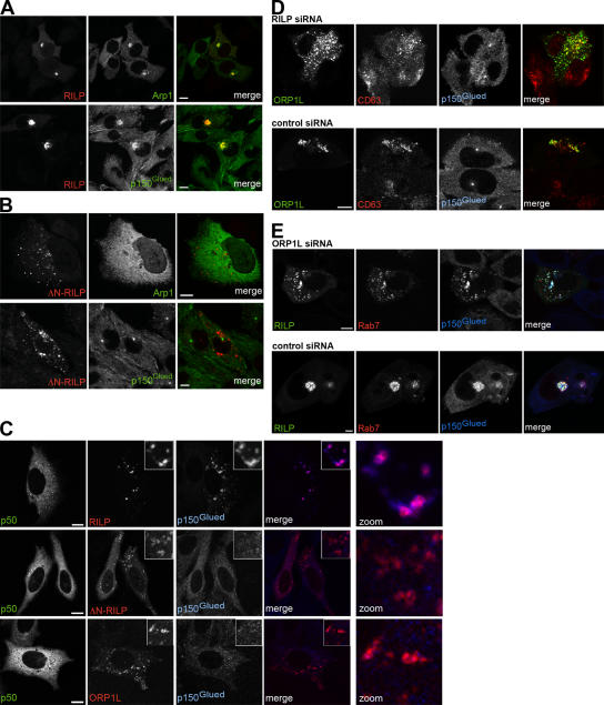
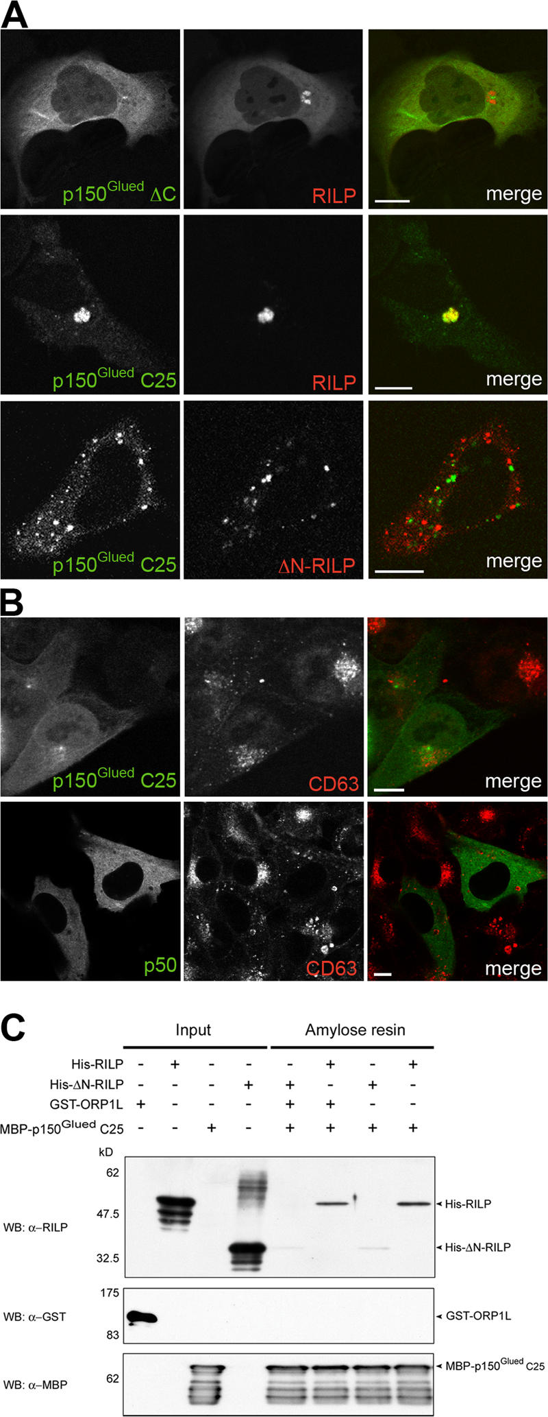
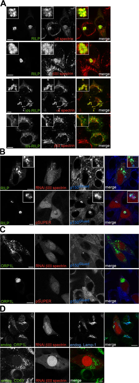
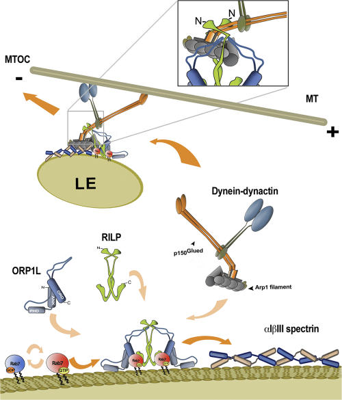
The small GTPase Rab7 controls late endocytic transport by the minus end-directed motor protein complex dynein-dynactin, but how it does this is unclear. Rab7-interacting lysosomal protein (RILP) and oxysterol-binding protein-related protein 1L (ORP1L) are two effectors of Rab7. We show that GTP-bound Rab7 simultaneously binds RILP and ORP1L to form a RILP-Rab7-ORP1L complex. RILP interacts directly with the C-terminal 25-kD region of the dynactin projecting arm p150(Glued), which is required for dynein motor recruitment to late endocytic compartments (LEs). Still, p150(Glued) recruitment by Rab7-RILP does not suffice to induce dynein-driven minus-end transport of LEs. ORP1L, as well as betaIII spectrin, which is the general receptor for dynactin on vesicles, are essential for dynein motor activity. Our results illustrate that the assembly of microtubule motors on endosomes involves a cascade of linked events. First, Rab7 recruits two effectors, RILP and ORP1L, to form a tripartite complex. Next, RILP directly binds to the p150(Glued) dynactin subunit to recruit the dynein motor. Finally, the specific dynein motor receptor Rab7-RILP is transferred by ORP1L to betaIII spectrin. Dynein will initiate translocation of late endosomes to microtubule minus ends only after interacting with betaIII spectrin, which requires the activities of Rab7-RILP and ORP1L.


Figures

Figure 1.
Figure 1.
The ORP1L, RILP, and p150Glued constructs and purified recombinant proteins. (A) Schematic representation of the constructs used in this study. The domains in ORP1L and p150Glued are indicated. RILP contains predicted coiled-coil regions only. Numbers indicate amino acid residue positions. (B) Purified fusion proteins were resolved by 10% SDS-PAGE and Coomassie stained. Positions of the molecular weight markers and the fusion proteins are indicated.
Figure 2.
Figure 2.
ORP1L and RILP colocalize and are part of a physical complex. (A) ORP1L and RILP colocalize on juxtanuclear late endocytic clusters. HeLa cells were transfected with GFP–RILP or GFP–ΔN-RILP, together with Xpress-tagged ORP1L or Xpress-tagged ORP1L–ANK domain (red), as indicated in the images. ANK and ORP1L colocalize with RILP or ΔN-RILP (right, merge). Bars, 10 μm. (B) ORP1L co-isolates with expressed RILP. HeLa cells were transfected with Xpress-tagged RILP and immunoprecipitated with anti-Xpress antibody (α-Xpress) or irrelevant mouse IgG (MIgG). The isolates were Western blotted with anti-ORP1L antibodies (WB: α-ORP1L). (bottom) Corresponding lanes probed with anti-Xpress antibody (WB: α-Xpress). (C) Coimmunoprecipitation of endogenous RILP and ORP1L. HeLa cell lysates were immunoprecipitated with rabbit anti-RILP serum (α-RILP) or irrelevant rabbit IgG (RIgG). The isolates were Western blotted with anti-ORP1L antibodies (WB: α-ORP1L).
Figure 3.
Figure 3.
FRET between ORP1L, Rab7, and RILP. (A) HeLa cells were transfected with GFP–RILP or GFP–ORP1L and cotransfected with mRFP–Rab7 or mRFP–RILP, as indicated. The transfected HeLa cells were cocultured with Mel JuSo cells stably expressing H2B–GFP (indicated by *) as an internal marker with a lifetime of 2.6 ns. (left) Wide-field image of the transfected and internal control cells. (right) FLIM image of the same cells, in which the fluorescence lifetime is depicted in false colors. The color scale with the respective lifetimes (in nanoseconds) is indicated. The fluorescence lifetime of GFP–RILP or GFP–ORP1L was determined on vesicles immobile during data acquisition (∼12 s). Bar, 10 μm. (B) The donor FRET efficiencies (ED) between the GFP- and mRFP-tagged proteins were determined and plotted in the bar diagram. Mean ± the SD. n > 20.
Figure 4.
Figure 4.
The ORP1L–Rab7–RILP heterotrimeric complex. (A) Proteins were pulled down from HeLa cell lysates with GST–ORP1L or GST and Western blotted with anti-RILP antiserum (WB: α-RILP). Pull-down assays were performed with purified proteins. (B) His6–RILP or His6–Rab7Q67L were immobilized on a matrix and used to pull down GST–ORP1L. Isolates were Western blotted with anti-ORP1L antibodies (WB: α-ORP1L). (C) GST–ORP1L ANK fragment (GST–ANK) was immobilized and incubated with His6–RILP, GTP-loaded His6–Rab7Q67L, or a mixture containing both His6–RILP and GTP-loaded His6– Rab7Q67L. The material pulled down by the ANK fragment was Western blotted with antibodies against Rab7 or RILP (indicated on the right). Duplicate samples are shown for each condition. (D) RILP enhances ORP1L binding to Rab7. GST–Rab7 was immobilized on a matrix, loaded with GTP, and incubated with in vitro–translated 35S-labeled ORP1L in the presence of increasing amounts of His6–RILP. 35S-labeled ORP1L protein pulled down by GST–Rab7 was visualized by SDS-PAGE and autoradiography (I). Similar pull-down experiments were performed with in vitro–translated 35S-labeled ORP1L ANK + PHD (II) or 35S-labeled ORP1L ΔORD fragments (III). Pull-down of full-length 35S-labeled ORP1L by active GST–Rab7 in the presence of increasing amounts of His6–ΔN-RILP (indicated above the lanes) (IV). A reciprocal pull-down experiment in which in vitro–translated 35S-labeled RILP was incubated with immobilized His6–Rab7–GTP in the presence of increasing amounts of GST–ORP1L (indicated above the lanes; V). (E) Quantification of the radioactive bands in D by phosphorimaging. The plotted data shows the relative increase in binding of in vitro–translated proteins to immobilized Rab7–GTP in the presence of a step-gradient of effector proteins. Binding of ORP1L to Rab7 was enhanced up to fourfold in the presence of RILP. The plotted data represents the mean of three independent experiments ± the SD (error bars).
Figure 5.
Figure 5.
Role of RILP and ORP1L in the recruitment of the p150Glued subunit of the dynein–dynactin motor. (A) HeLa cells were transfected with myc-tagged RILP (red), and the location of GFP–Arp1 and endogenous p150Glued (green) was determined. (B) HeLa cells were transfected with VSV-tagged ΔN-RILP (red), and the location of GFP–Arp1 or endogenous p150Glued (green) was determined. (C) HeLa cells were transfected with p50dynamitin (green) and HA-tagged RILP, ΔN-RILP, or ORP1L (red), and subsequently stained for endogenous p150Glued (blue), as indicated. The merge and the magnified merge (zoom) images only show the stainings in the red and blue images. (D) HeLa cells were transfected with GFP–ORP1L (green) and RILP siRNA or scrambled siRNA, as indicated. At 72 h after transfection, cells were fixed and stained for endogenous CD63 (red) and p150Glued (blue). The merge shows only the ORP1L and CD63 labeling. (E) HeLa cells were transfected with GFP–RILP (green), mRFP–Rab7 (red), and ORP1L siRNA or a scrambled siRNA, as indicated, and fixed at 48 h after transfection. Cells were stained for endogenous p150Glued (blue). Bars, 10 μm.
Figure 6.
Figure 6.
RILP interacts with the C-terminal 25-kD fragment of p150Glued. (A) HeLa cells were transfected with mRFP–RILP or mRFP-tagged ΔN-RILP, along with GFP-tagged ΔC and C25 fragments of p150Glued. (B) HeLa cells were transfected with GFP–p150Glued C25 or GFP–p50dynamitin, fixed, and stained with anti-CD63 antibodies. (C) Purified GTPγS-loaded Rab7Q67L coupled to GST-beads was incubated with combinations of purified RILP, ΔN-RILP, ORP1L, or p150Glued C25 (aa 1,049–1,278), as indicated above the gels. Proteins bound in a GTP-dependent manner were eluted with EDTA and the C-terminal MBP–p150Glued fragments (C25) were captured on amylose resin. The input and resin-bound proteins were Western blotted with anti-RILP, -GST, or -MBP antibodies. Bars, 10 μm.
Figure 7.
Figure 7.
αlβlll spectrin localizes to LEs for ORP1L-mediated RILP–p150Glued–driven minus-end transport. (A) Mel JuSo cells were transfected with GFP–RILP or GFP–ΔN-RILP for 48 h before fixation and staining for αI spectrin or βIII spectrin. αI and βIII spectrin (red) are located on RILP- and ΔN-RILP–labeled vesicles, as well as other subcellular structures. Note that βIII spectrin labeling localizes differently within the GFP–RILP– or GFP–ΔN-RILP–labeled vesicles. (insets) Magnification of vesicles showing the localization of RILP or ΔN-RILP and αI spectrin or βIII spectrin. (B) HeLa cells were transfected with GFP–RILP and pSUPER vector coexpressing mRFP and a control or βIII spectrin shRNA. Cells were fixed at 72 h after transfection and stained for endogenous p150Glued (blue). (C) HeLa cells were transfected with GFP–ORP1L and pSUPER vector coexpressing mRFP and a control or βIII spectrin shRNA. At 72 h after transfection, cells were fixed and stained for endogenous p150Glued (blue). (D) Endogenous ORP1L and spectrin. HeLa cells were transfected with a βIII spectrin shRNA pSUPER construct coexpressing mRFP. At 72 h after transfection, cells were fixed and stained for endogenous ORP1L (green) or endogenous CD63 (green) and the late endocytic marker Lamp-1 (blue). Bars, 10 μm.
Figure 8.
Figure 8.
Model for function of ORP1L in the translocation of the Rab7–RILP–p150Glued–dynein motor complex to βIII spectrin for minus-end transport. Active GTP-loaded Rab7 localizes to membranes of LEs and recruits the effectors RILP and ORP1L to form a dimer of a heterotrimeric complex. The C-terminal domain of RILP interacts with the switch and interswitch (RabSF1 and RabSF4) regions of Rab7, and the N-terminal half of RILP binds to the C-terminal domain of p150Glued (shown in detail in the box). Subsequently, the 2.4-MD dynein–dynactin motor is recruited by the 0.35-MD ORP1L–Rab7–RILP complex on LEs. ORP1L is required to direct the entire complex to the 0.6-MD αIβIII spectrin. The interaction is mediated by Arp1 binding to βIII spectrin. αIβIII spectrin localizes to several different compartments, but it is not sufficient on its own to recruit dynein motors. Microtubule-based, dynein motor–driven minus-end transport of LEs can only occur after the specific late endocytic dynein receptor Rab7–RILP has interacted with the general dynein membrane receptor βIII spectrin, a process that is facilitated by ORP1L.

Similar articles

Cited by

References

    1. Ahmad, F.J., C.J. Echeverri, R.B. Vallee, and P.W. Baas. 1998. Cytoplasmic dynein and dynactin are required for the transport of microtubules into the axon. J. Cell Biol. 140:391–401. - PMC - PubMed
    1. Bastiaens, P.I., and A. Squire. 1999. Fluorescence lifetime imaging microscopy: spatial resolution of biochemical processes in the cell. Trends Cell Biol. 9:48–52. - PubMed
    1. Berger, B., D.B. Wilson, E. Wolf, T. Tonchev, M. Milla, and P.S. Kim. 1995. Predicting coiled coils by use of pairwise residue correlations. Proc. Natl. Acad. Sci. USA. 92:8259–8263. - PMC - PubMed
    1. Bergink, S., F.A. Salomons, D. Hoogstraten, T.A. Groothuis, H. de Waard, J. Wu, L. Yuan, E. Citterio, A.B. Houtsmuller, J. Neefjes, et al. 2006. DNA damage triggers nucleotide excision repair-dependent monoubiquitylation of histone H2A. Genes Dev. 20:1343–1352. - PMC - PubMed
    1. Burkhardt, J.K., C.J. Echeverri, T. Nilsson, and R.B. Vallee. 1997. Overexpression of the dynamitin (p50) subunit of the dynactin complex disrupts dynein-dependent maintenance of membrane organelle distribution. J. Cell Biol. 139:469–484. - PMC - PubMed

Publication types

MeSH terms